Horticulture & Landscape Architecture
Biochemistry
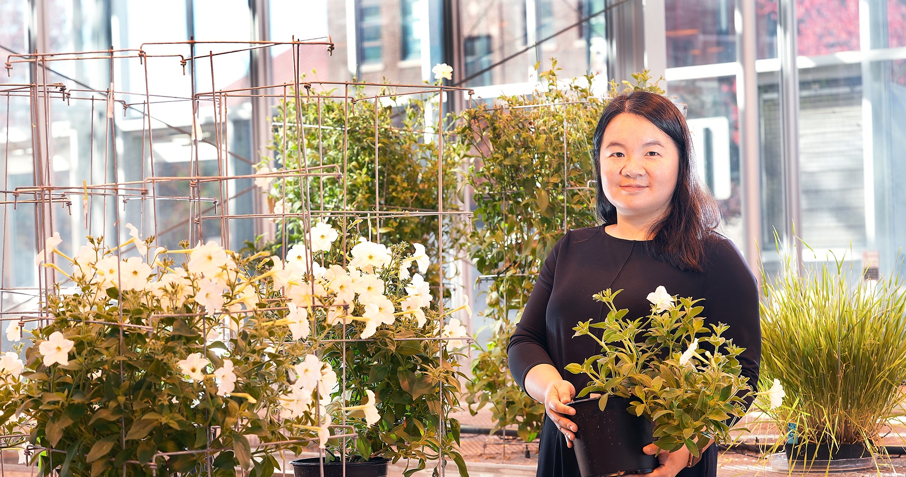
Decoding the plant world’s complex biochemical communication networks
A Purdue University-led research team has begun translating the complex molecular language of petunias. Their grammar and vocabulary are well...