

cantaloupe diseases
Alternaria Leaf Blight
Alternaria leaf blight-Alternaria leaf blight has mostly round lesions with a bulls-eye or concentric ring structure, as do many Alternaria diseases on other crops. Fortunately, lesions do not occur on stems or fruit. However, in severe cases sufficient defoliation can occur to cause loss of fruit of quality either by lower soluble sugars and/or sunscald on fruit. Alternaria leaf blight doesn’t seem to be an important problem most years. Fungicide schedules designed to control gummy stem blight or anthracnose seem to also control Alternaria leaf blight.
 Figure 1. Alternaria leaf blight on cantaloupe leaf. Note large, mostly circular lesions with concentric rings.
 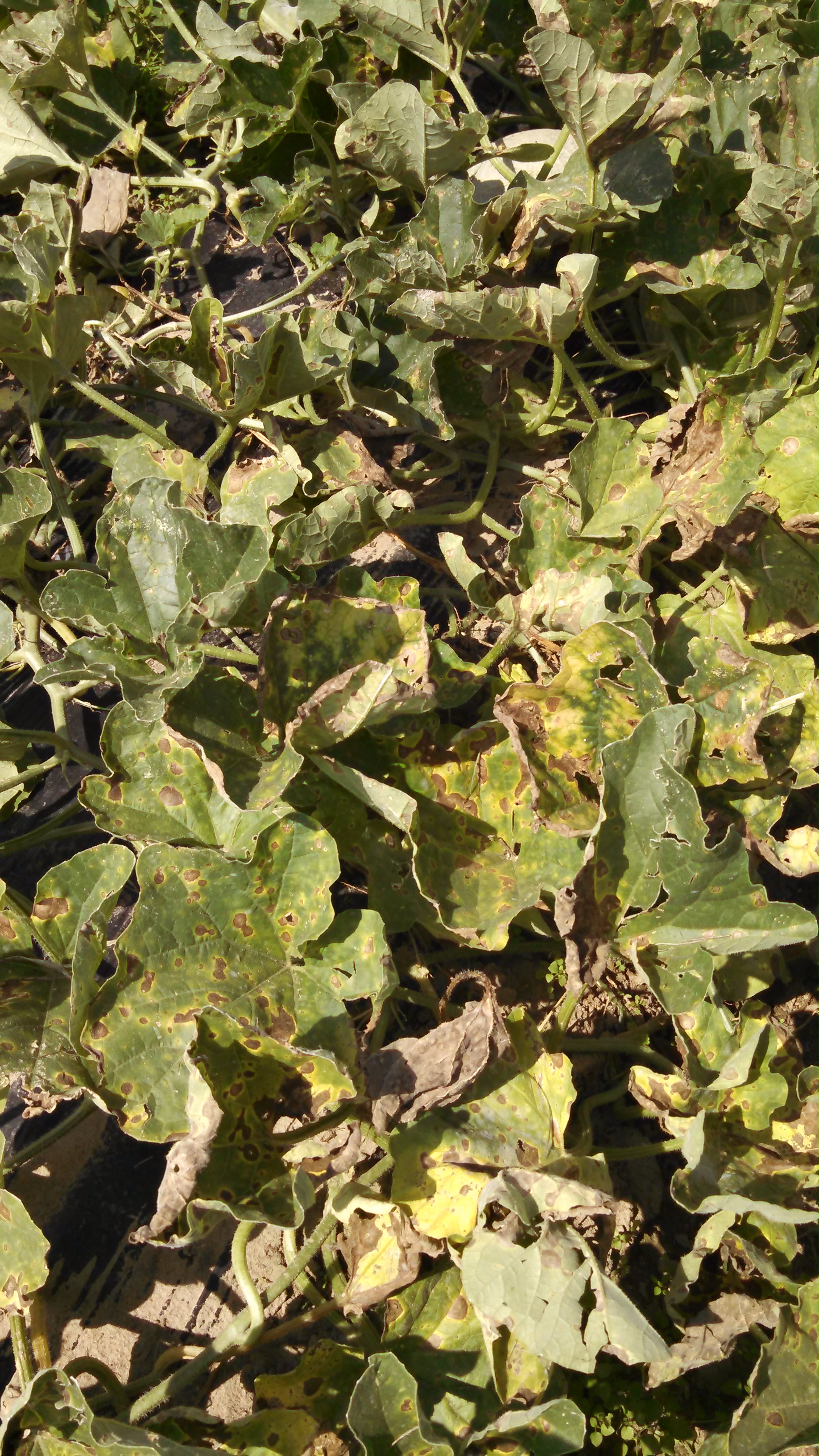 Figure 2. Image of several cantaloupe leaves with Alternaria leaf blight lesions.
 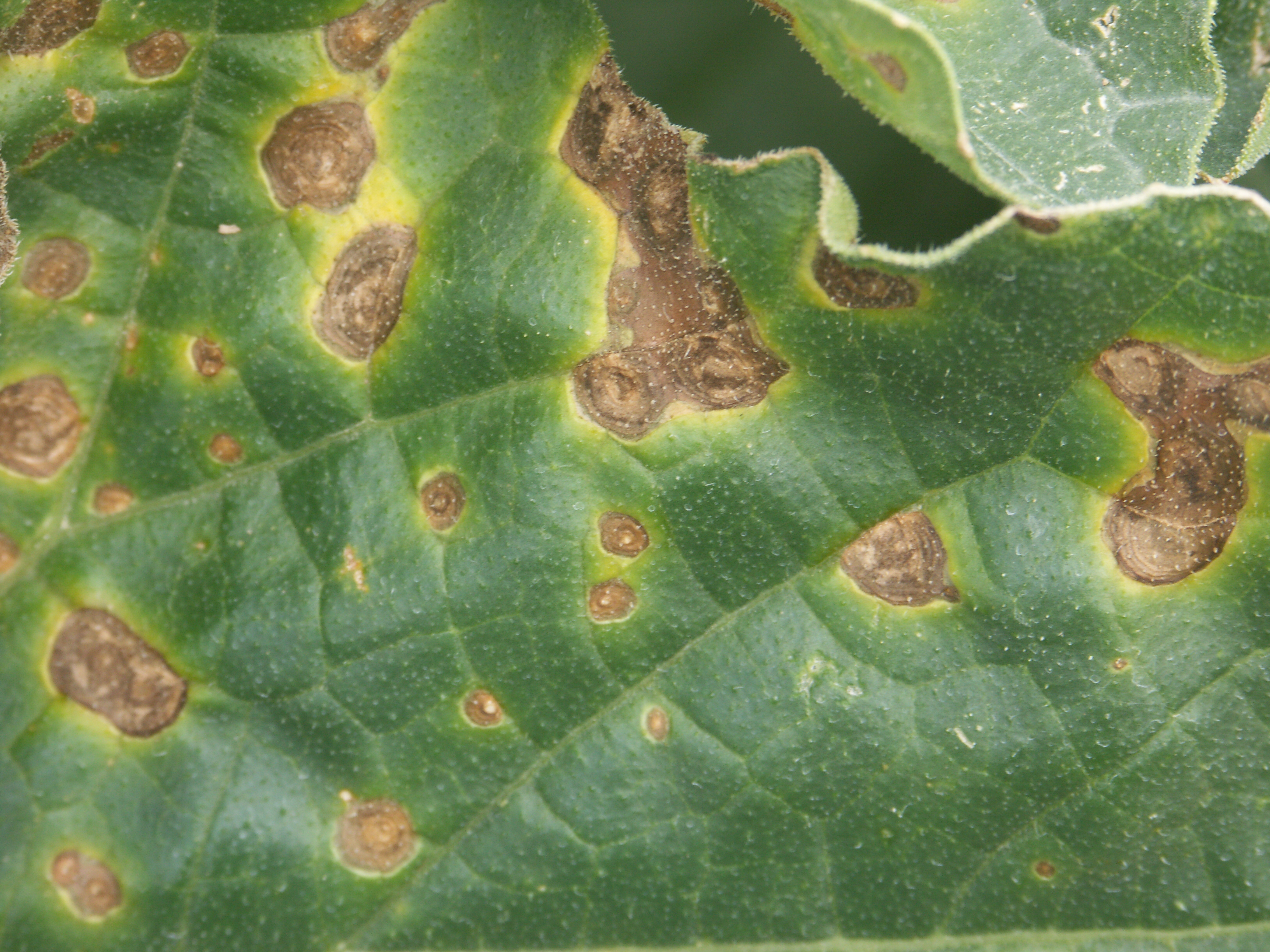 Figure 3. Close-up of a leaf with Alternaria leaf blight. Note concentric ring structure of lesions.
 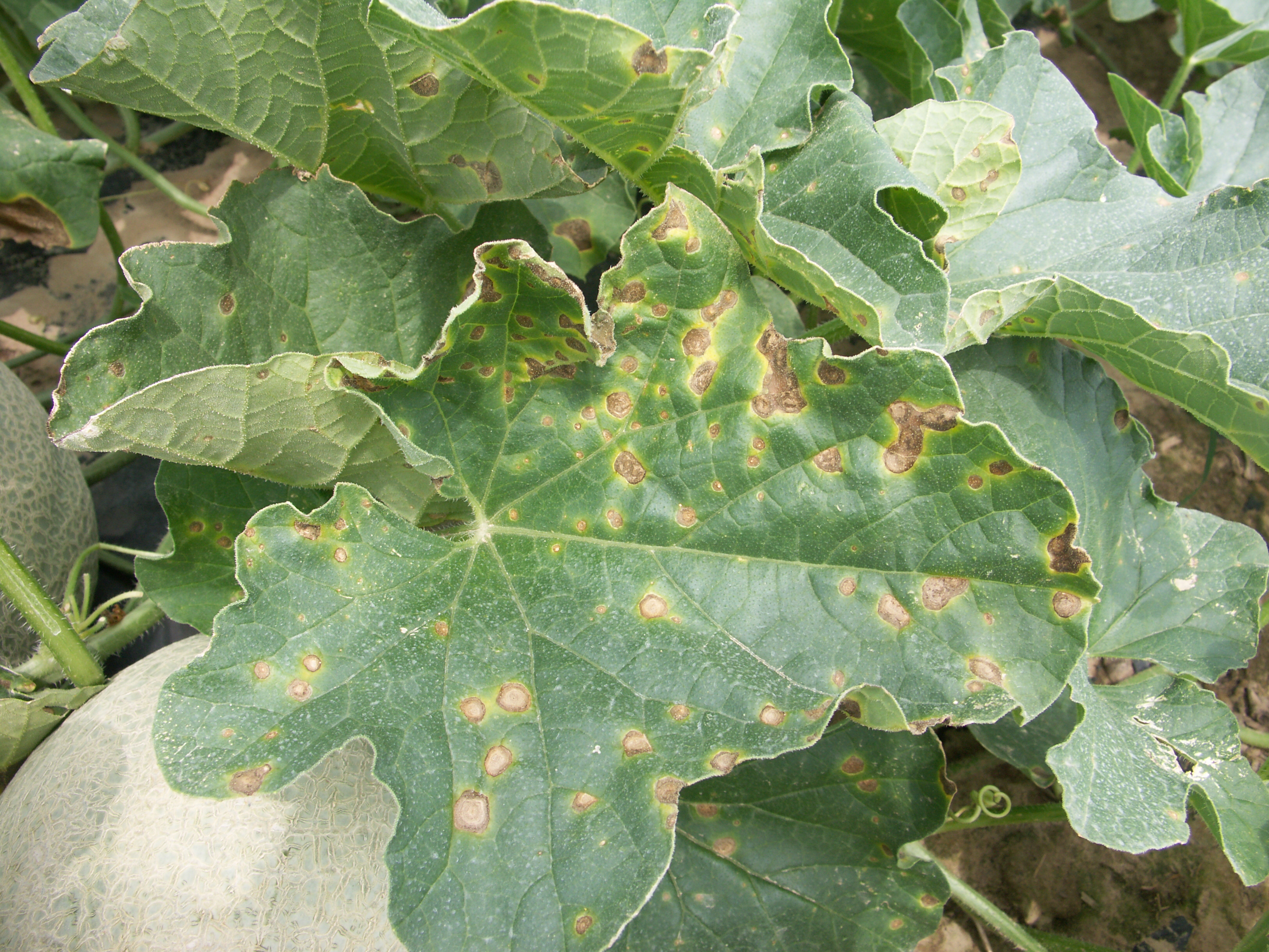 Figure 4. Image of a leaf with lesions of Alternaria leaf blight just beginning to coalesce. Note chlorotic halos.
 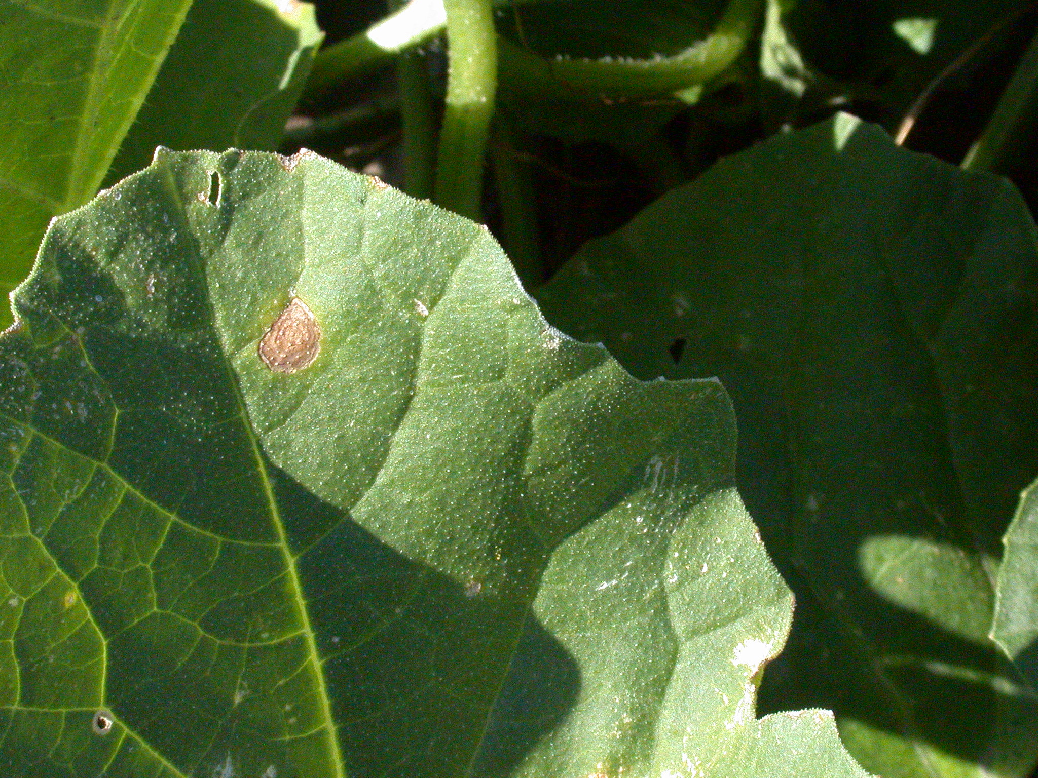 Figure 5. A close-up of an individual lesion of Alternaria leaf blight.
 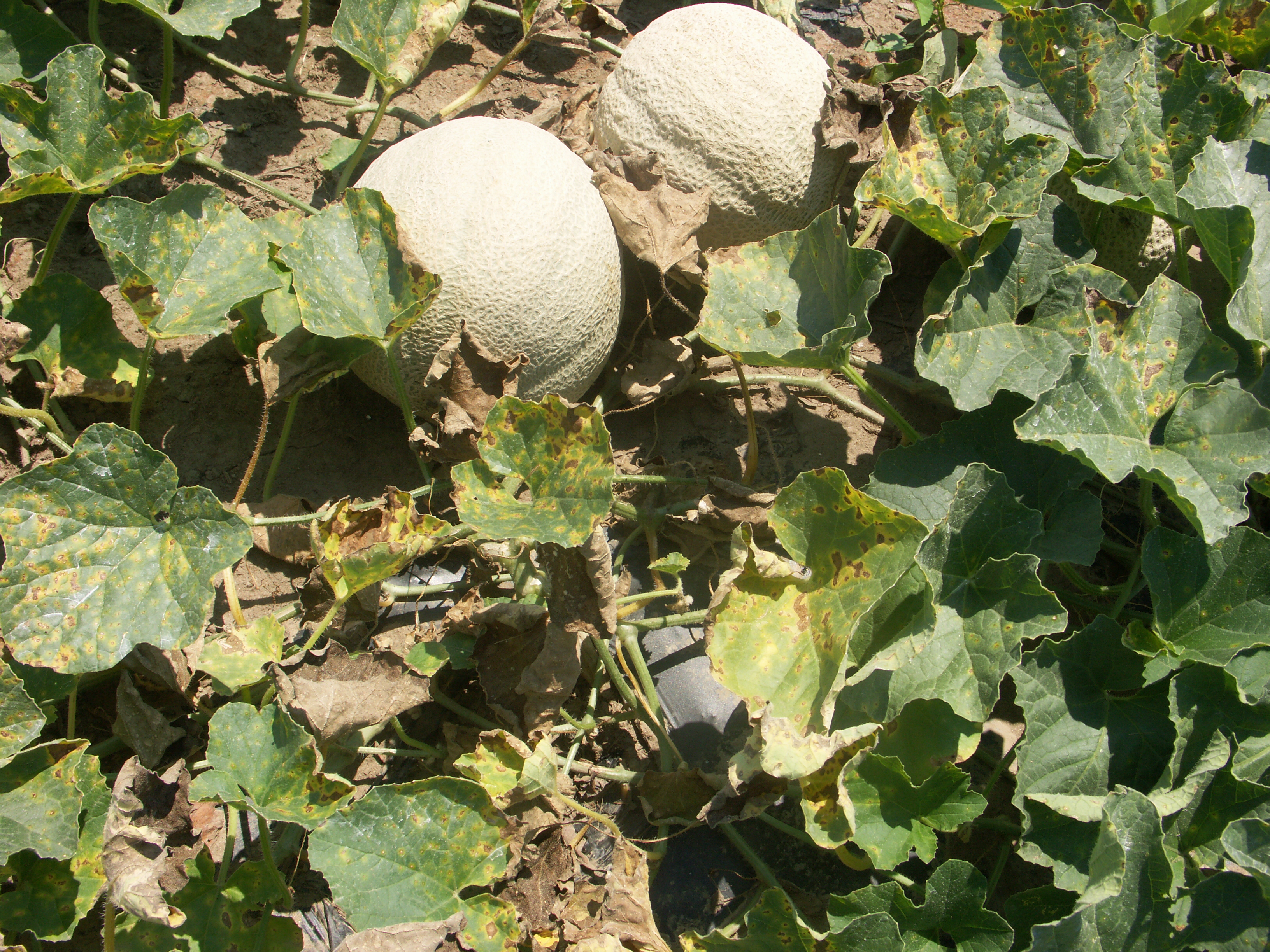 Figure 6. Cantaloupe fruit exposed to possible sunburn due to Alternaria leaf blight.
 Angular leaf spot
Angular leaf spot-Lesions of angular leaf spot may be irregular in shape and water-soaked. Lesions may be present on the margin of the leaf, perhaps due to the presence of hydathodes which may provide a mode of entry. Chlorosis may be minor. Angular leaf spot prefers cool temperatures. This is mostly a disease of transplant facilities: I rarely observe this disease in the field. Angular leaf spot is not usually an economic problem; however, it may be confused with bacterial fruit blotch.
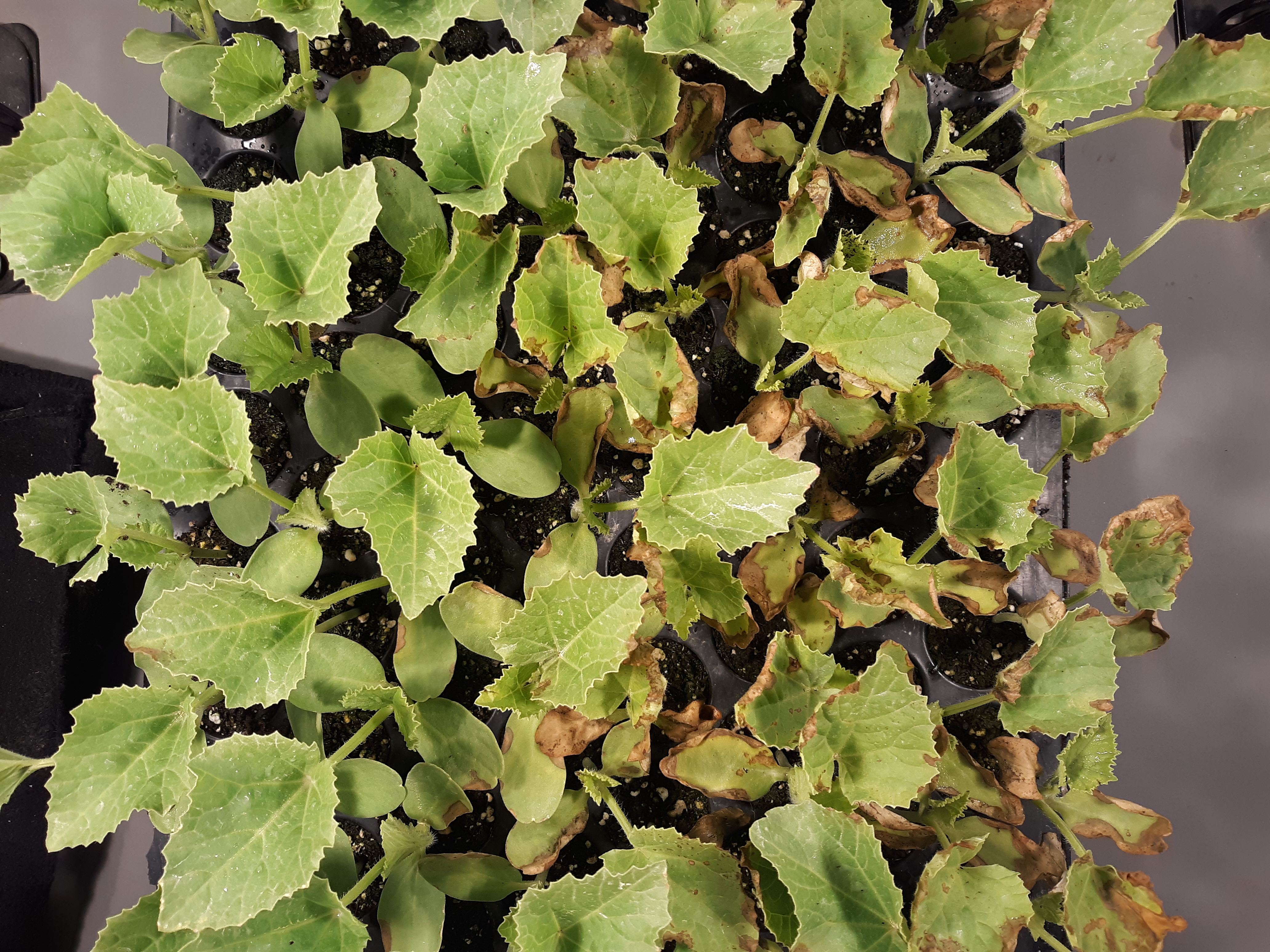 Figure 1. Angular leaf spot of cantaloupe. Note necrotic lesions, primarily marginal, on a large proportion of the seedlings.
 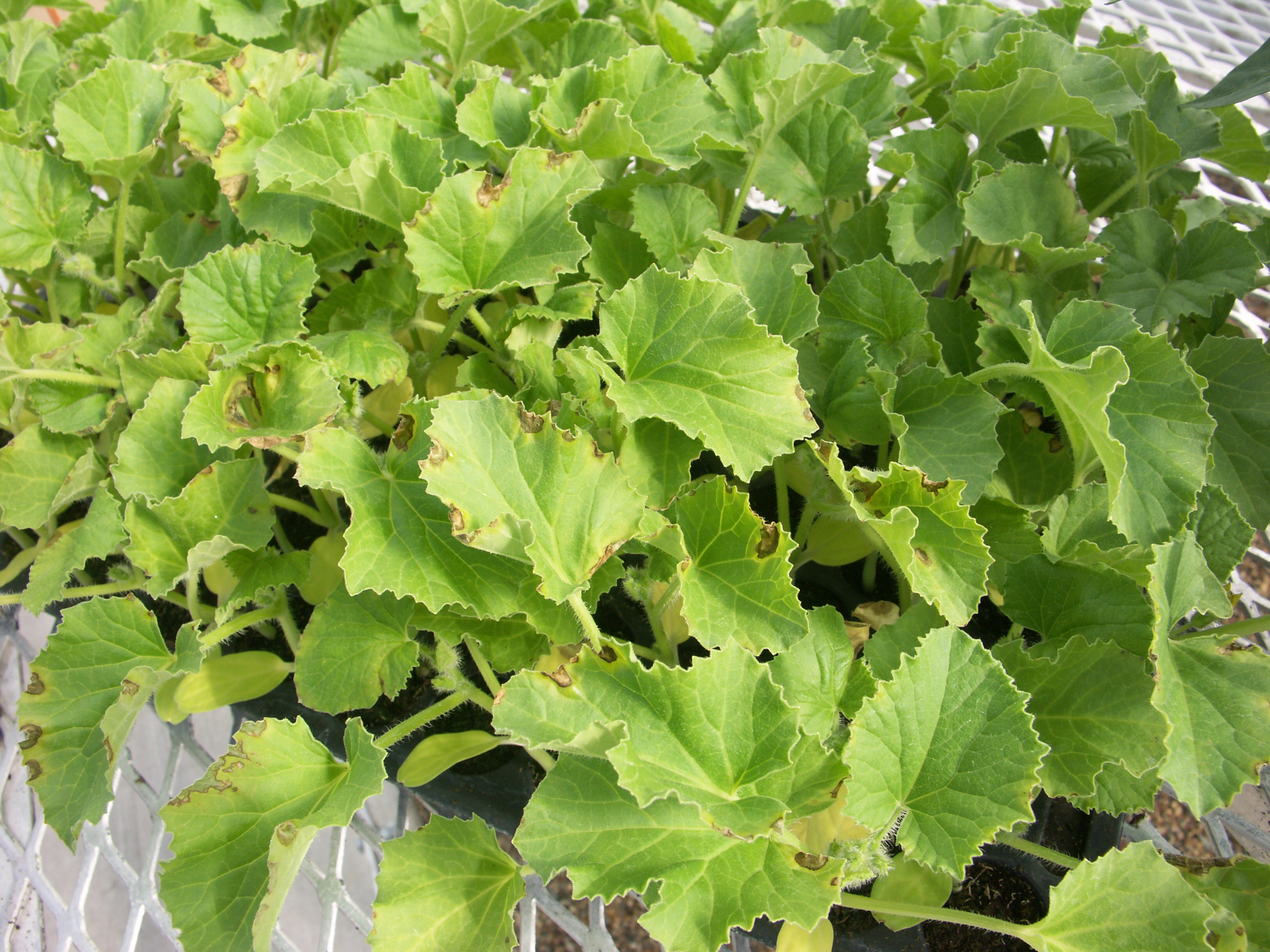 Figure 2. Angular leaf spot of cantaloupe. Note water-soaked necrotic lesions, primarily marginal.
 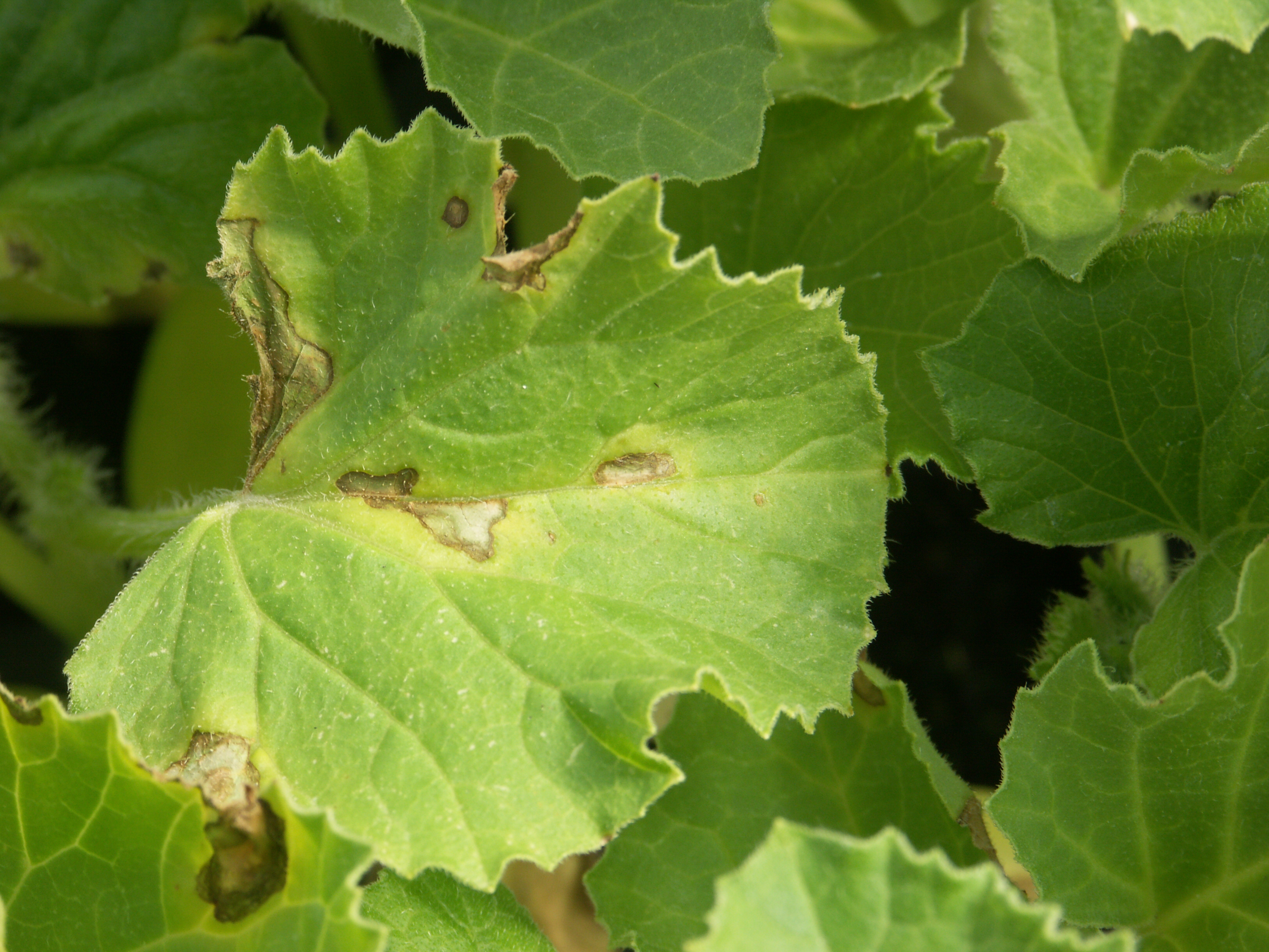 Figure 3. Angular leaf spot of cantaloupe. Note irregular, necrotic lesions.
 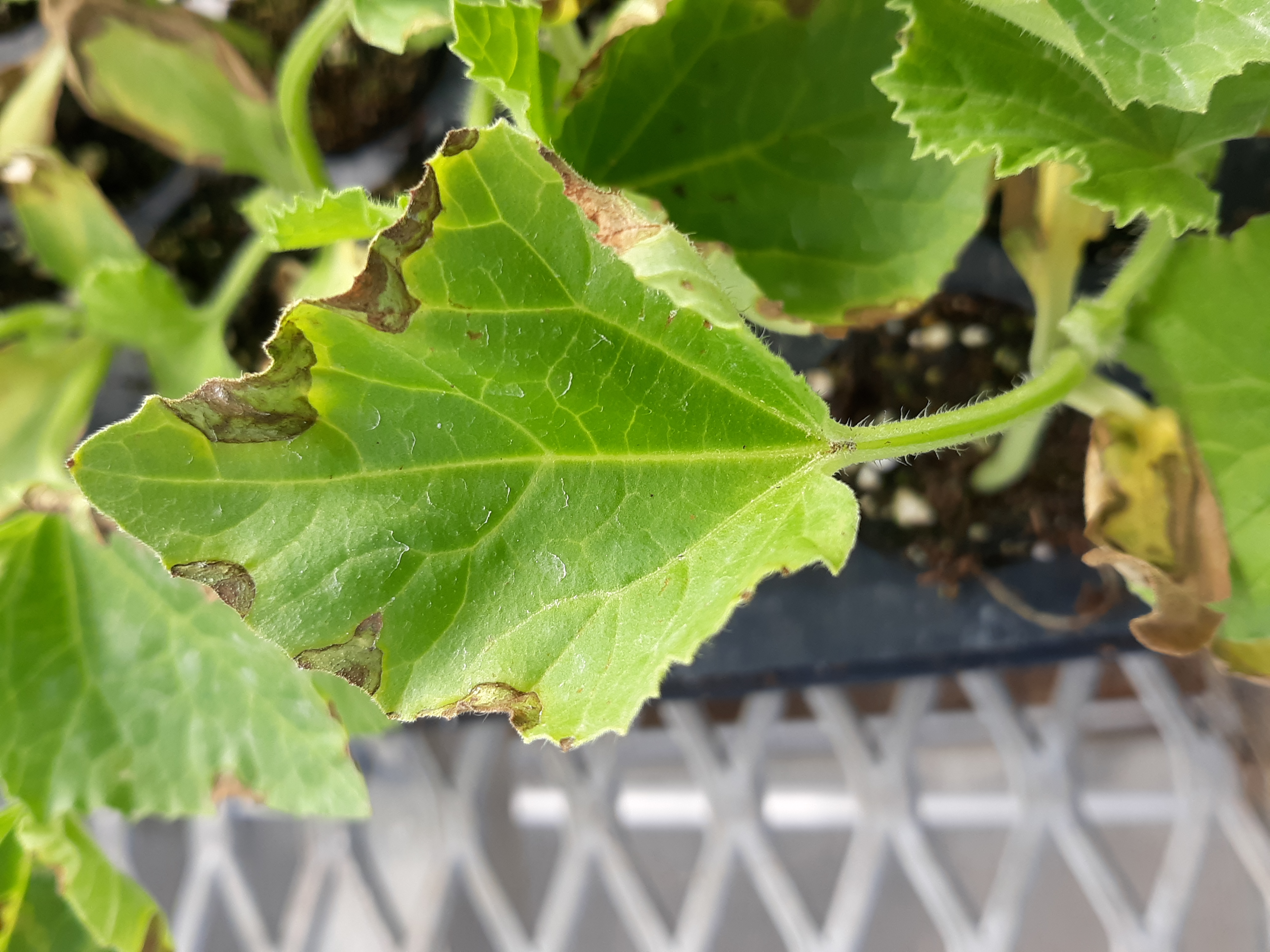 Figure 4. Angular leaf spot of cantaloupe.
 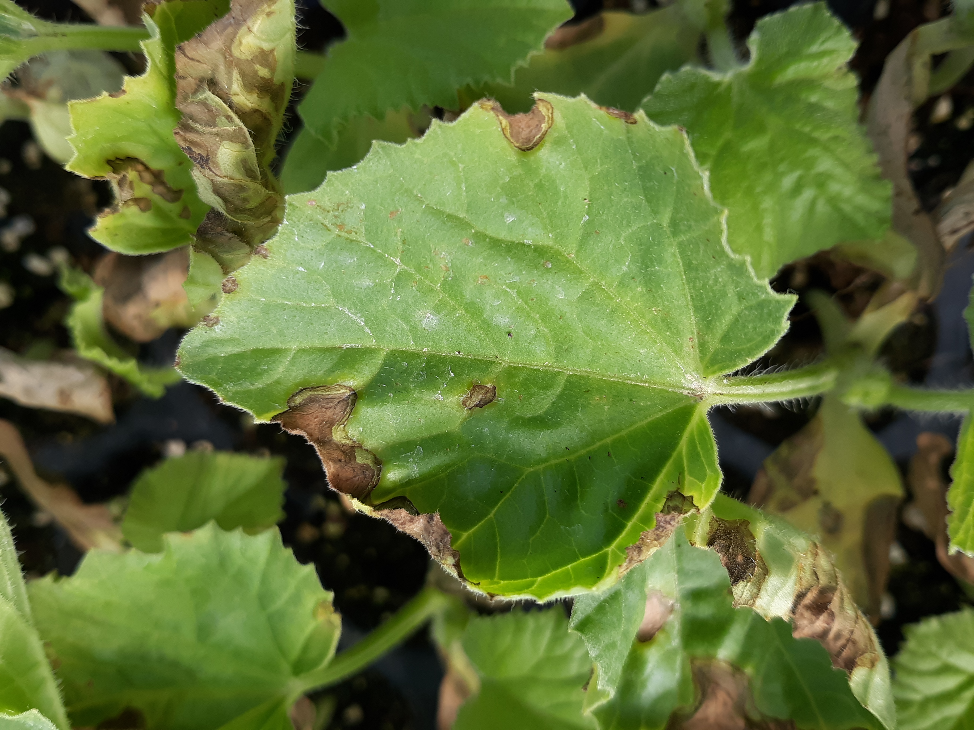 Figure 5. Angular leaf spot of cantaloupe. Note marginal lesions, perhaps infecting hydathodes.
 Anthracnose
Anthracnose of cantaloupe-The lesions of anthracnose of cantaloupe on leaves appear generally round with zig-zag margins. However, the lesions do not usually appear as jagged as those of anthracnose of watermelon. I don’t observe as many cases of anthracnose of cantaloupe as I do on watermelon. This may simply be due to the larger acreage of watermelon in Indiana. However, it is important to remember that anthracnose of cantaloupe is usually caused by race 2 of Colletotrichum orbiculare, the same race that affects cucumber. Anthracnose on watermelon is caused by race 1.
 Figure 1. Anthracnose lesions on cantaloupe fruit appear sunken while lesions on nearby leaves appear jagged.
  Figure 2. Anthracnose lesions on cantaloupe fruit.
 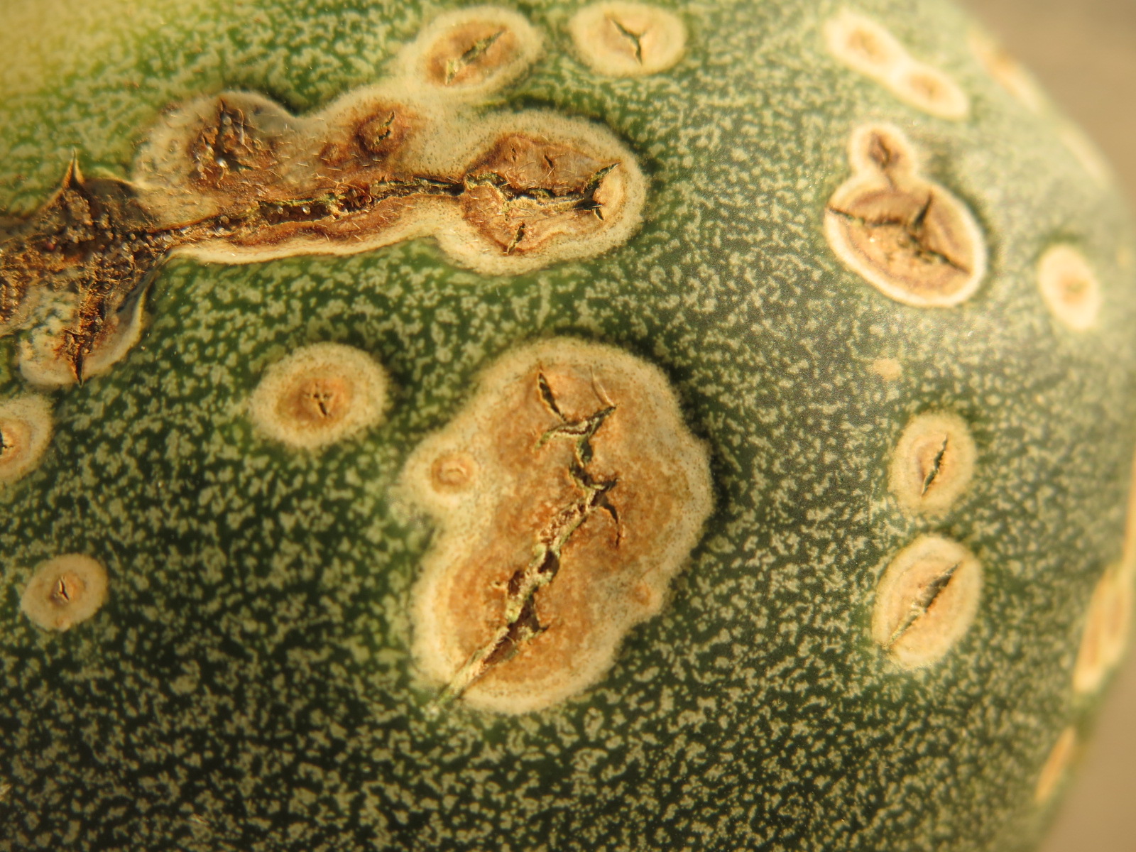 Figure 3. Advanced lesions of anthracnose on cantaloupe fruit. Note cracked appearance.
 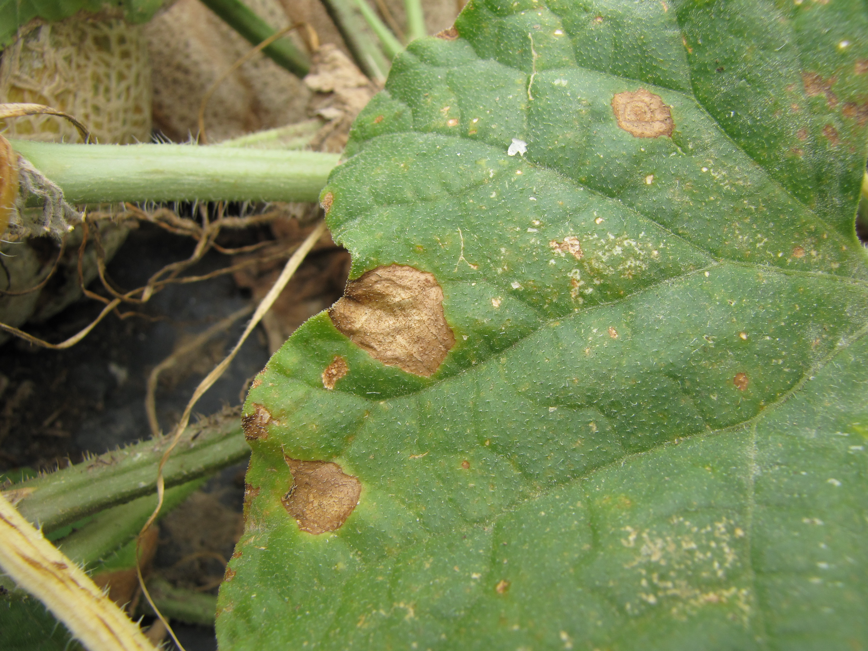 Figure 4. Close-up of anthracnose lesions on cantaloupe leaves.
 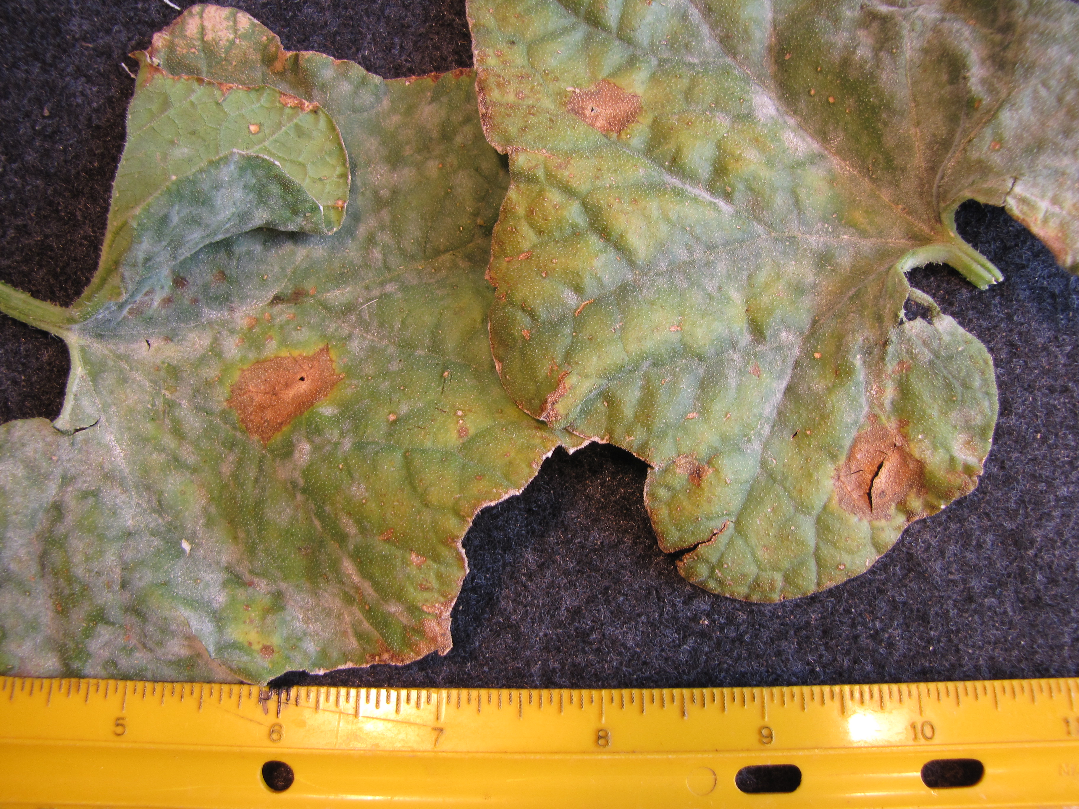 Figure 5. Lesions of anthracnose on cantaloupe leaves.
 Bacterial Wilt
Bacterial wilt of cantaloupe-The complex biology of bacterial wilt makes managing this pest difficult. Bacterial wilt is caused by a bacterium vectored by the striped or spotted cucumber beetle. Bacterial wilt is quite common every year and can be recognized by the wilt and decline of affected plants.
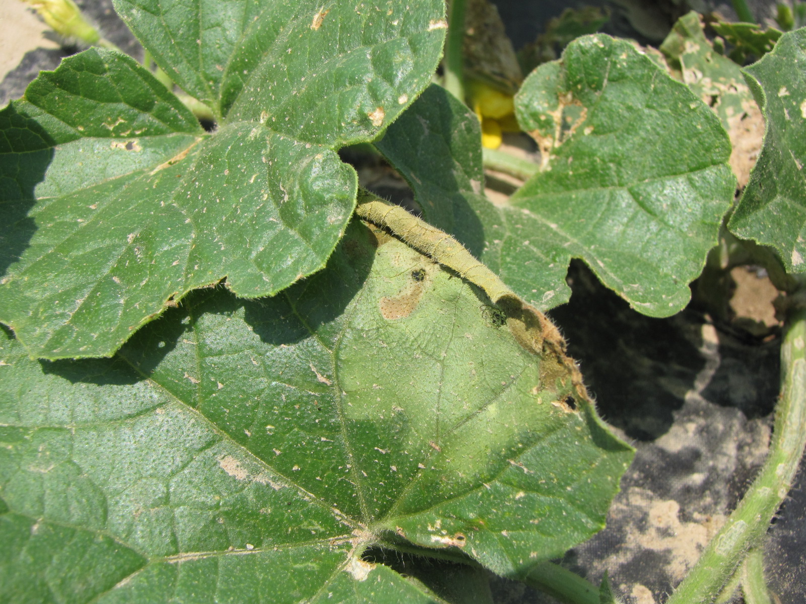 Figure 1. The wilted and collapsed area on the margin of this leaf is due to bacterial wilt of cantaloupe. Note also the areas of the leaf eaten by cucumber beetles.
 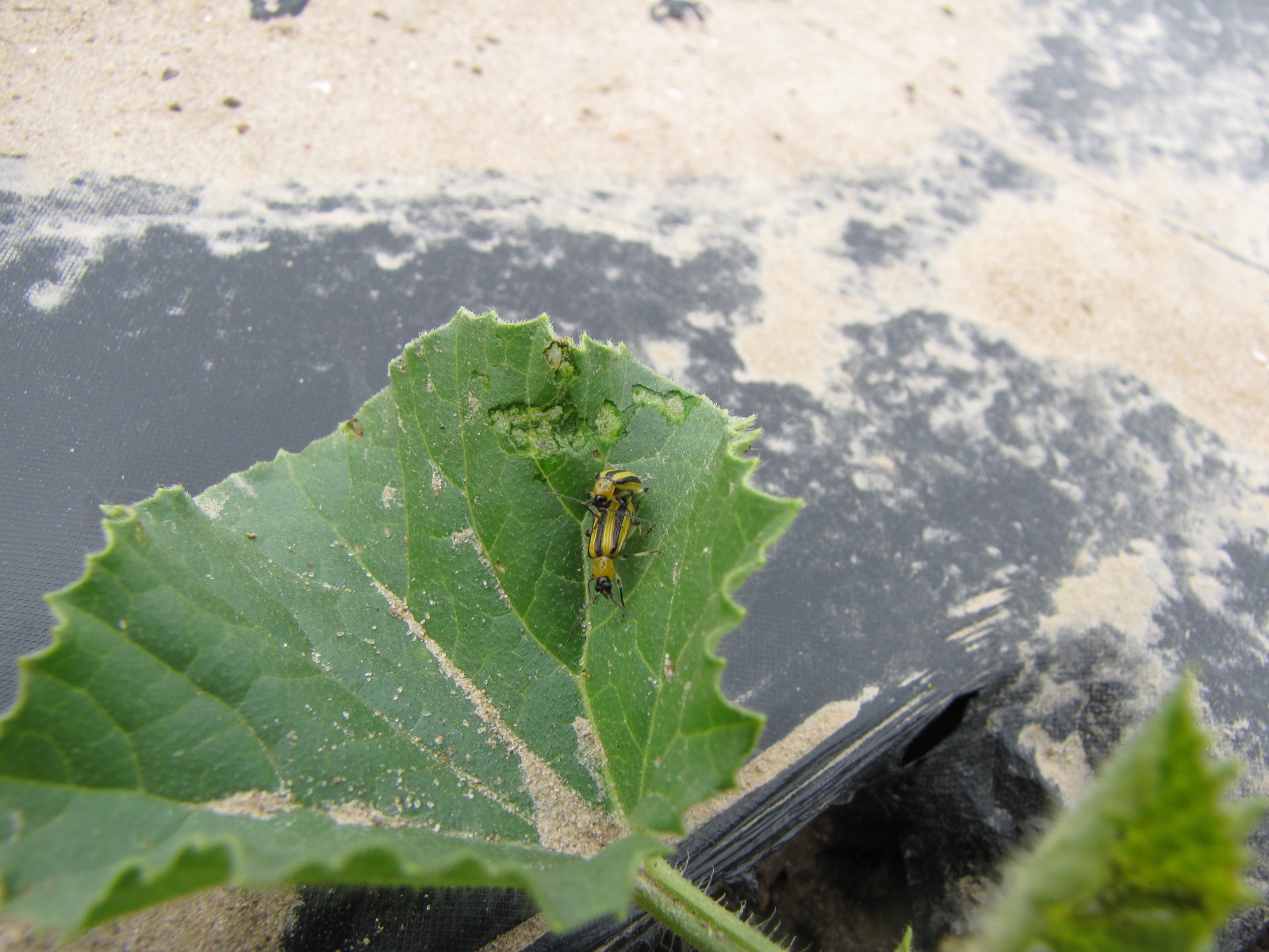 Figure 2. Cucumber beetle feeding can be observed on this cantaloupe leaf. If insect frass enters the area that has been fed upon, bacterial wilt may result.
 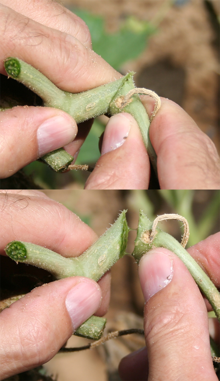 Figure 3. Quick field test for possible bacterial wilt. Stringy, viscous sap may indicate presence of Erwinia tracheiphila, causal pathogen of bacterial wilt.
 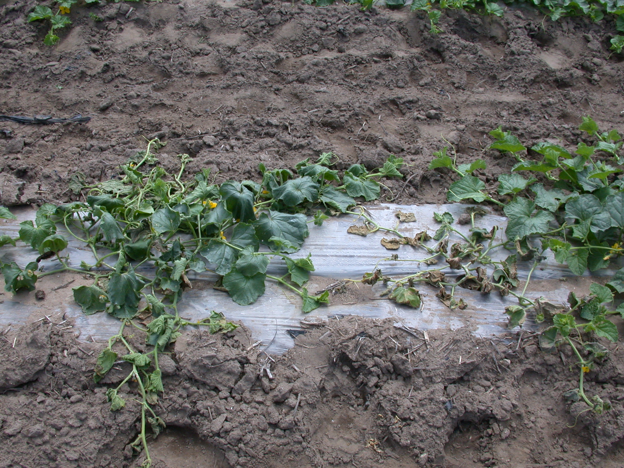 Figure 4. General wilt of cantaloupe caused by bacterial wilt.
 Fusarium fruit rot
Fusarium fruit rot-This disease was more common when cantaloupe varieties with deep sutures were frequently grown. The disease often starts with a soft area at the terminus of a suture. A white mold will often be observed in this area.
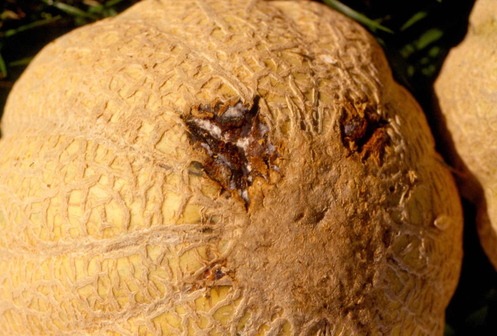 Figure 1. Fusarium fruit rot of cantaloupe.
 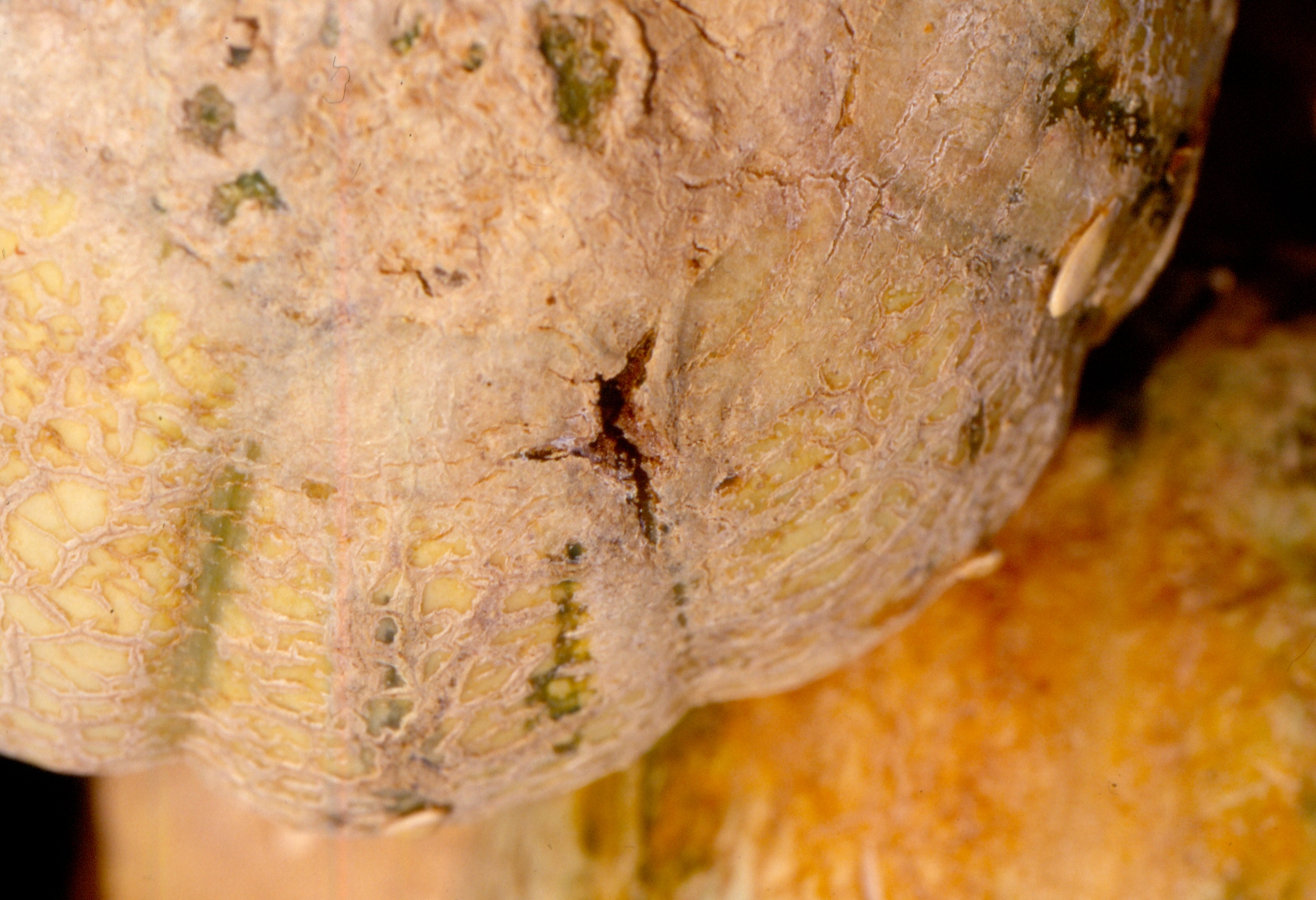 Figure 2. Fusarium fruit rot of cantaloupe. Note lesion has started in suture area. Varieties with deep sutures seem relatively more susceptible to this disease.
 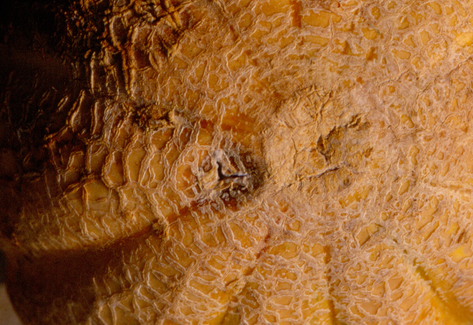 Figure 3. Fusarium fruit rot of cantaloupe.
 Gummy stem blight
Gummy stem blight of cantaloupe-Lesions on leaves tend to appear drier than gummy stem blight lesions on watermelon leaves. Occasionally, lesions occur on fruit, in which case the disease name is black rot. Gummy stem blight has also been observed as an important disease in transplant houses. Management of gummy stem blight depends, in part, on a good knowledge of fungicide insensitivity among isolates of gummy stem blight in Indiana.
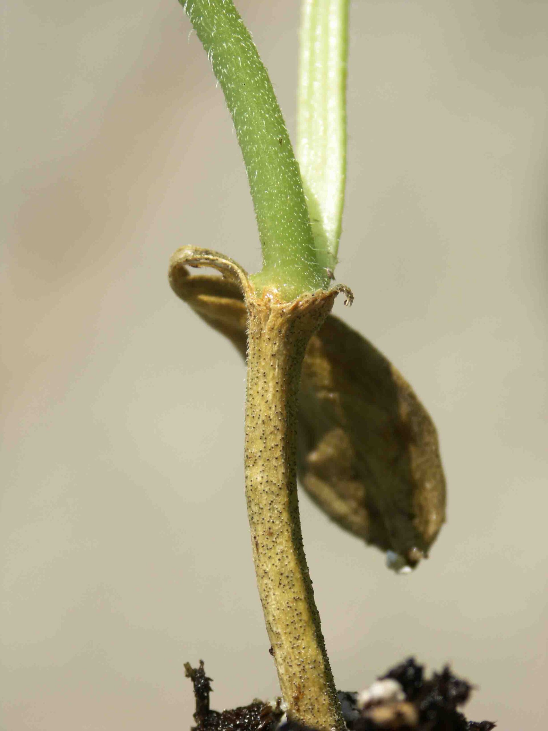 Figure 1. Gummy stem blight lesion on the hypocotyl of a cantaloupe transplant. Note presence of dark pycnidia.
 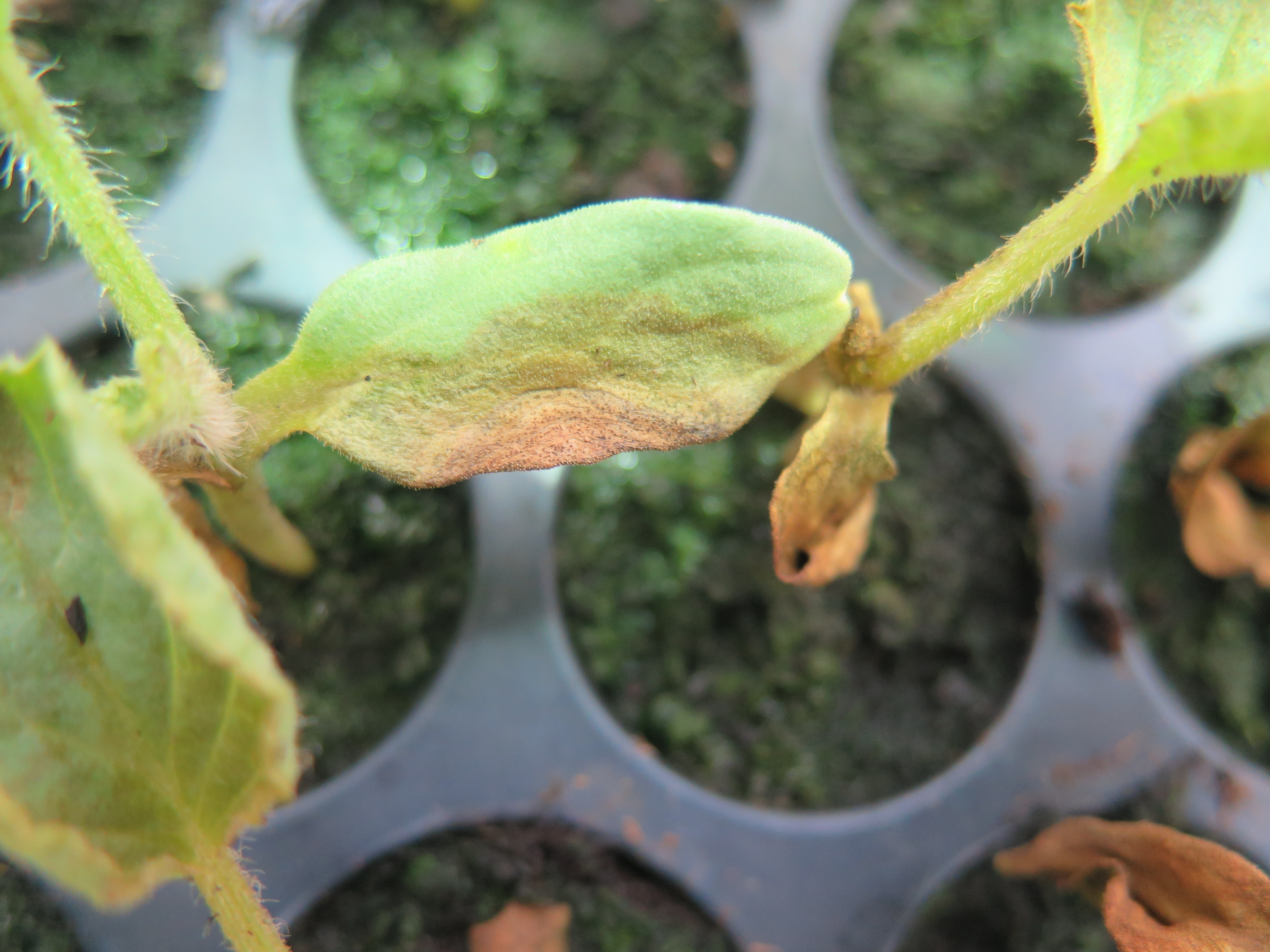 Figure 2. Gummy stem blight lesion on cotyledon of cantaloupe transplant. Note pycnidia.
 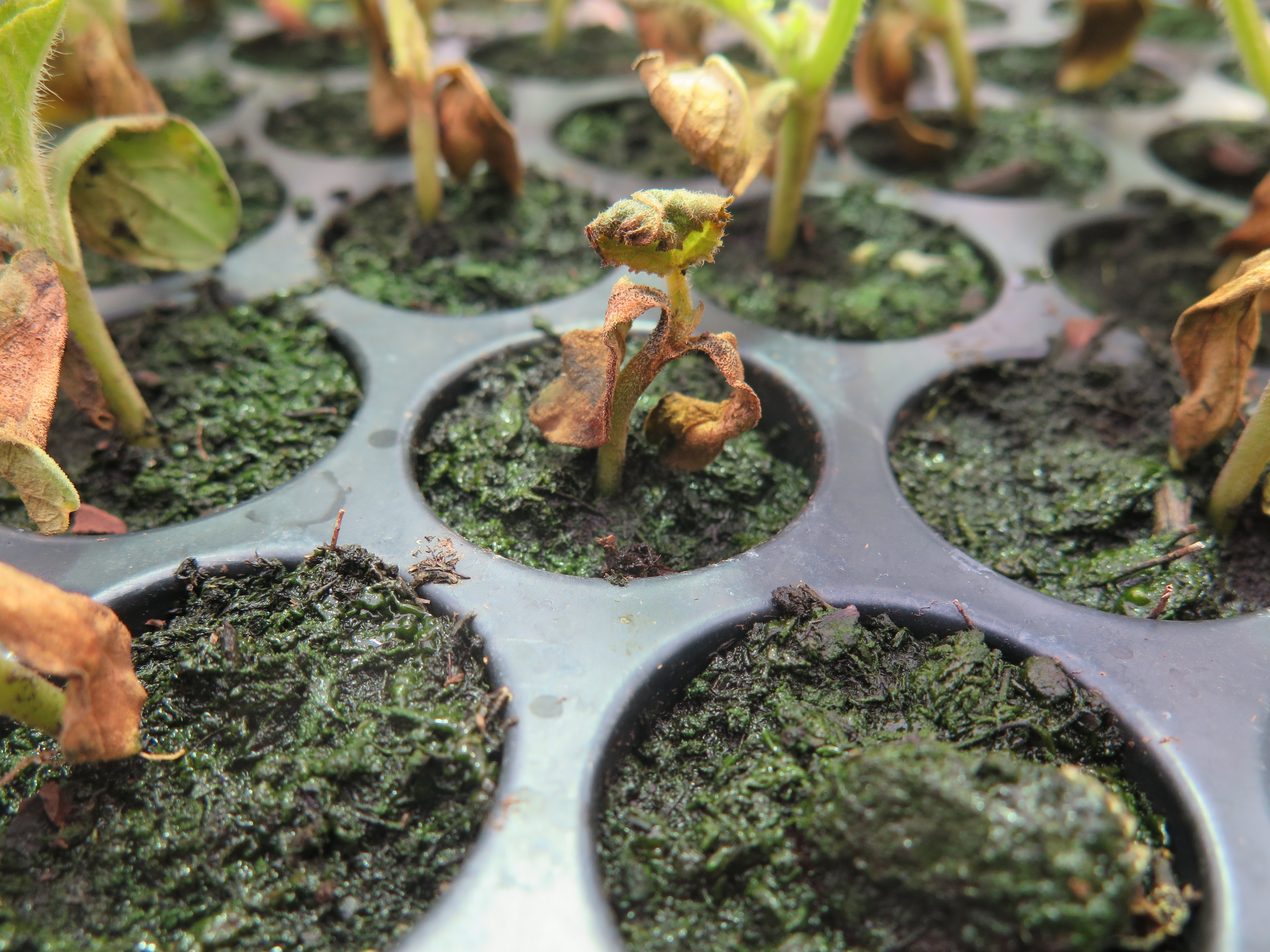 Figure 3. The gummy stem blight fungus has completely colonized this cantaloupe transplant.
 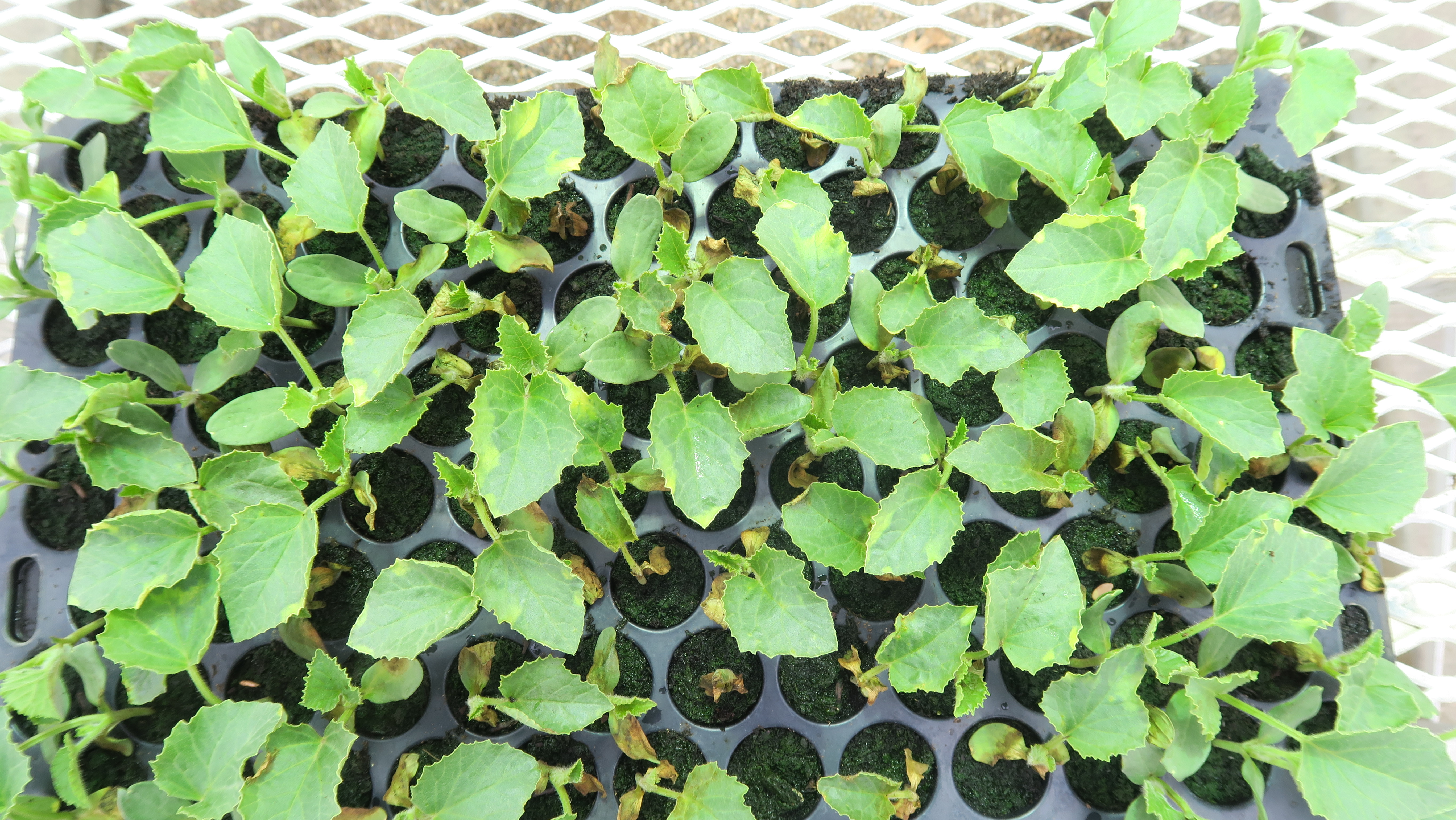 Figure 4. Gummy stem blight on multiple cantaloupe seedlings in a transplant tray.
 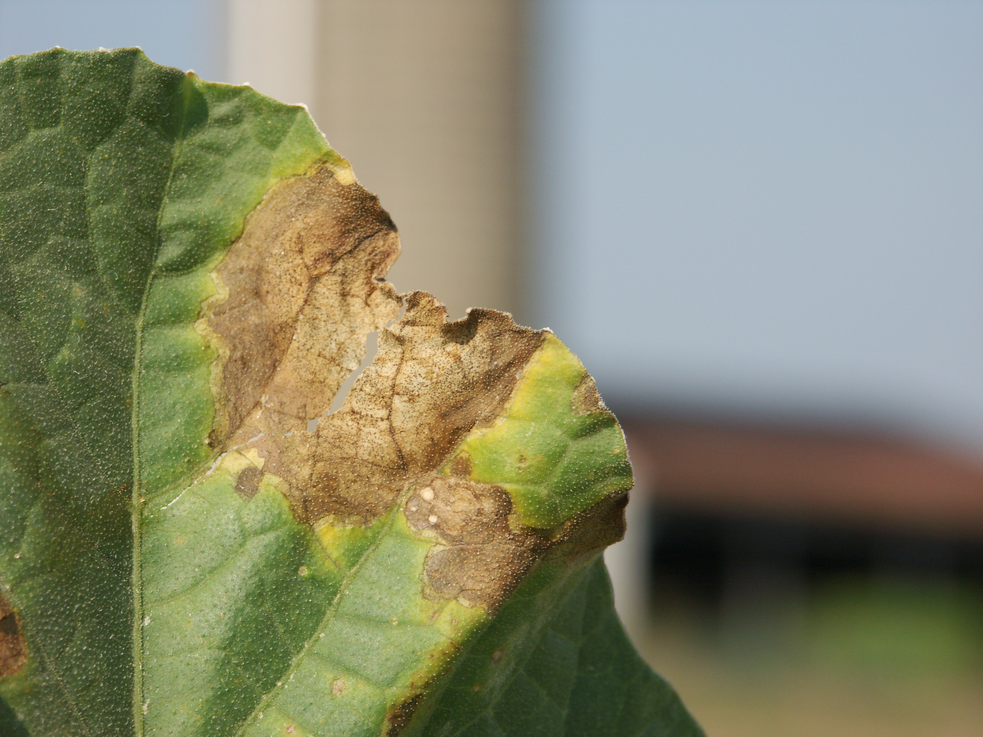 Figure 5. Gummy stem blight lesion on cantaloupe leaf. Note dark pycnidia in lesion and chlorotic margin of lesion.
 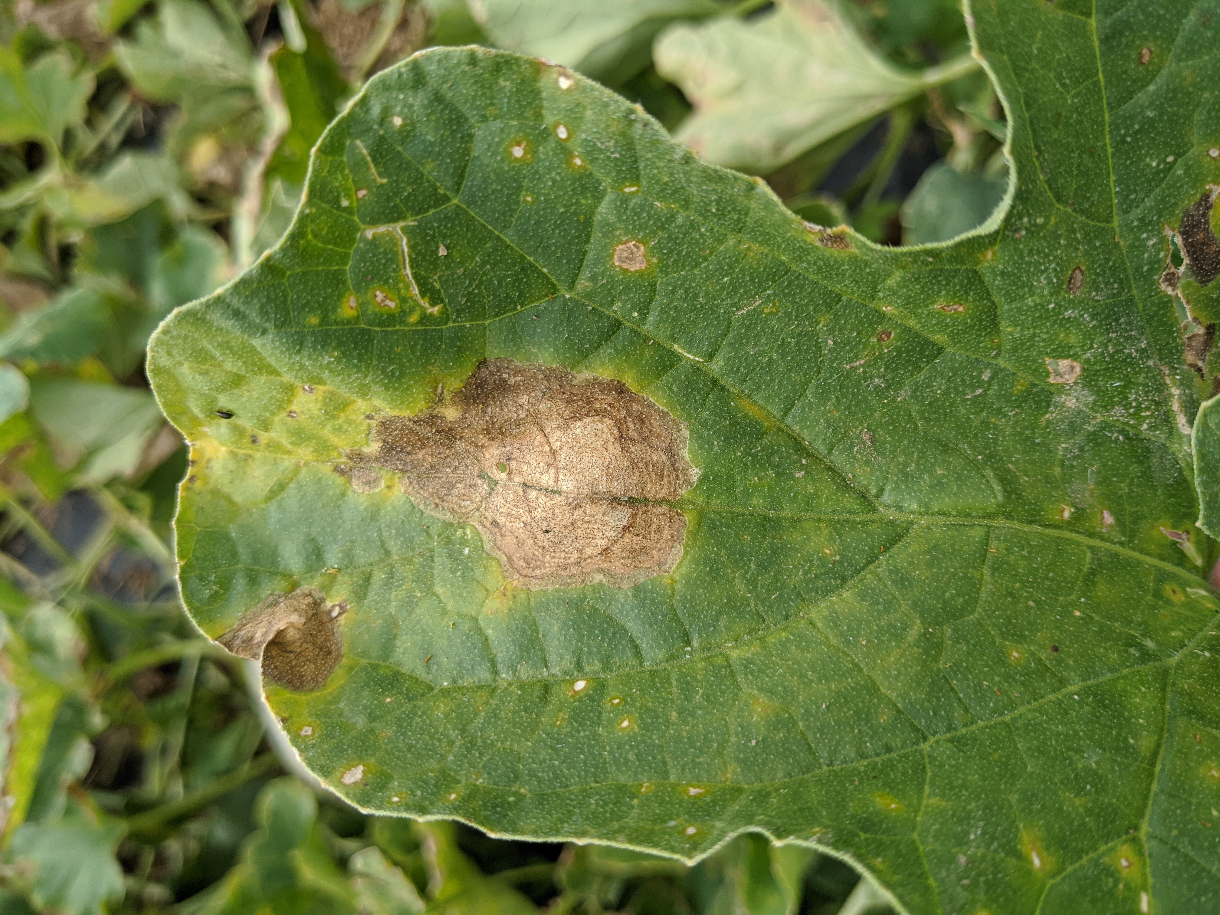 Figure 6. Gummy stem blight of cantaloupe.
 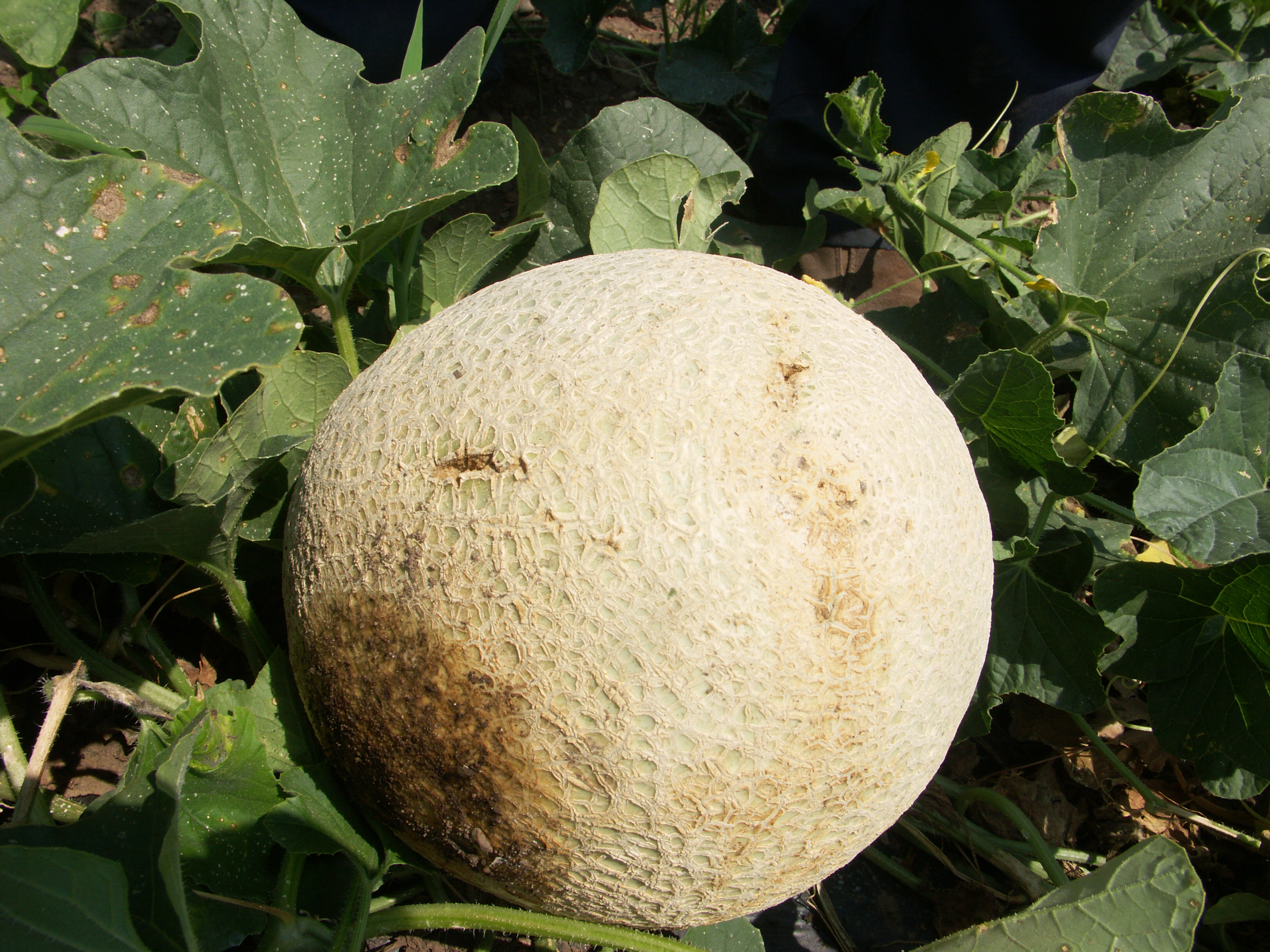 Figure 7. Gummy stem blight lesion on cantaloupe fruit.
 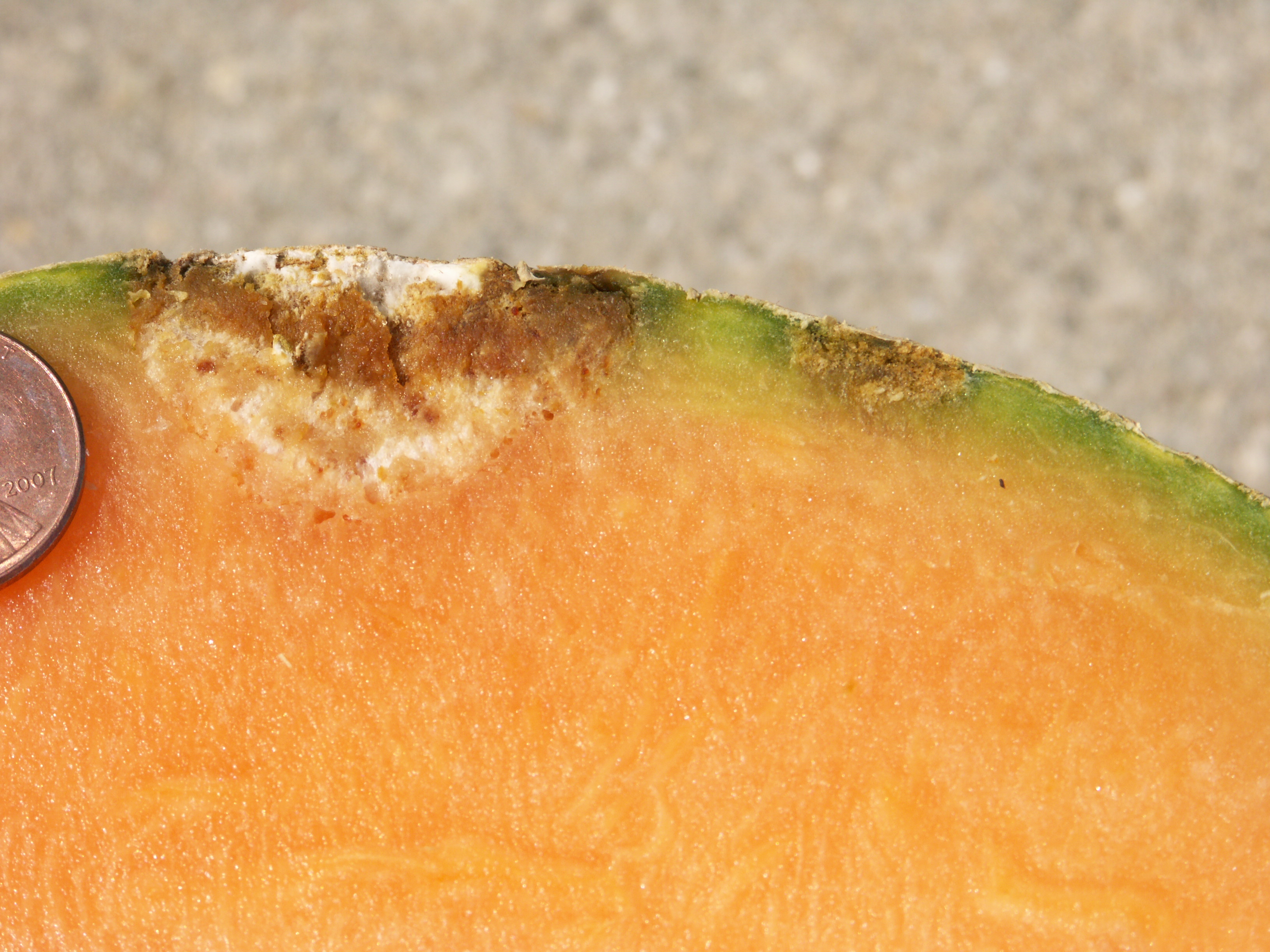 Figure 8. Cross section of cantaloupe with gummy stem blight lesion (black rot).
 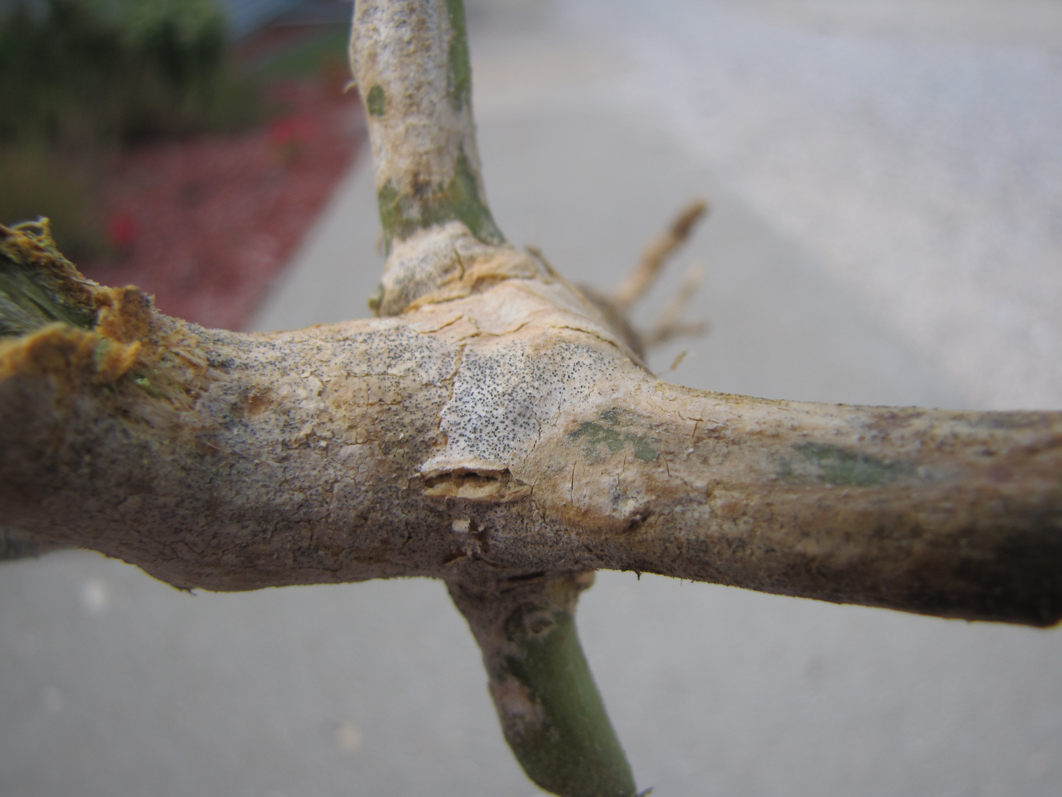 Figure 9. Crown of cantaloupe plant with necrosis caused by gummy stem blight. Note pycnidia.
 Magnesium deficiency of Cantaloupe
Magnesium deficiency-Symptoms are usually interveinal and include necrotic areas. Plants with symptoms are often clustered in areas where the soil pH is low, usually much lower than 6.0. Areas affected by Magnesium deficiency are usually high and well drained.
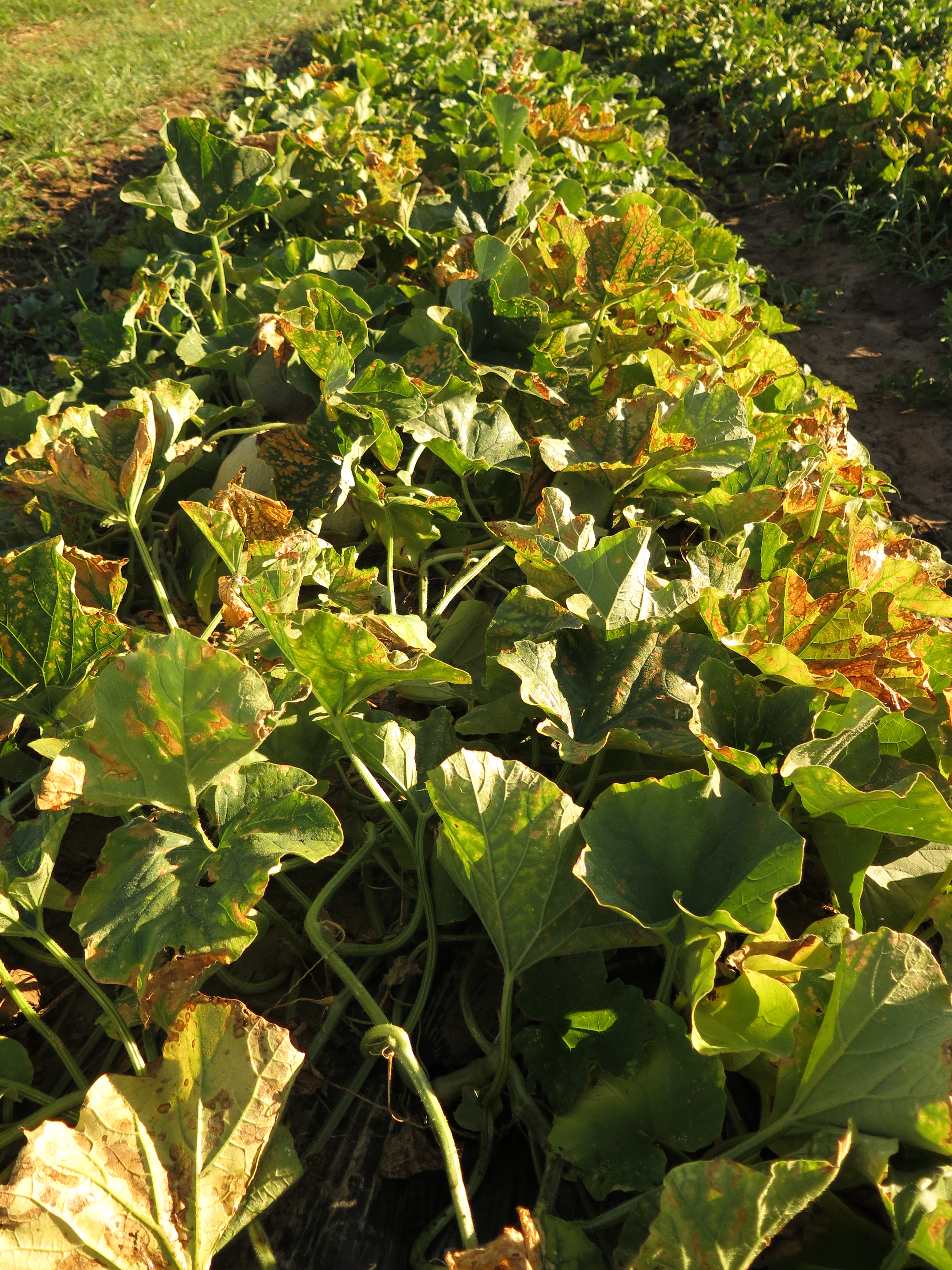 Figure 1. Magnesium deficiency of cantaloupe.
 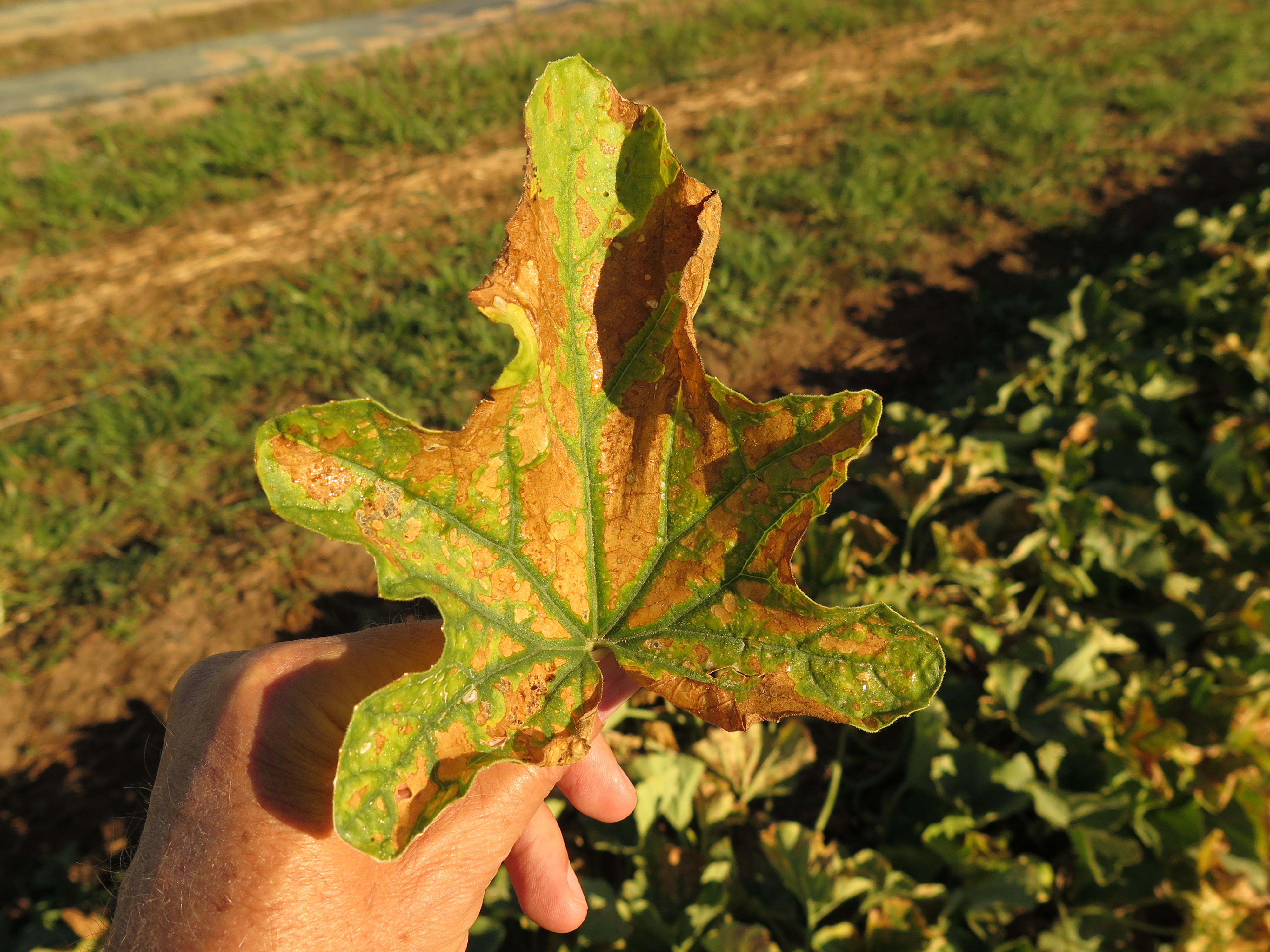 Figure 2. Magnesium deficiency of cantaloupe.
 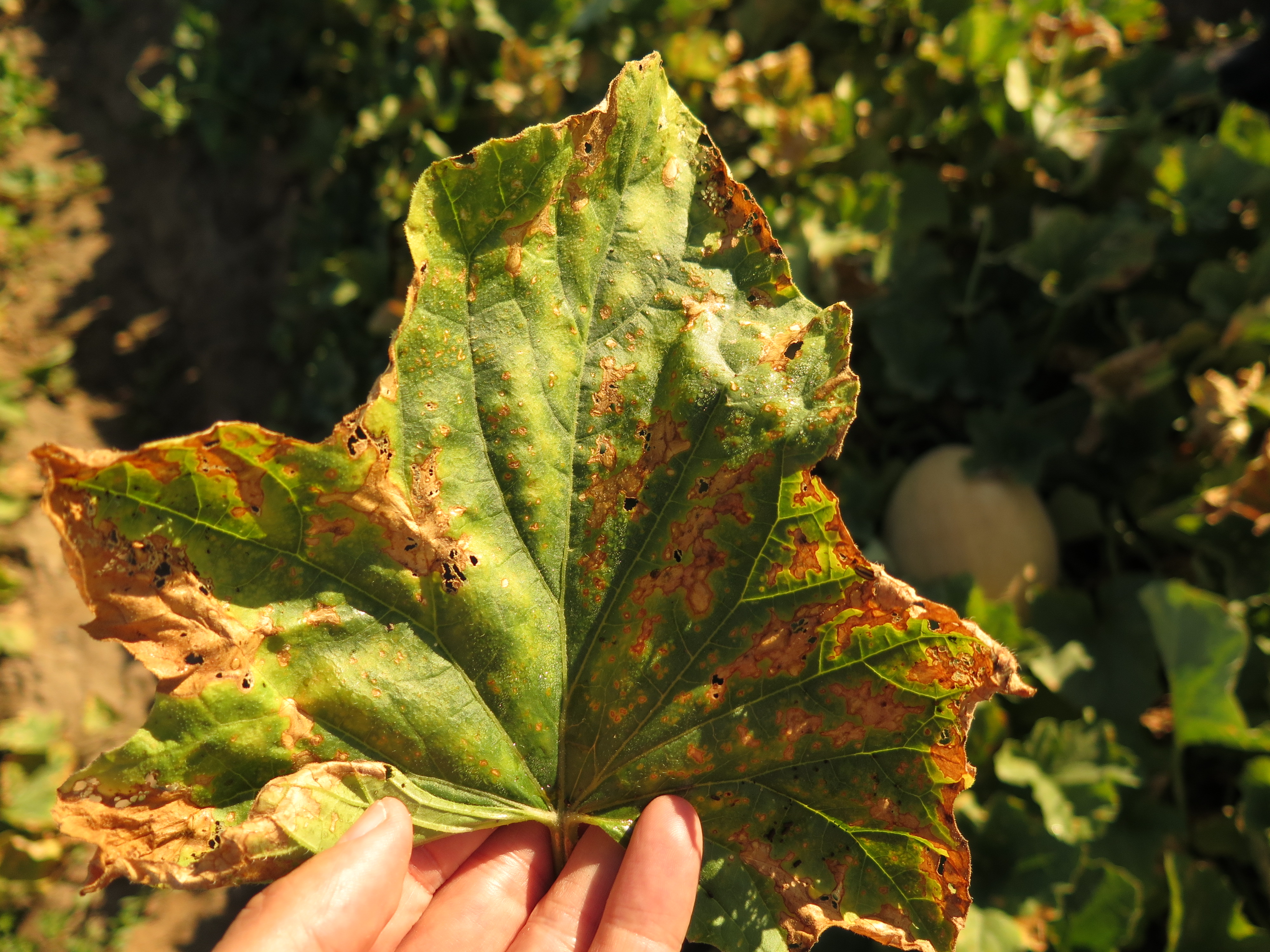 Figure 3. Magnesium deficiency of cantaloupe.
 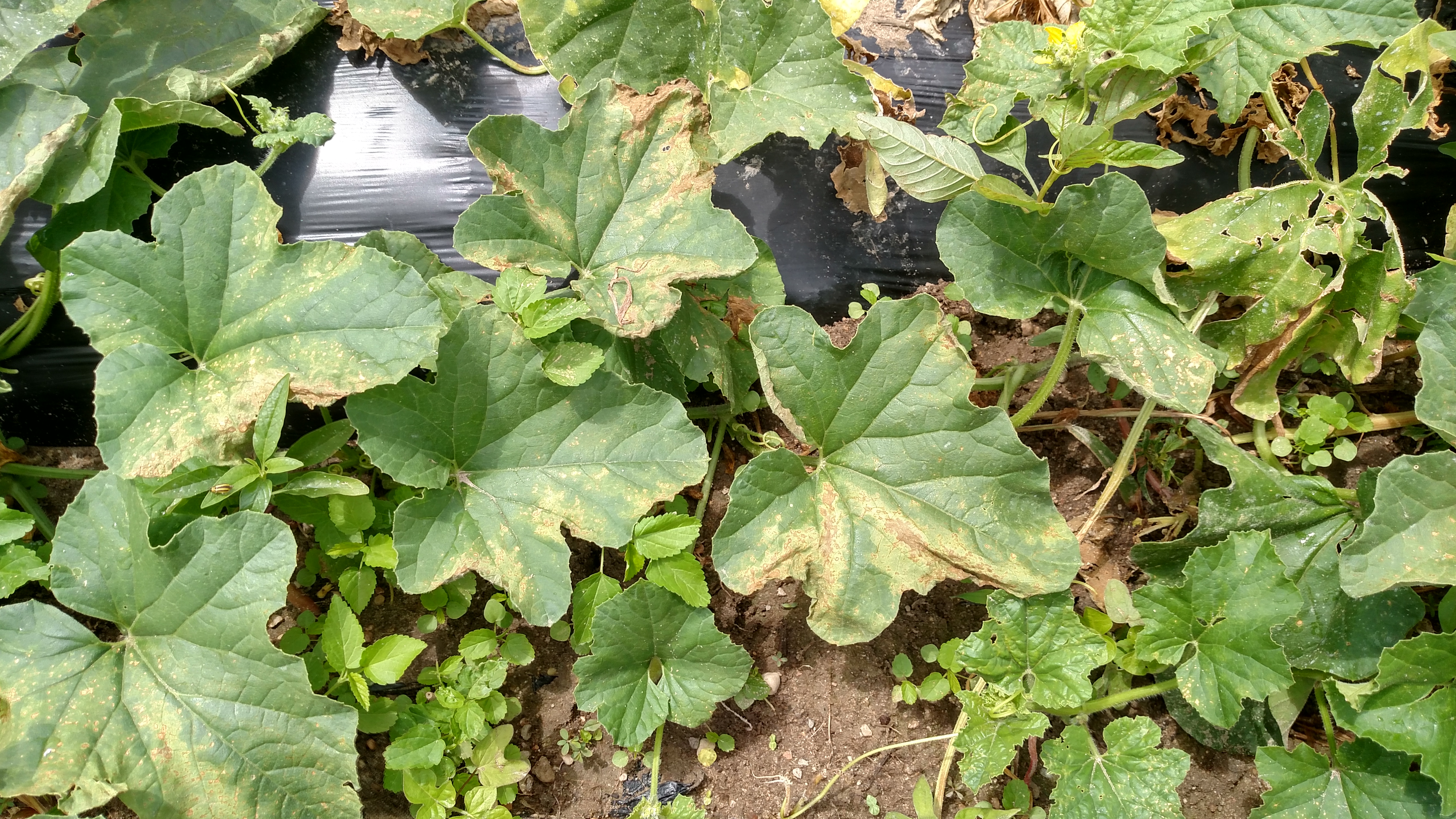 Figure 4. Magnesium deficiency of cantaloupe.
 Manganese toxicity of Cantaloupe
Manganese toxicity-Leaves affected with Manganese toxicity, when held up the light, will exhibit minute chlorotic pinpoint lesions. Aggregated symptoms appear as necrotic areas between the veins. Plants with symptoms are often clustered in areas where the soil pH is much lower than 6.0. Plants affected by manganese toxicity are often in relatively low areas of the field.
 Figure 1. Manganese toxicity of cantaloupe. Symptoms usually cluster together in a field where soil pH is low.
 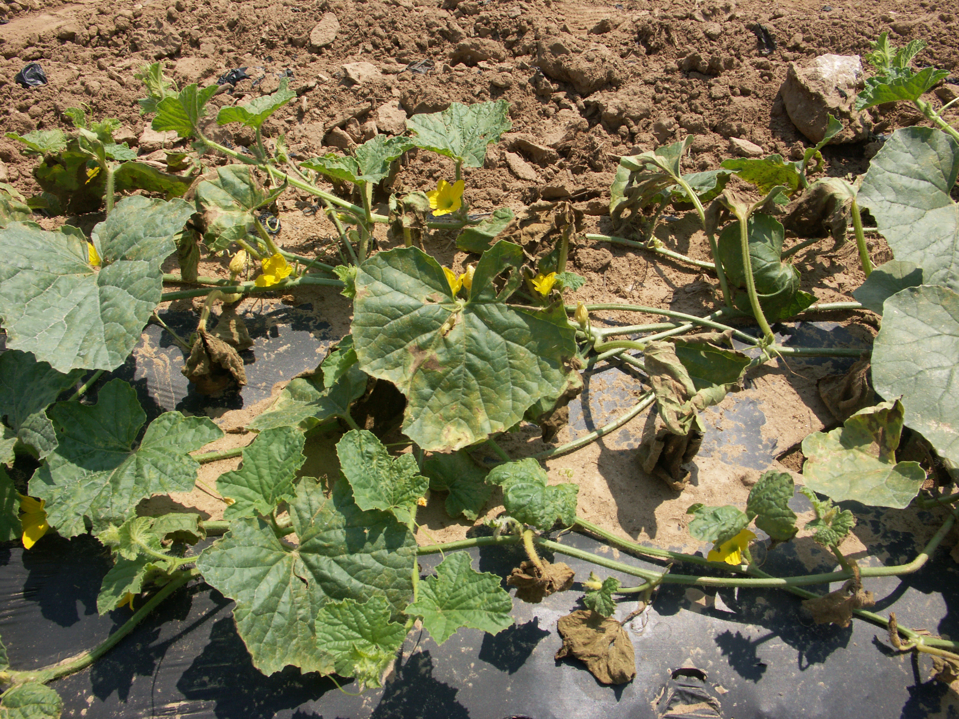 Figure 2. Manganese toxicity of cantaloupe.
 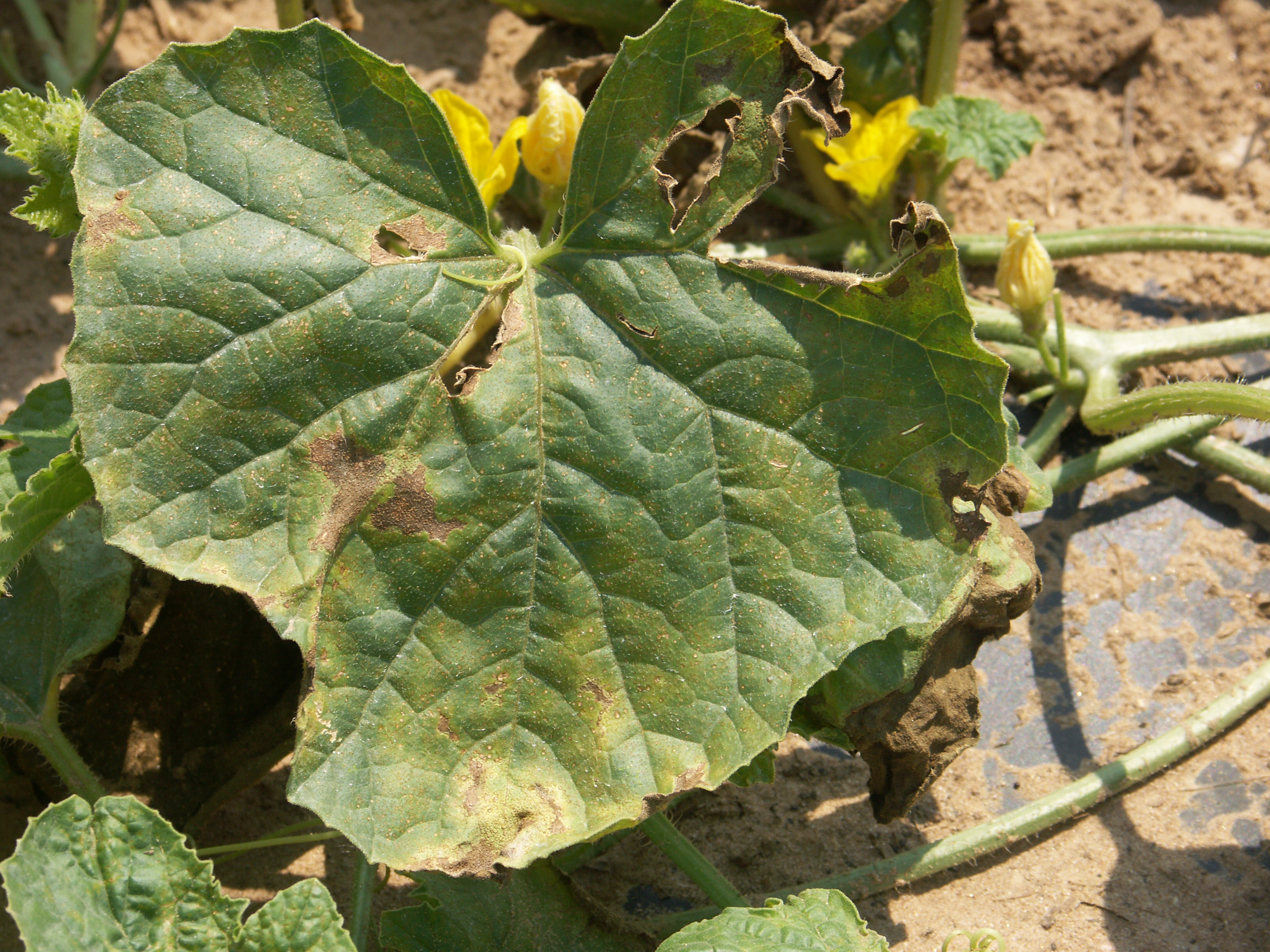 Figure 3. Manganese toxicity in cantaloupe.
 Phytophthora blight
Phytophthora blight-For the most part, cantaloupe is not as susceptible to Phytophthora blight as, for example, pumpkin and watermelon. However, under conducive conditions, Phytophthora blight can infect any plant part of cantaloupe. In some of the photos below, Phytophthora blight has caused a lesion on the crown of the plants which has caused the plant to wilt.
 Figure 1. Phytophthora blight of cantaloupe. Note collapsed vines.
  Figure 2. Phytophthora blight of cantaloupe. Note collapsed vine.
 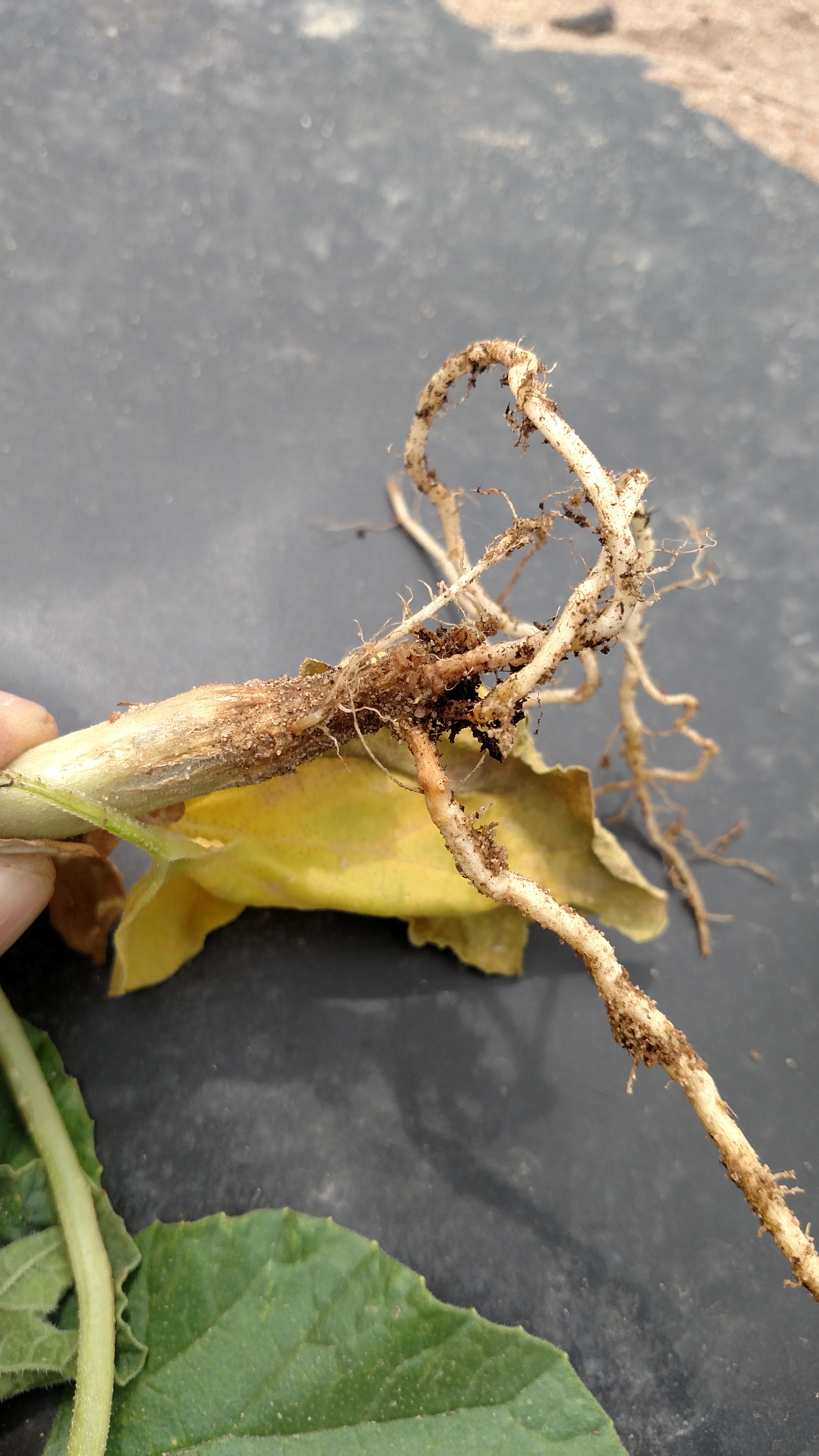 Figure 3. Phytophthora blight of cantaloupe. Note rotten and necrotic area at collar.
  Figure 4. Phytophthora blight of cantaloupe. Note lesion on stem.
  Figure 5. Phytophthora blight of cantaloupe. Note sporulation on fruit.
 Powdery mildew
This disease is relatively common, but host resistance and systemic fungicides are available for management. The disease may be recognized by the talc-like growth on the upper and lower surfaces of leaves.
 Figure 1. Colonies of powdery mildew are visible on the upper surface of this cantaloupe leaf.
 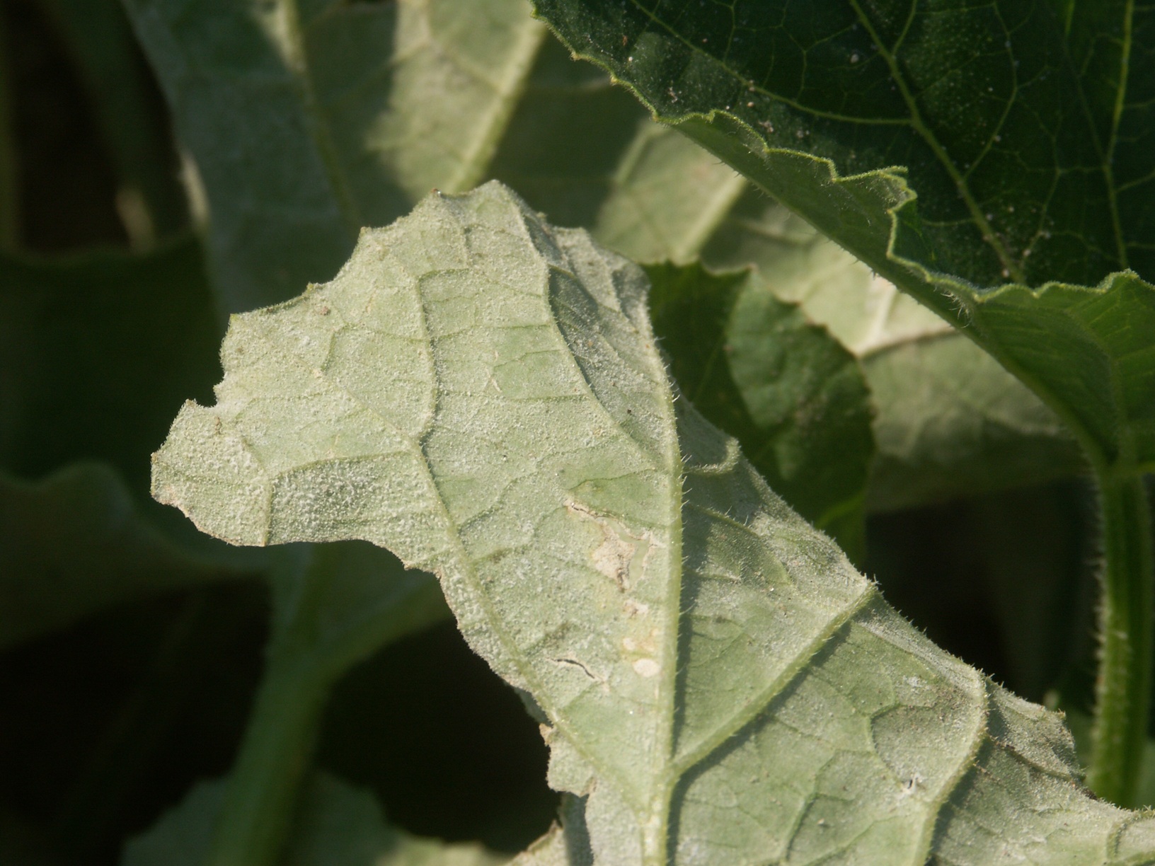 Figure 2. Sporulation of the powdery mildew fungus is visible on the lower surface of a cantaloupe leaf.
 Root knot nematode of Cantaloupe
Root knot nematode-Initial symptoms due to root knot nematode of cantaloupe may be decline or stunt. Under severe conditions, the plant may actively wilt. The roots of affected plants exhibit galls as shown here.
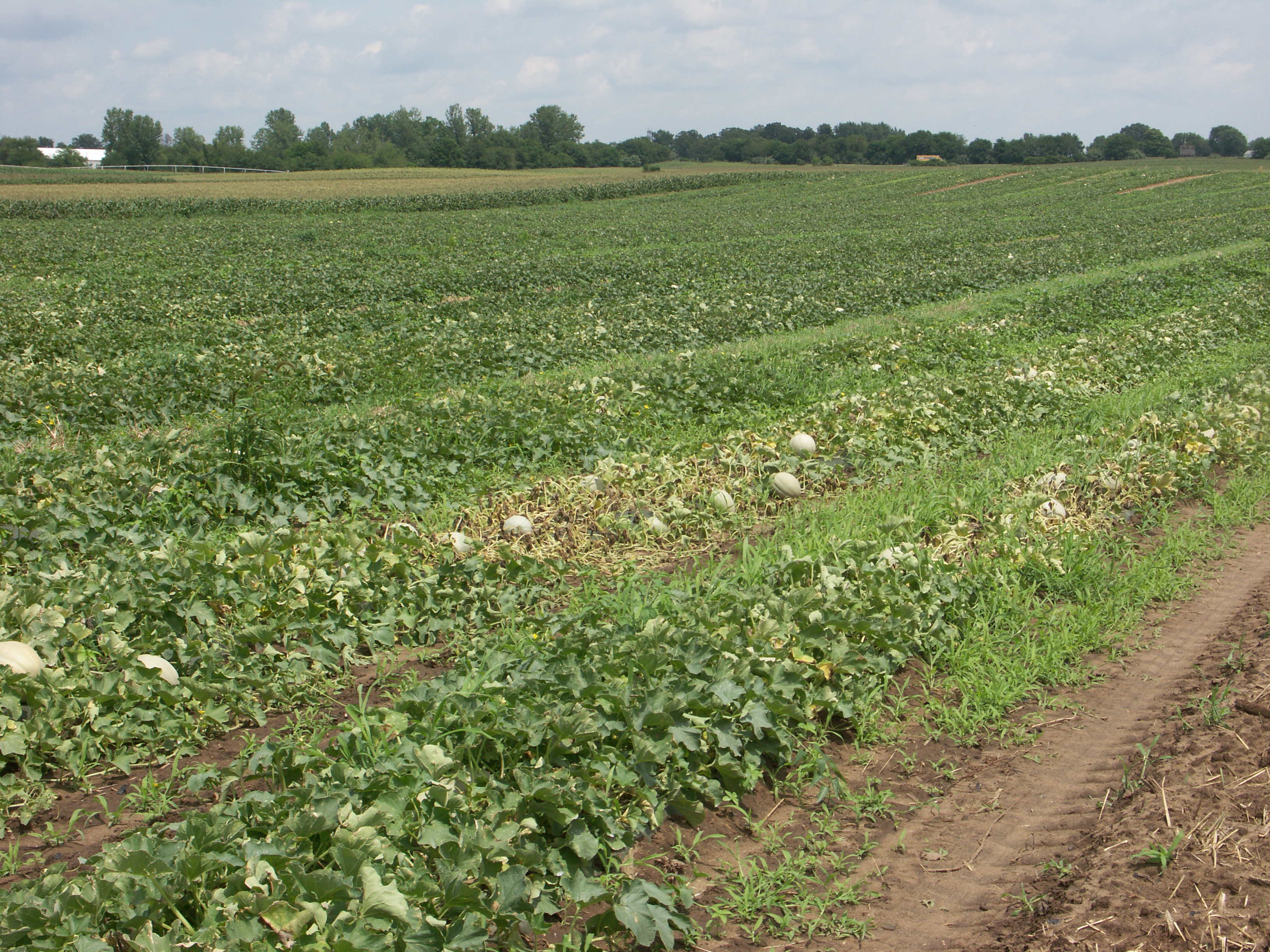 Figure 1. Root knot nematode of cantaloupe. Note wilt of affected plants.
 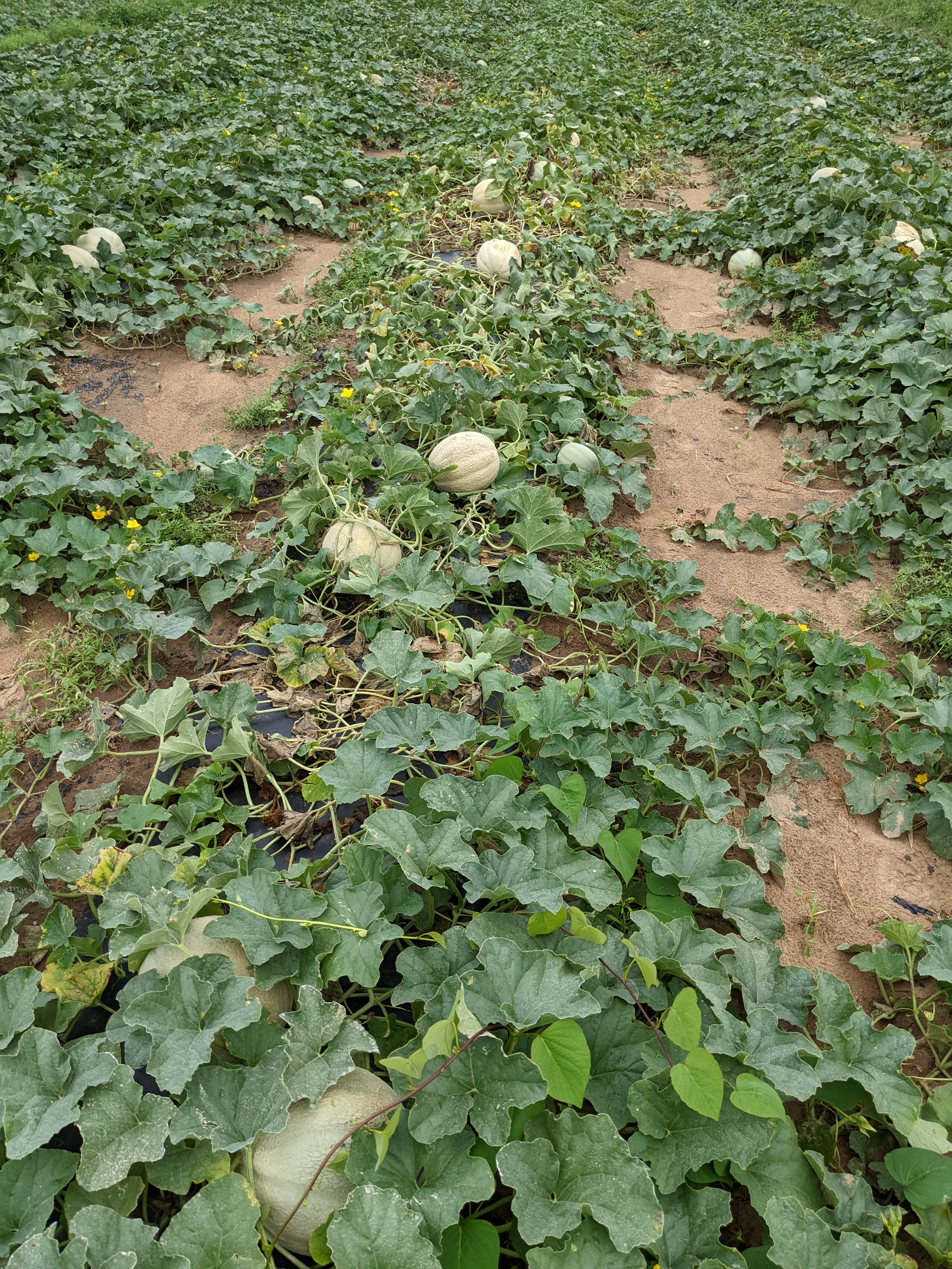 Figure 2. Root knot nematode of cantaloupe. Note wilt of affected plants.
 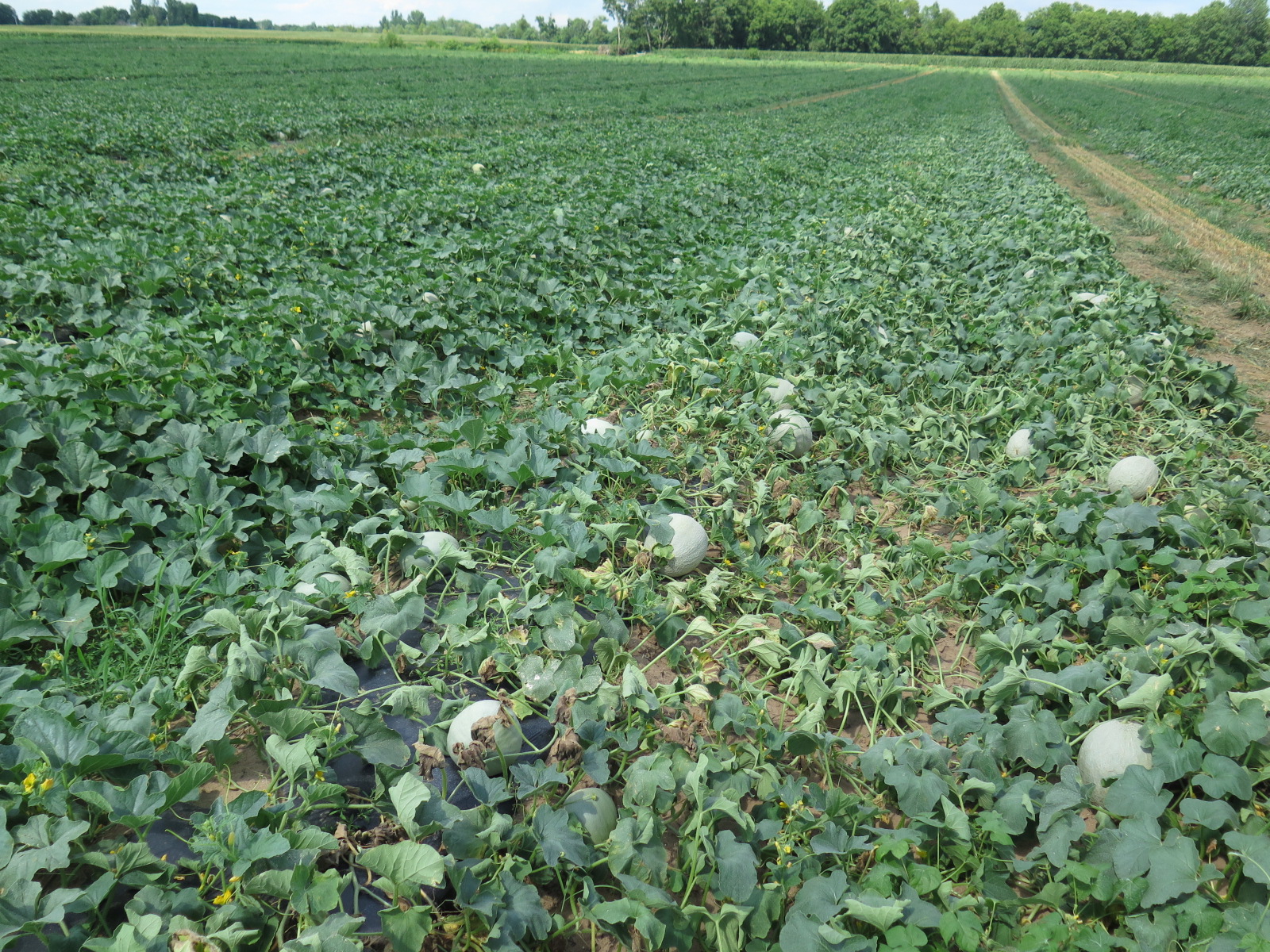 Figure 3. Root knot nematode of cantaloupe. Note wilt of affected plants is on hill.
 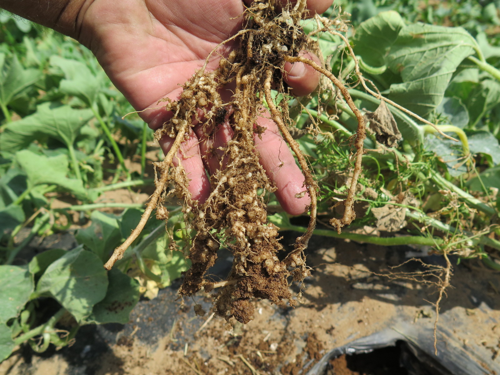 Figure 4. Root knot nematode of cantaloupe. Note galls on roots.
 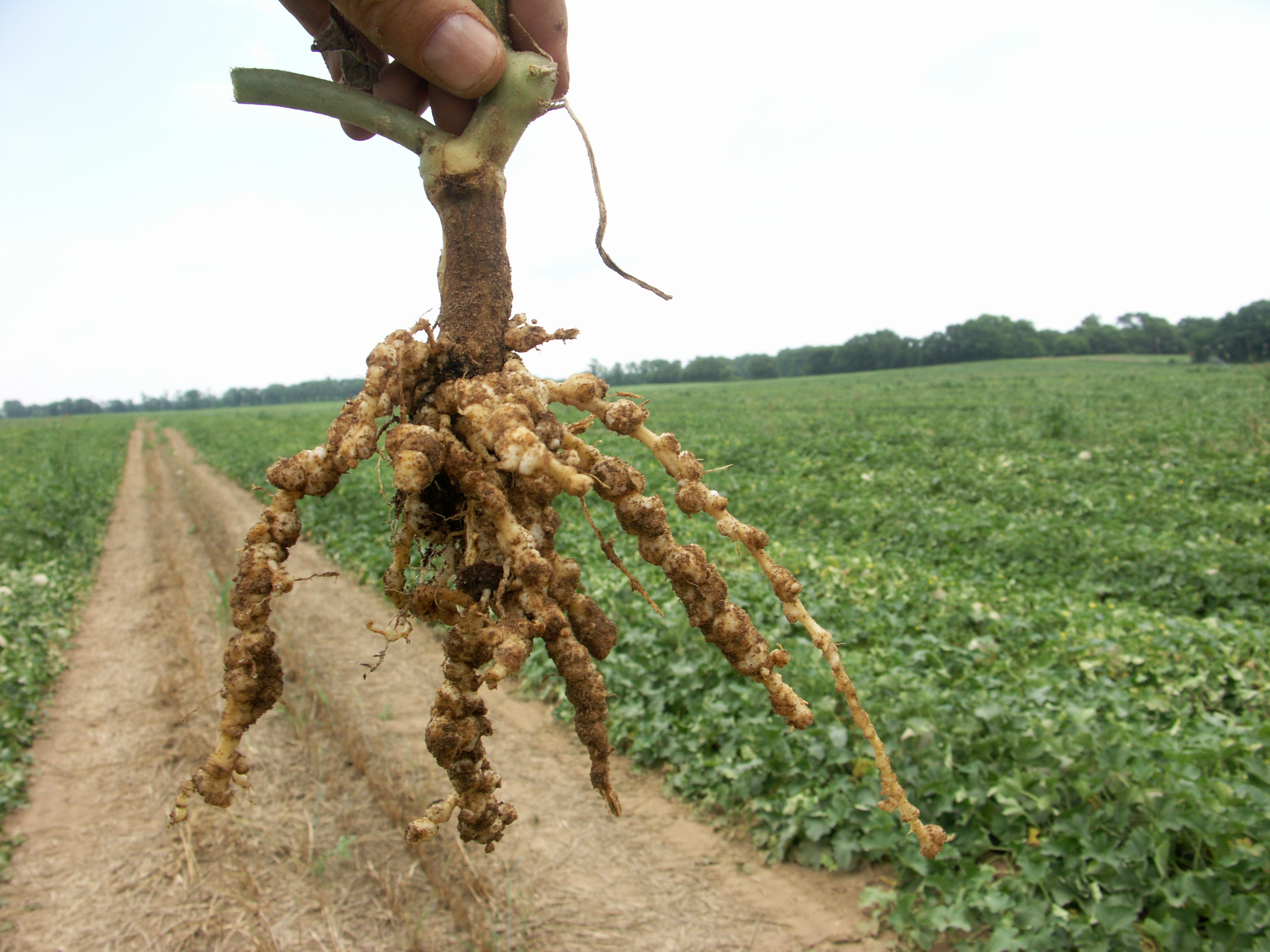 Figure 5. Root knot nematode of cantaloupe. Affected roots with galls is held up against background of wilted plants.