Figure 5. Anthracnose on pepper fruit. This is the same fruit as in figure 4. However, this was taken 7 days later.
Figure 6. Anthracnose of pepper. Penny is for size comparison.
Figure 7. Anthracnose of pepper fruit. Penny is for size comparison.
Figure 8. Anthracnose of pepper fruit.
Bacterial spot of pepper
Figure 1. Bacterial spot of pepper.
Figure 2. Bacterial spot of pepper.
Figure 3. Bacterial spot of peppers.
Figure 4. Bacterial spot of pepper.
Figure 5. Bacterial spot on pepper transplants. These symptoms are quite severe, perhaps because they have occurred under greenhouse conditions.
Figure 6. Bacterial spot on pepper transplants.
Figure 7. Bacterial spot of peppers.
Figure 8. Bacterial spot of peppers.
Figure 9. Bacterial spot on pepper fruit.
Figure 10. Bacterial spot on pepper fruit.
Phytophthora blight of pepper
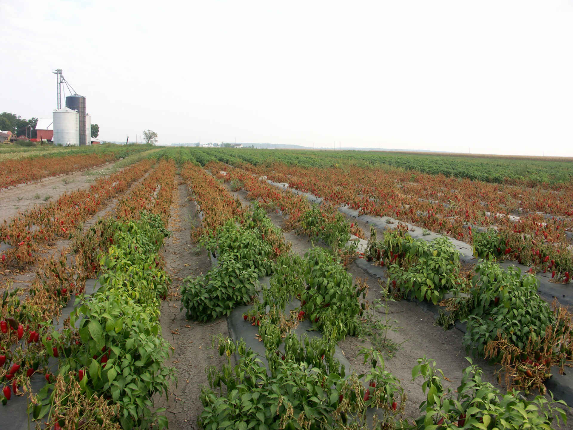
Figure 1. Pepper plants in the low area of this field have been killed outright by Phytophthora blight.
Figure 2. Pepper plants are wilted or dead as a result of Phytophthora blight of pepper.
Figure 3. Stem lesion from Phytophthora blight of pepper.
Figure 4. Pepper plants with Phytophthora blight. Cucumber culls with Phytophthora blight were discarded in the field, resulting in the infection of the peppers. The remains of the cucumber culls can be observed in the foreground of the photo.
Figure 5. Close up of pepper plant in Figure 4.
Figure 6. Pepper plant wilted due to Phytophthora blight of pepper.
Figure 7. Phytophthora blight on pepper fruit showing sporulation on surface of fruit.
Figure 8. Lesion on stem of pepper plant due to Phytophthora blight.
Southern blight of pepper
Figure 1. Pepper plant wilted due to southern blight.
Figure 2. Southern blight of pepper. Note white fungus on stem.
Tomato spotted wilt virus
Figure 1. Lesion with ring-like structure due to tomato spotted wilt virus on pepper.
Figure 2. Pepper leaves with tomato spotted wilt virus. Note positive test strip for TSWV.