

tomato diseases
Processing tomato production in Indiana is usually second in the US—a distant second to California. However, processing tomato is an important crop throughout Indiana. The most important diseases of processing tomatoes are bacterial spot and bacterial canker. White mold can also be a problem. Many of the fresh market tomatoes produced in Indiana are grown in high tunnels or greenhouses. The tomato diseases common in high tunnels are leaf mold, white mold and gray mold. Tomato spotted wilt virus, transmitted by thrips, can also be important.
Anthracnose
Anthracnose-This disease often affects tomato fruit that are overripe and close to the ground. But this is not always the case. Sometimes caused by same organism responsible for anthracnose of pepper.
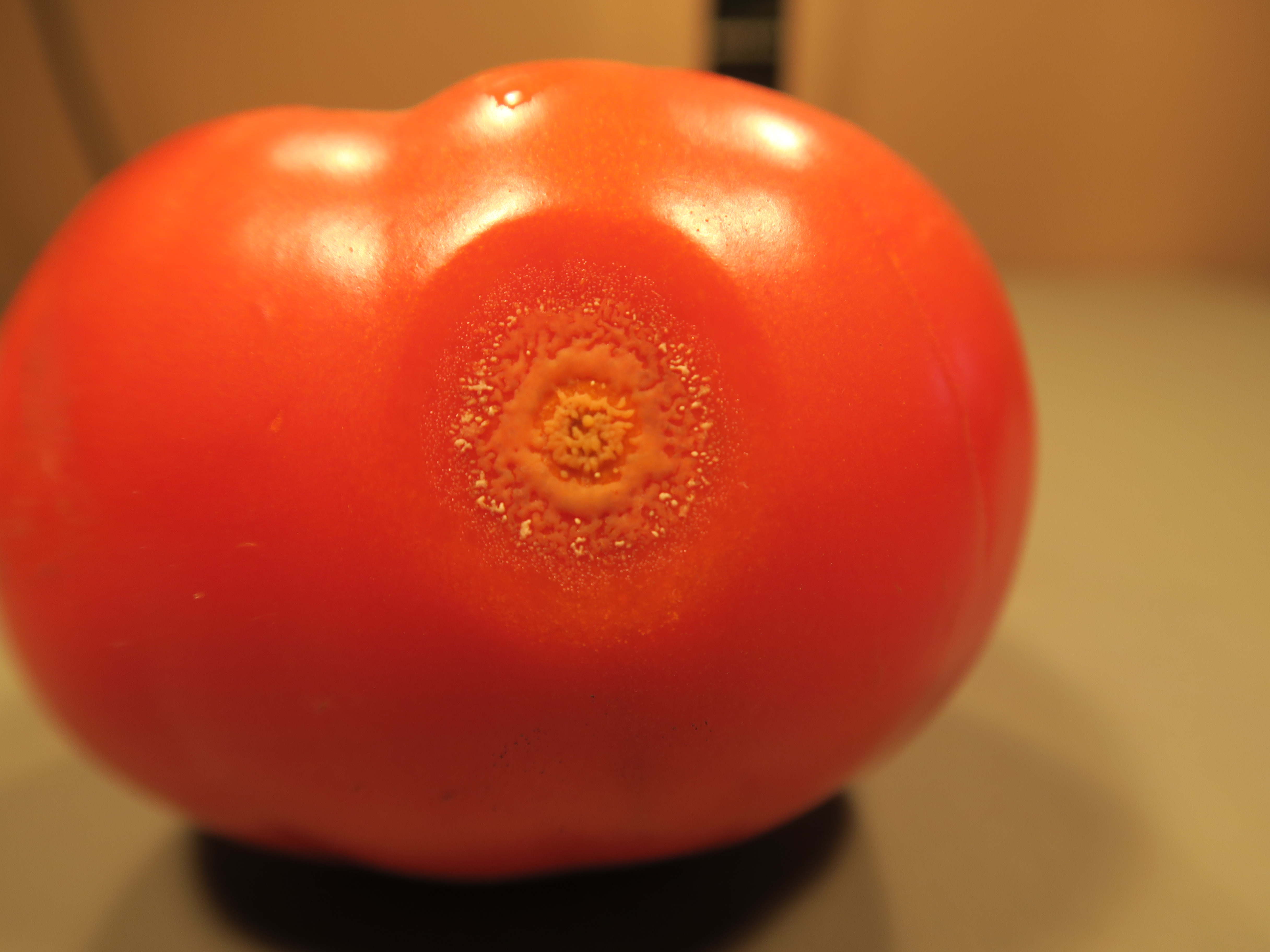 Figure 1. Anthracnose of tomato. Note orange sporulation of the pathogen on the lesion.
 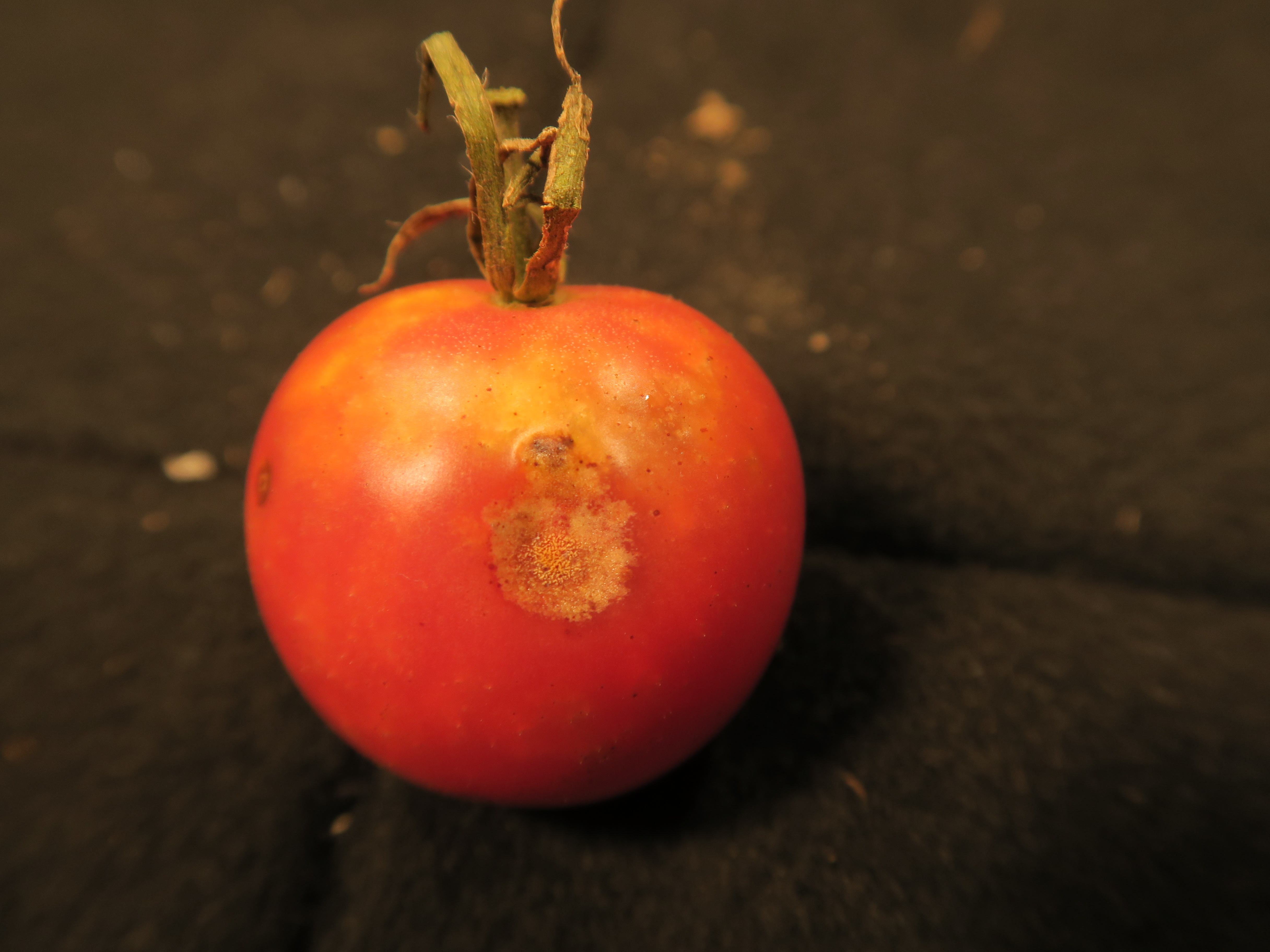 Figure 2. Anthracnose of tomato. Note sunken lesion and orange sporulation.
 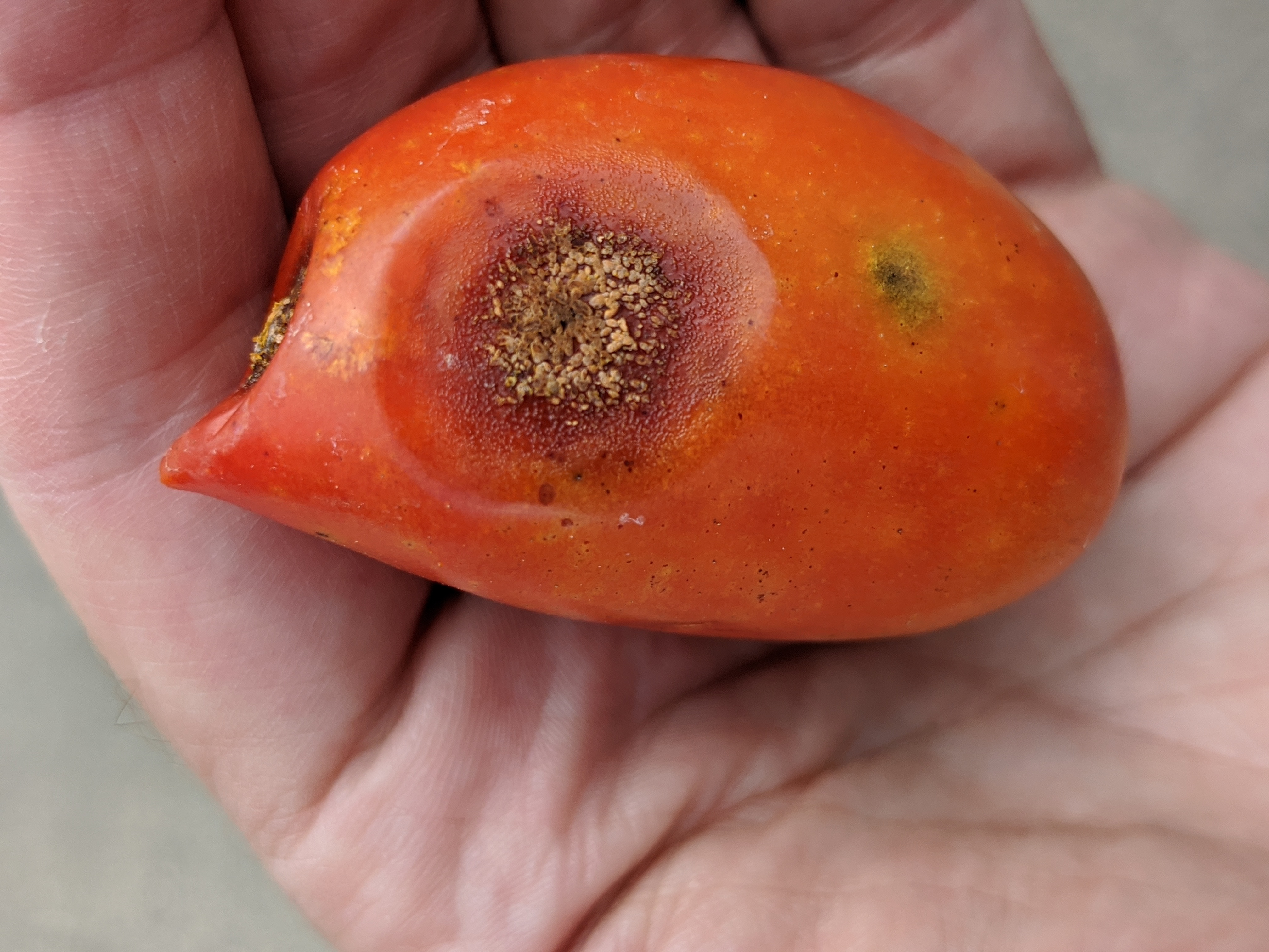 Figure 3. Anthracnose of tomato.
 Bacterial canker
Bacterial canker-the first symptom observed is often the marginal necrosis and chlorosis on leaves. If stems are cut low to the ground, an internal discoloration is often observed. Fruit may also have a distinctive birds-eye lesion. This may be our most important tomato disease. May occur in field or greenhouse tomatoes.
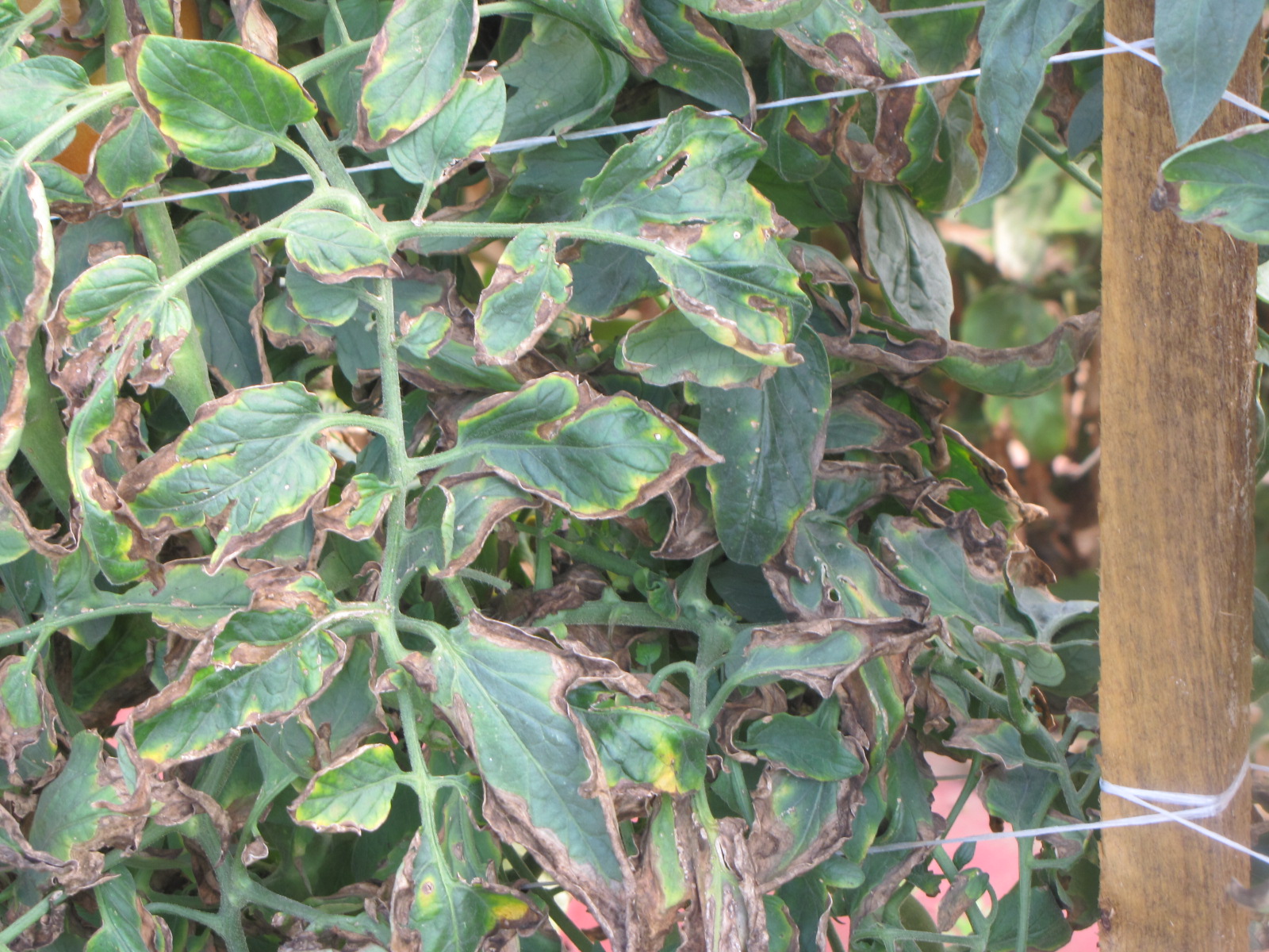 Figure 1. Necrosis and chlorosis on leaf margin, also known and ‘firing’, due to bacterial canker. This is a very common symptom.
 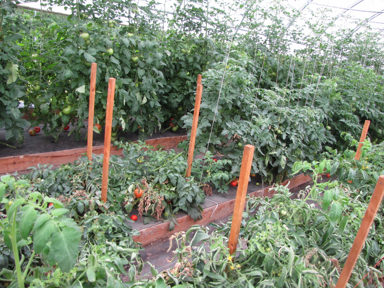 Figure 2. Bacterial canker in greenhouse has resulted in stunting and necrosis.
 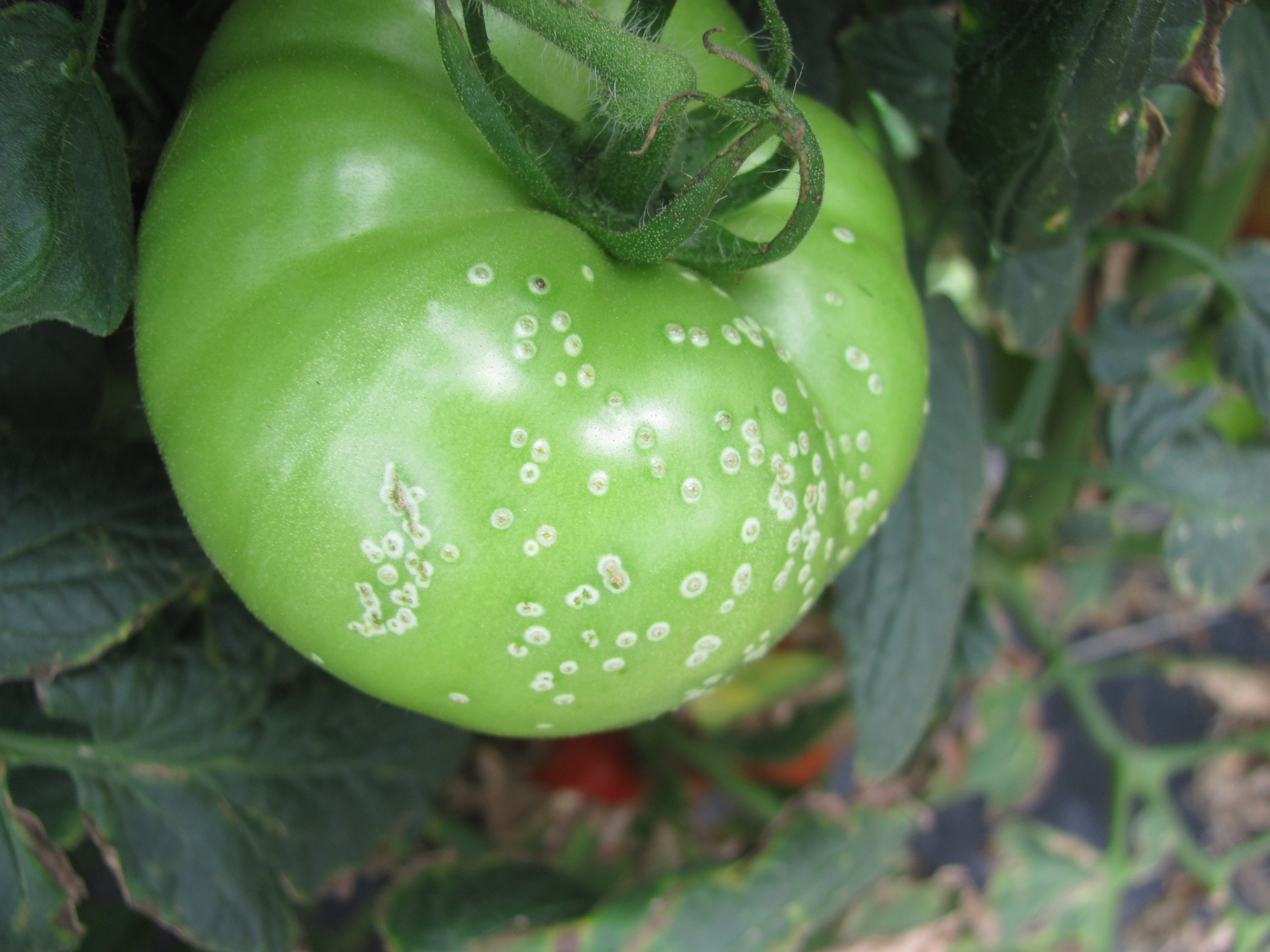 Figure 3. Bird’s eye spot infection on tomato fruit as a result of infection with bacterial canker. This symptom may not necessarily occur.
 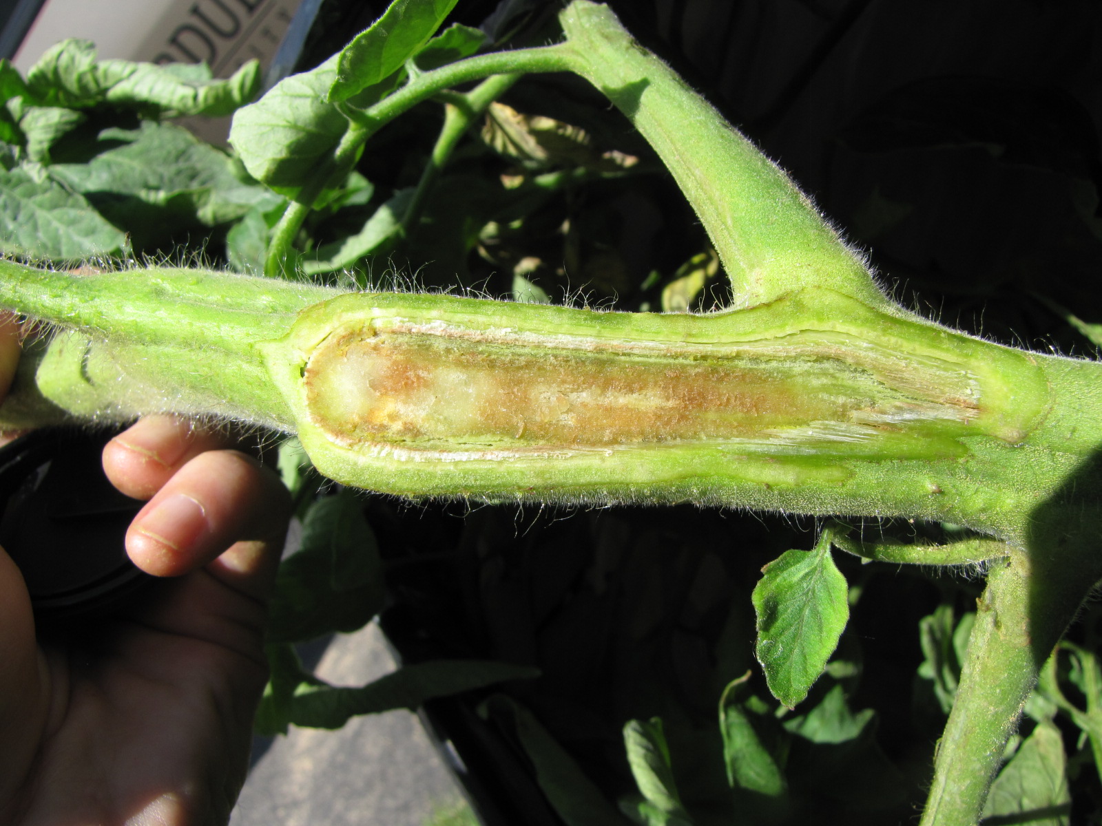 Figure 4. Vascular discoloration of tomato stem due to bacterial canker. This symptoms indicates that the infection is systemic.
 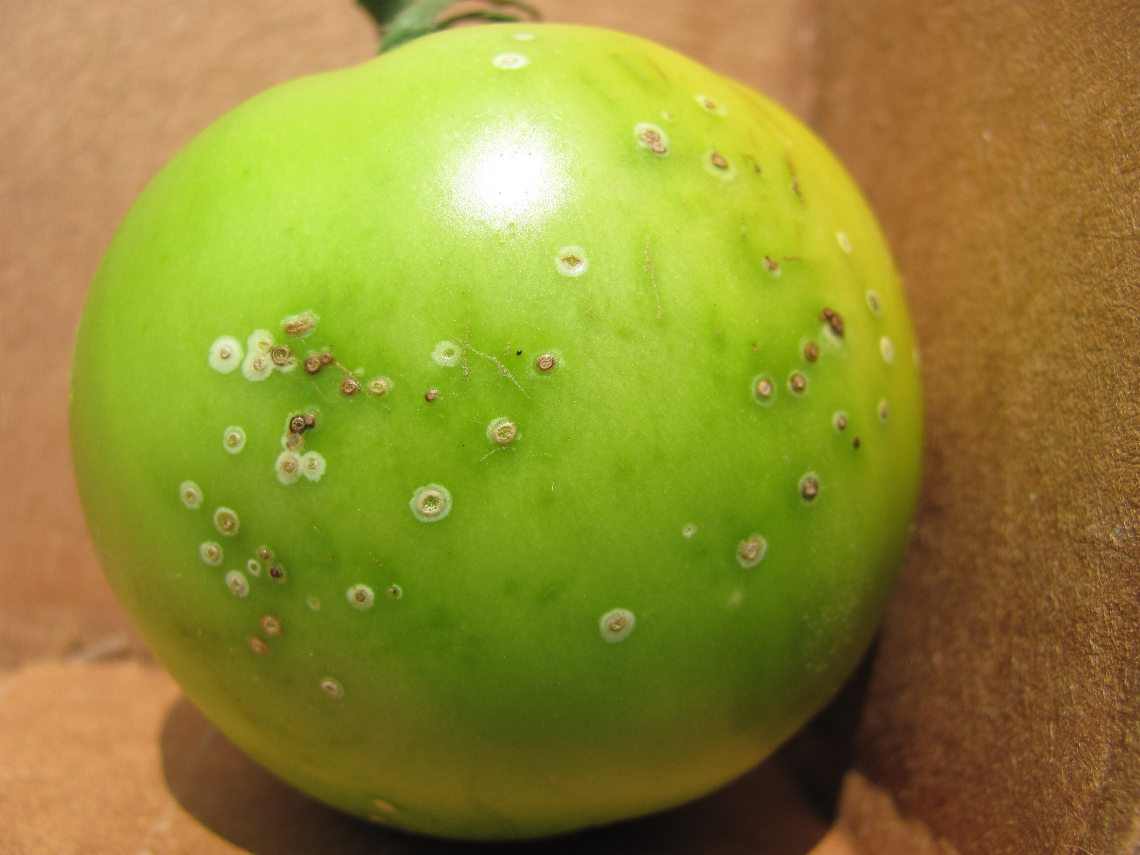 Figure 5. Fruit symptoms of bacterial canker.
 Bacterial speck
Bacterial speck of tomato-This disease prefers cooler weather than bacterial spot. Often an early season disease. Typically, each lesion is associated with chlorosis, whereas bacterial spot lesions are often only associated with chlorosis when several lesions coalesce. I have only observed this in field tomatoes. Not usually as important as bacterial spot.
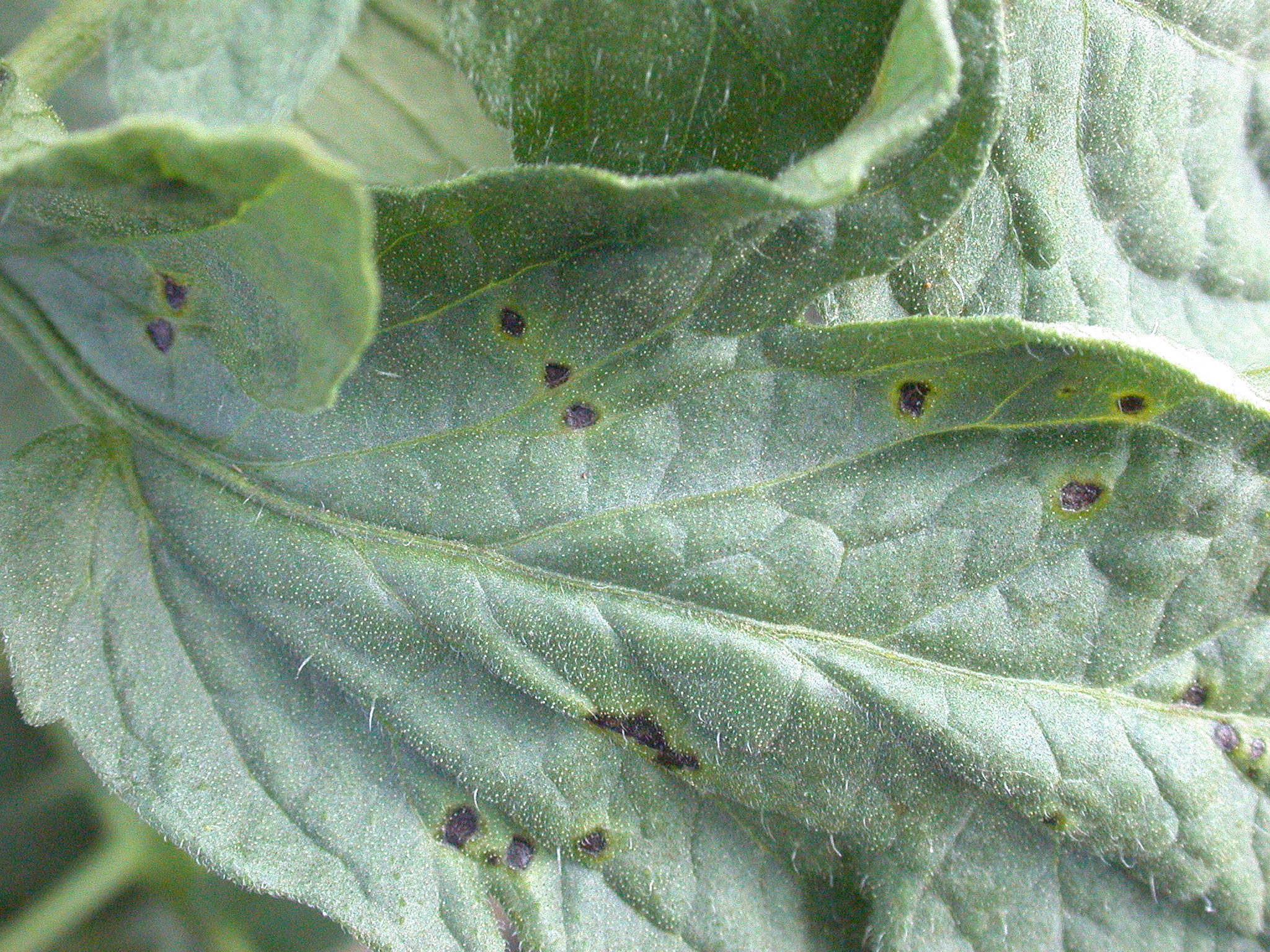 Figure 1. Bacterial speck of tomato. Note that each lesion on the leaf has a small chlorotic halo in contrast to bacterial spot where chlorosis usually does not occur until a large amount of lesions are in one small area.
 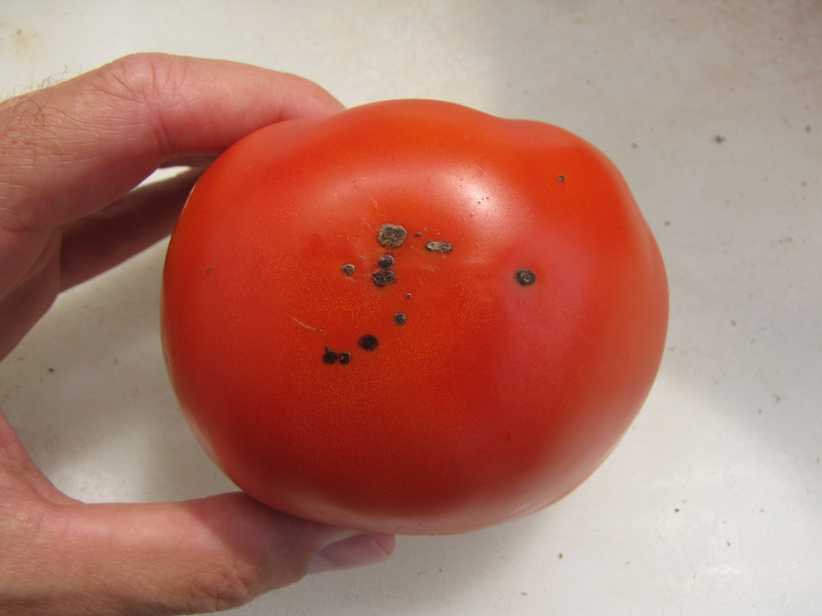 Figure 2. Bacterial speck of tomato fruit lesions. For the most part, fruit lesions of bacterial speck or smaller than lesions of bacterial spot.
 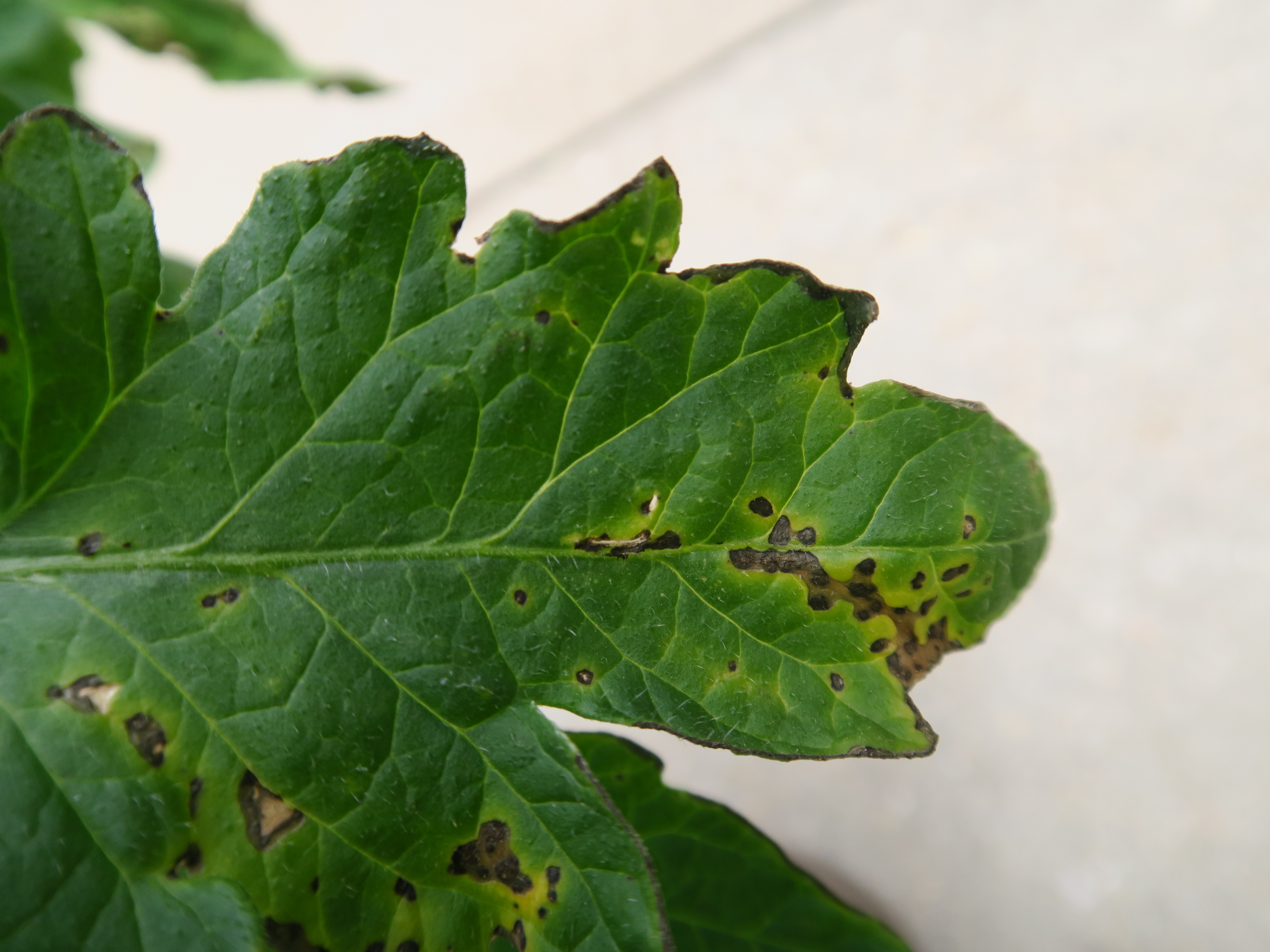 Figure 3. Lesions of bacterial speck of tomato on leaf.
 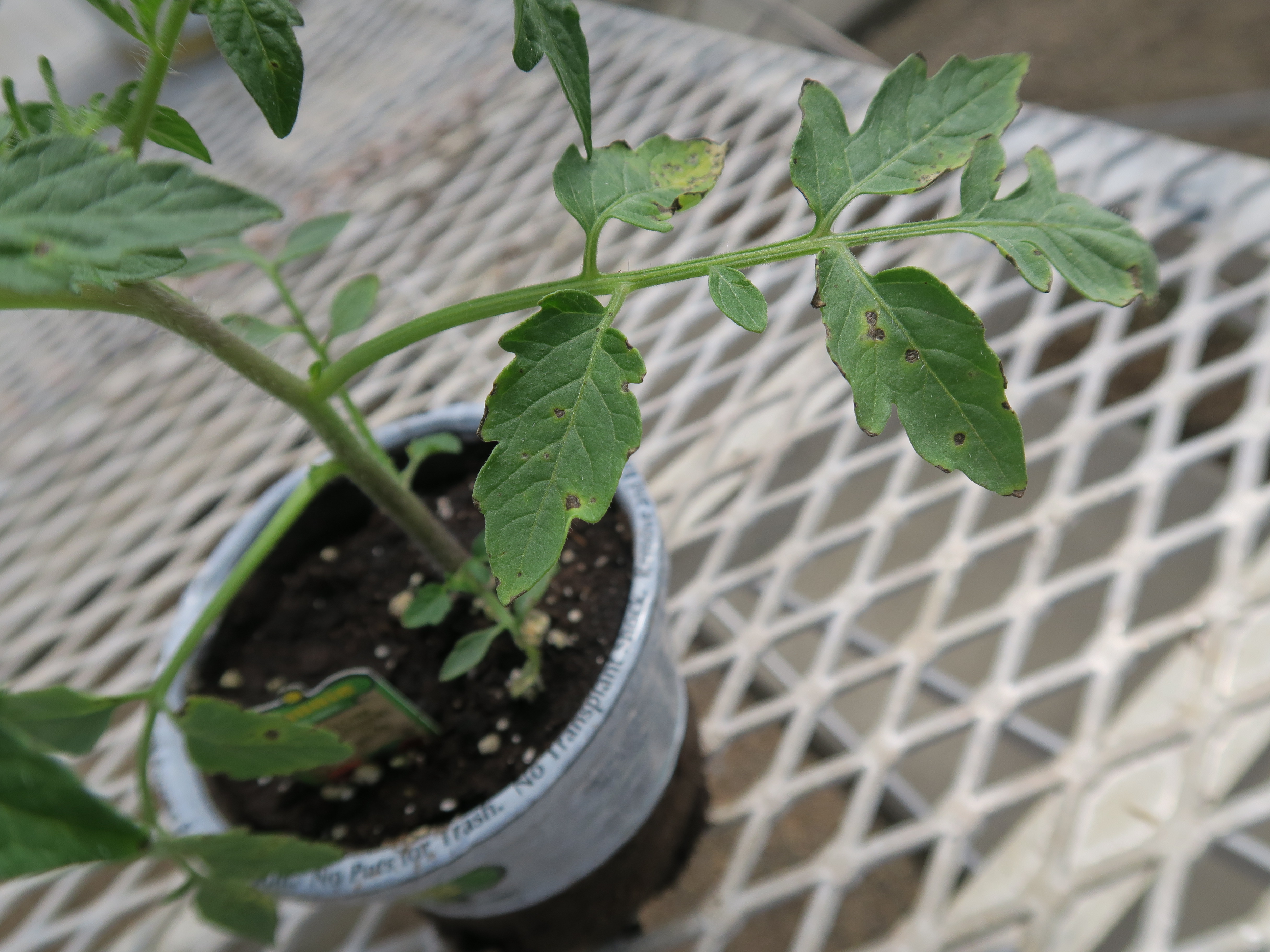 Figure 4. Leaf lesions of bacterial speck on a retail tomato transplant.
 Bacterial spot
Bacterial spot of tomato-Important disease of field tomatoes. Water-soaked lesions are associated with chlorosis only when several lesions occur in close proximity. Lesions on fruit may be large and scab-like. Copper resistance is an important factor in managing this disease.
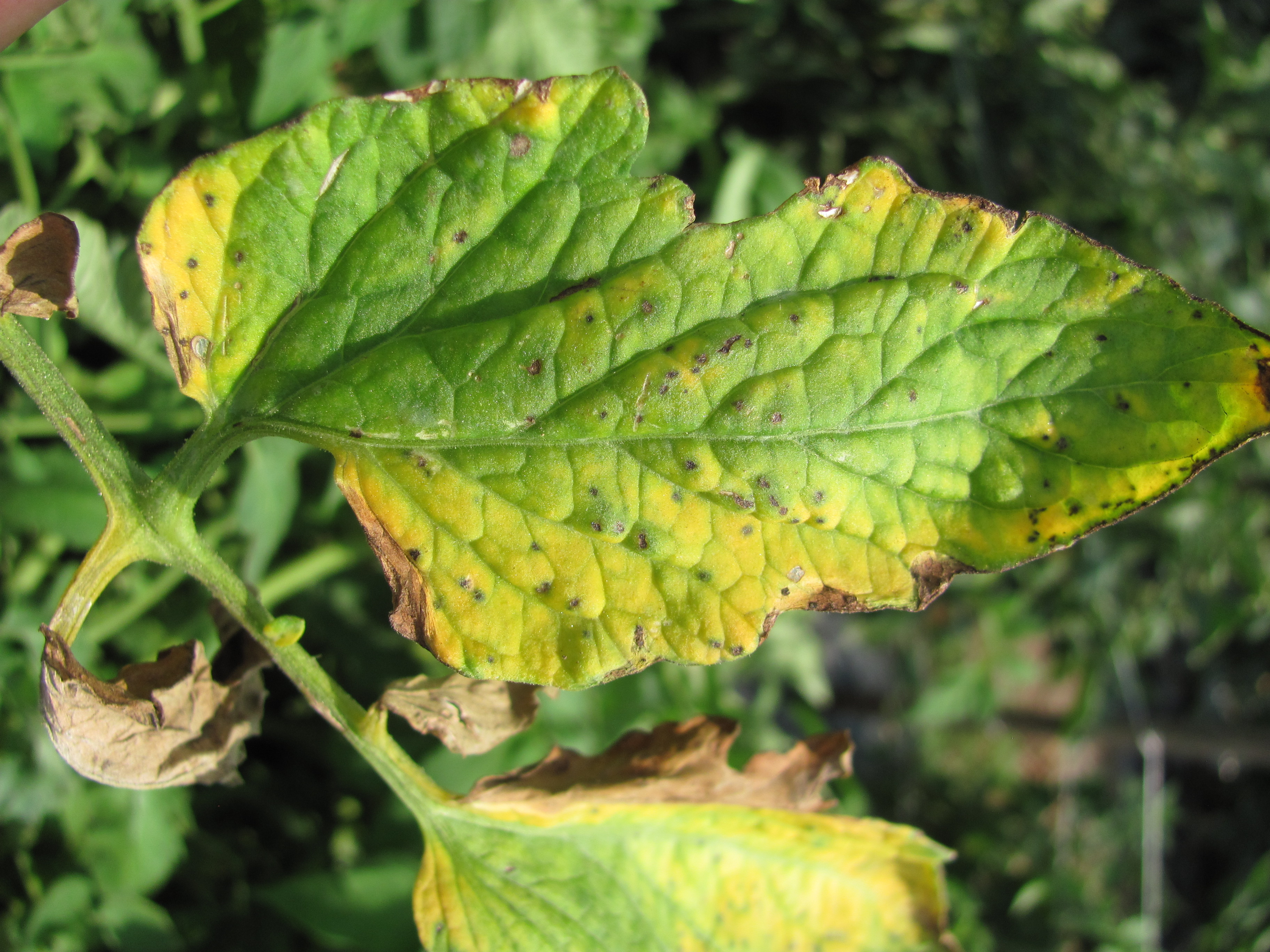 Figure 1. A tomato leaf with lesions of bacterial spot.
 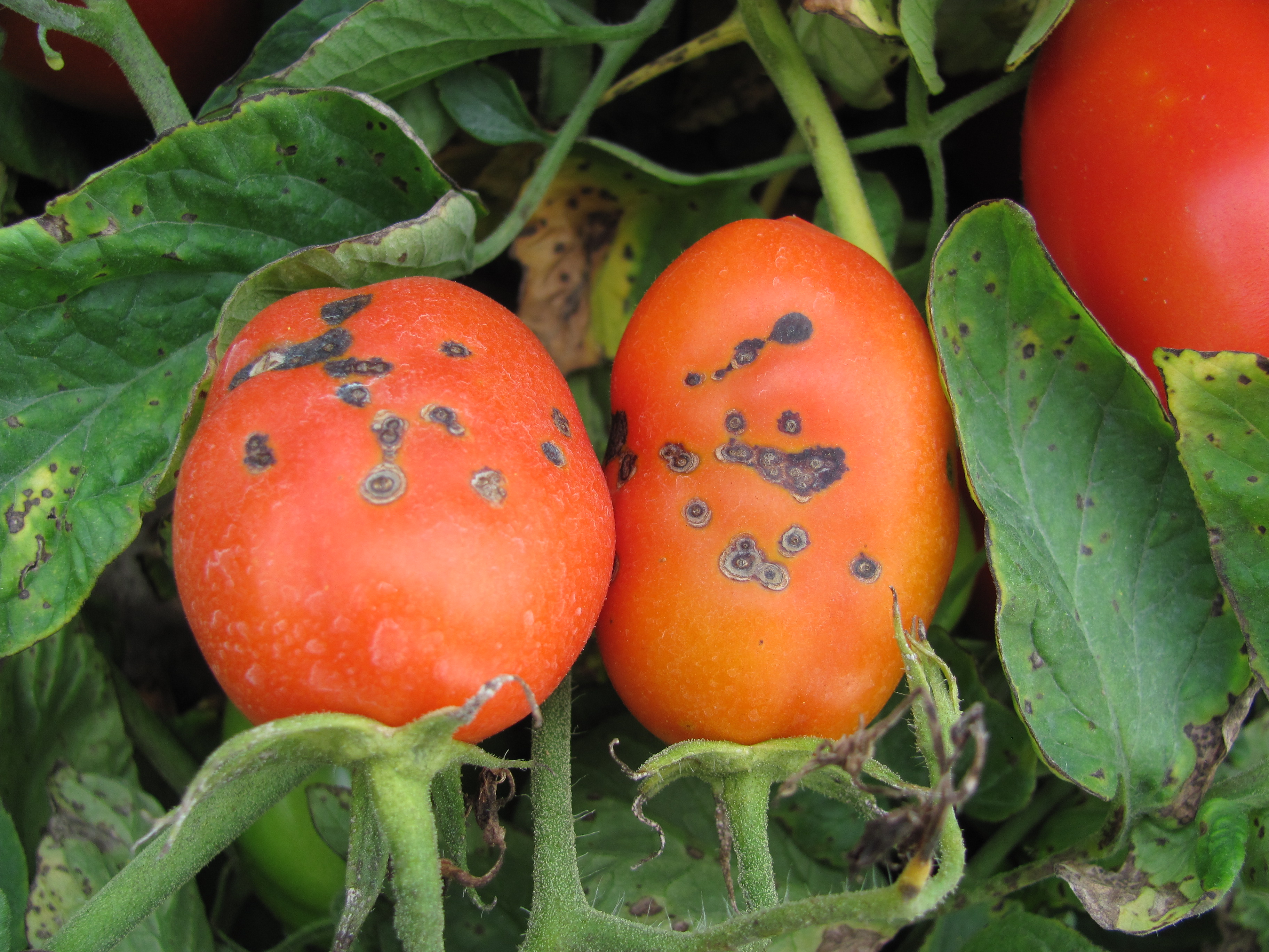 Figure 2. Bacterial spot on leaves and fruit.
 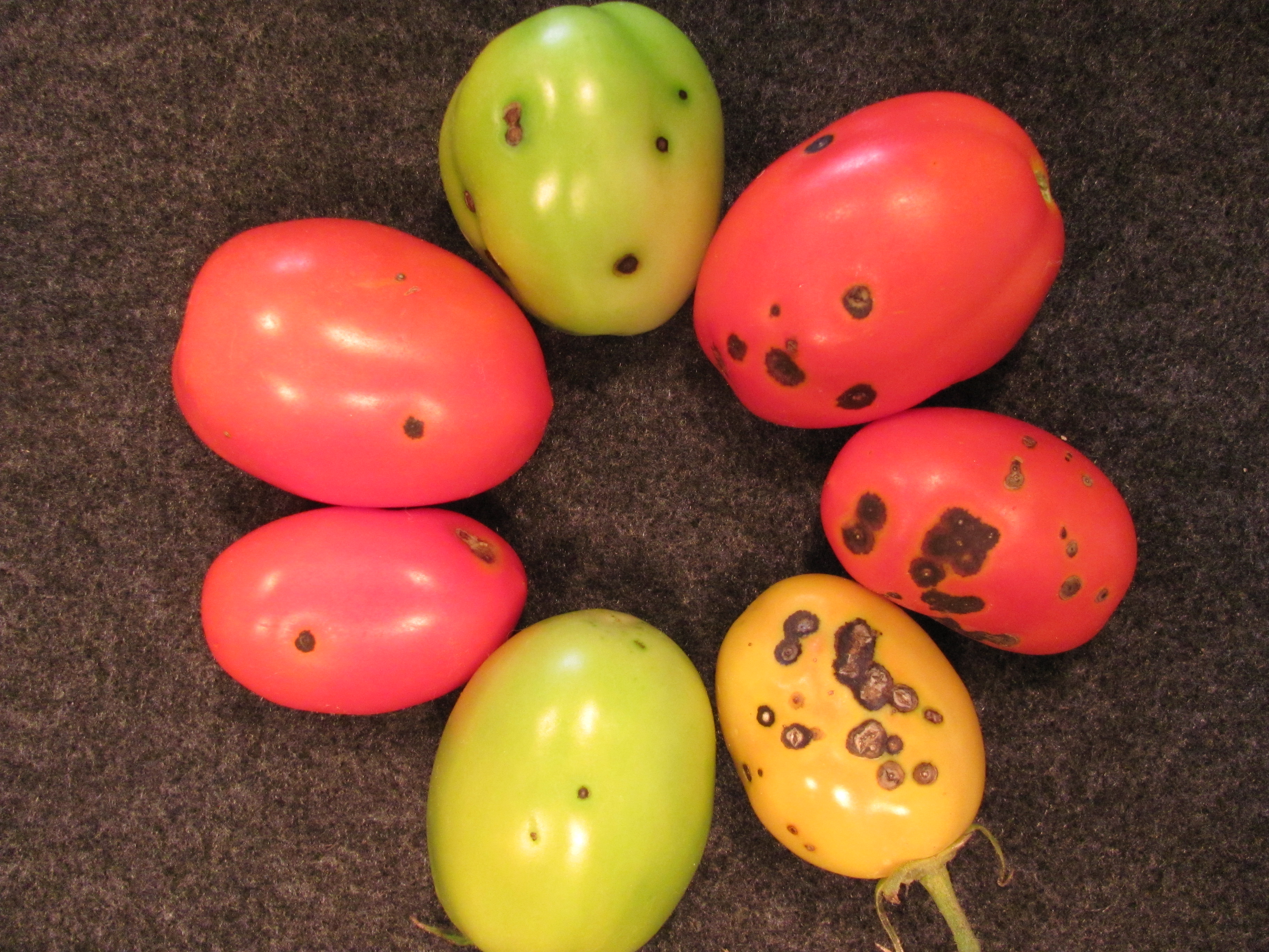 Figure 3. A range of symptoms of bacterial spot on fruit.
 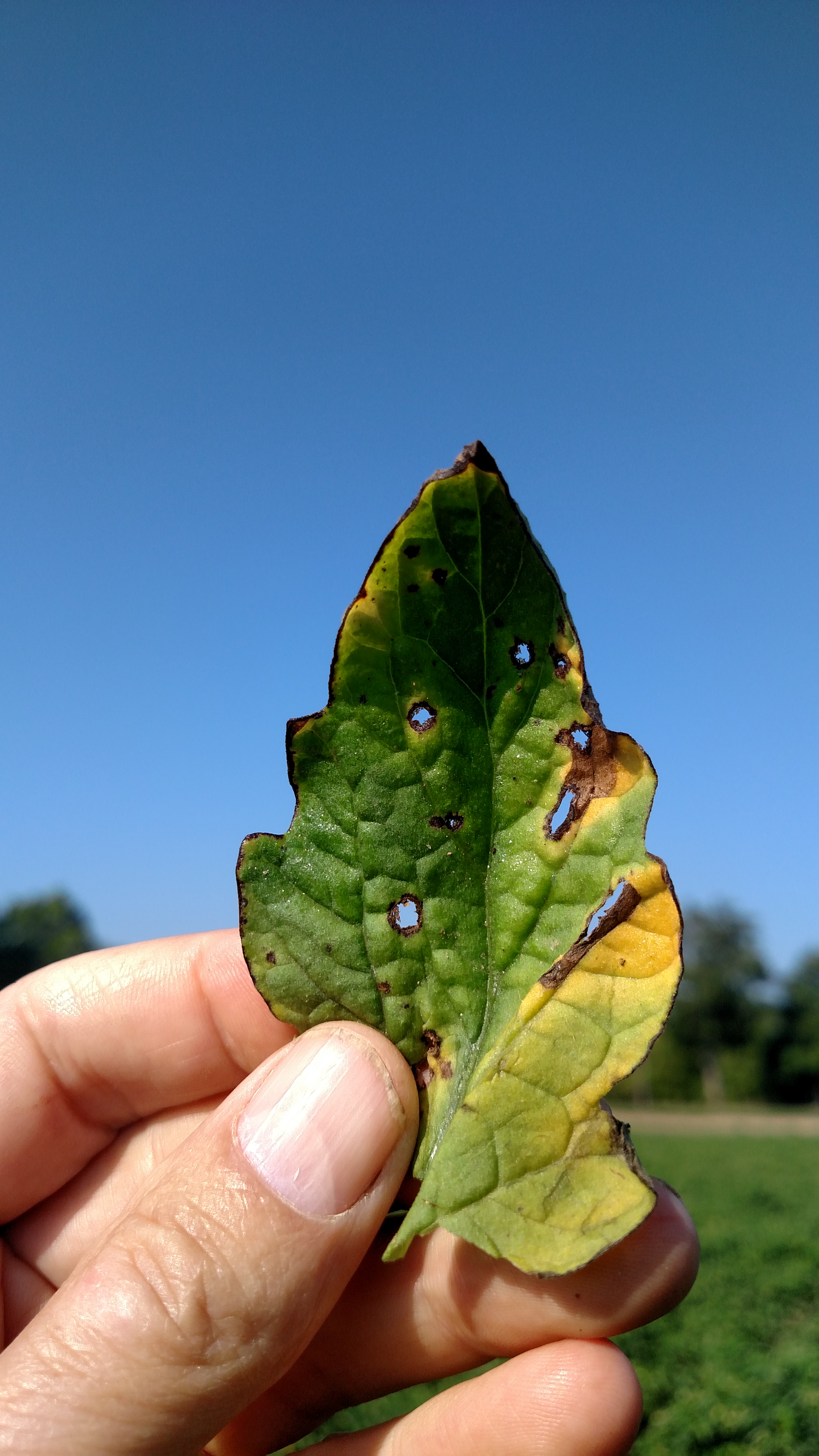 Figure 4. Symptoms caused by Xanthomonas perforans on a tomato leaf. The species name ‘perforans’ comes from the ability of the pathogen to perforate the leaf, although other symptom types are possible.
 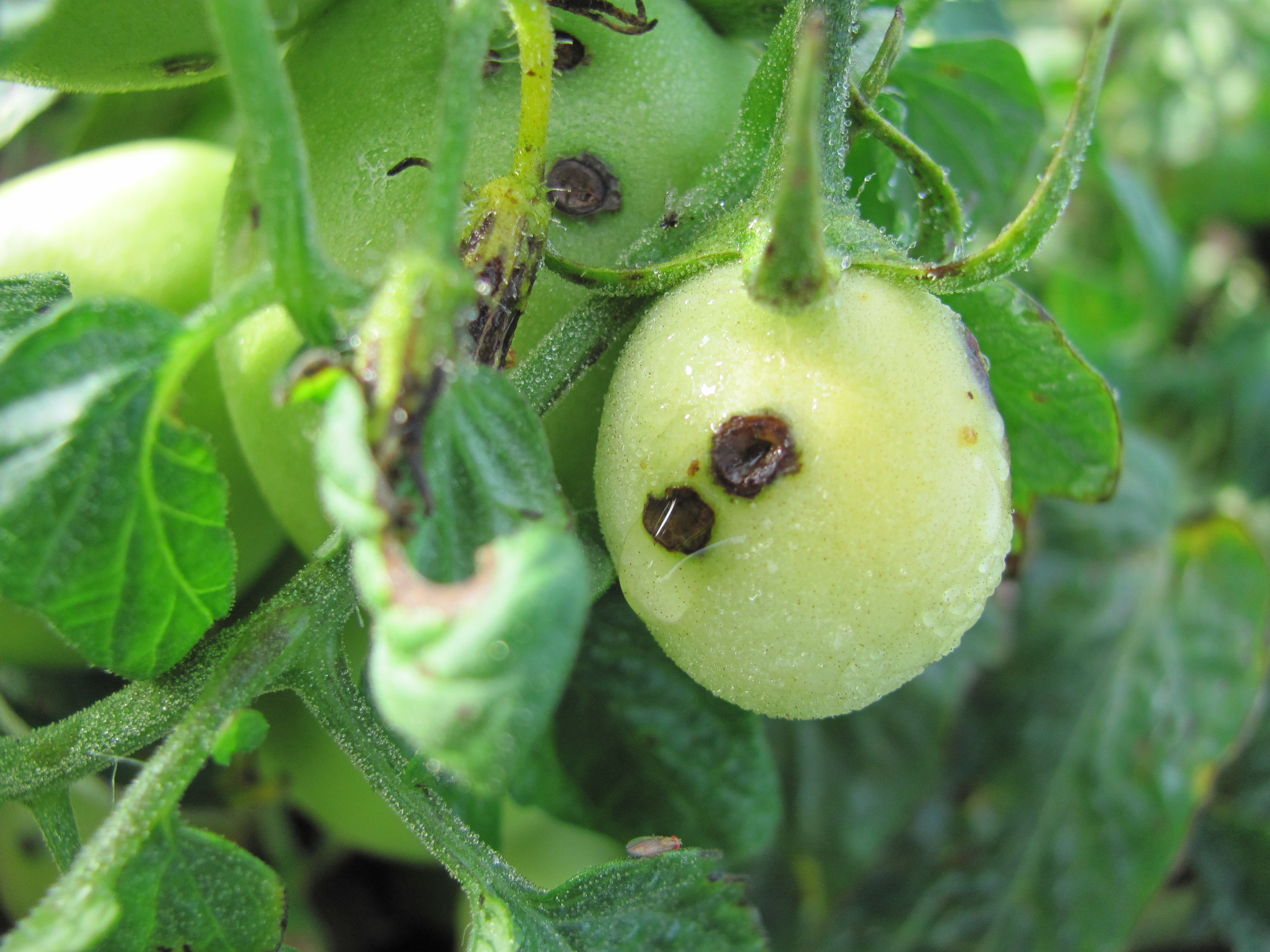 Figure 5. Bacterial spot lesions on fruit that is still wet with dew. Note the symptoms on flower bud.
 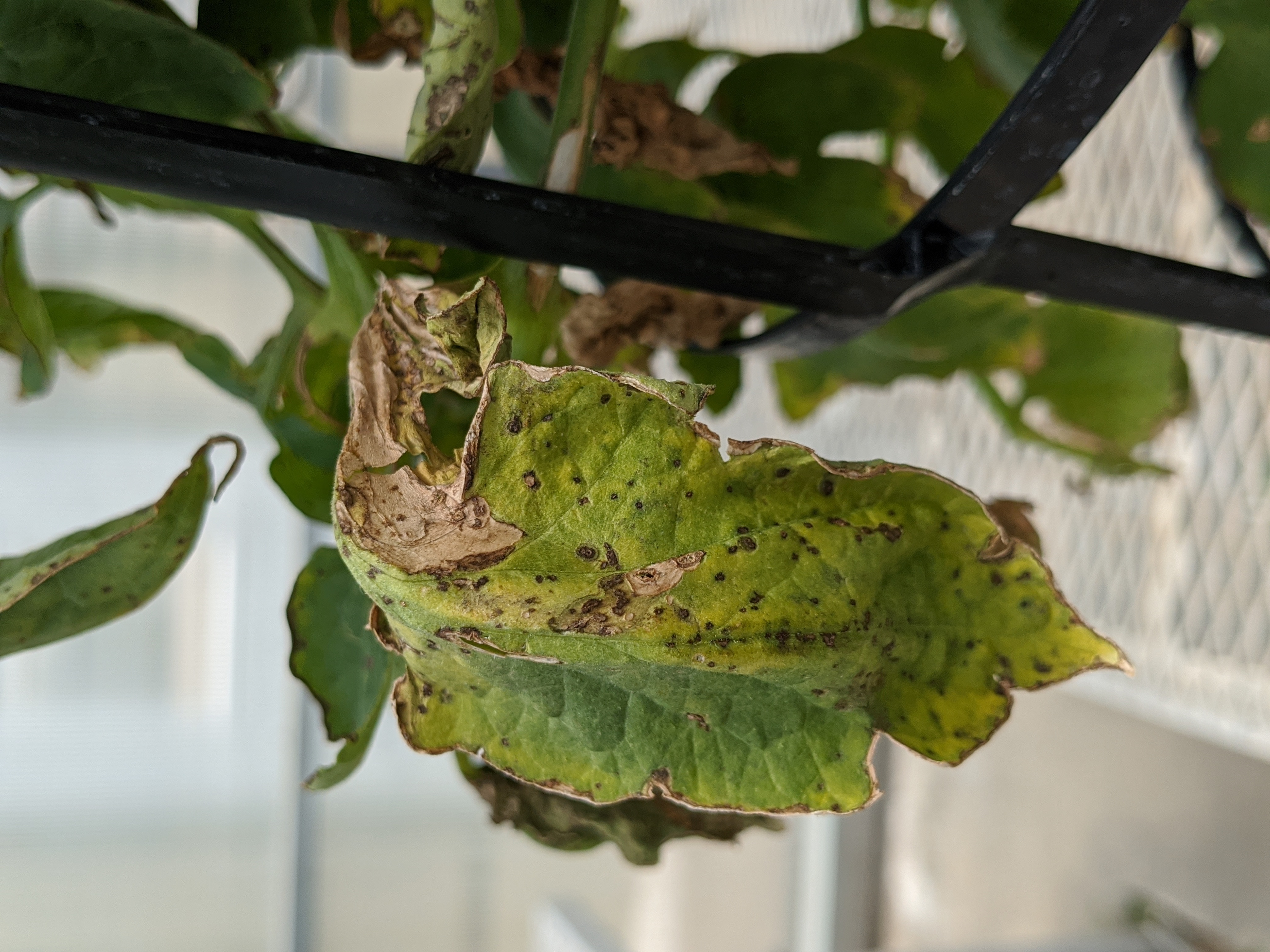 Figure 6. Lesions of bacterial spot on a tomato leaf.
 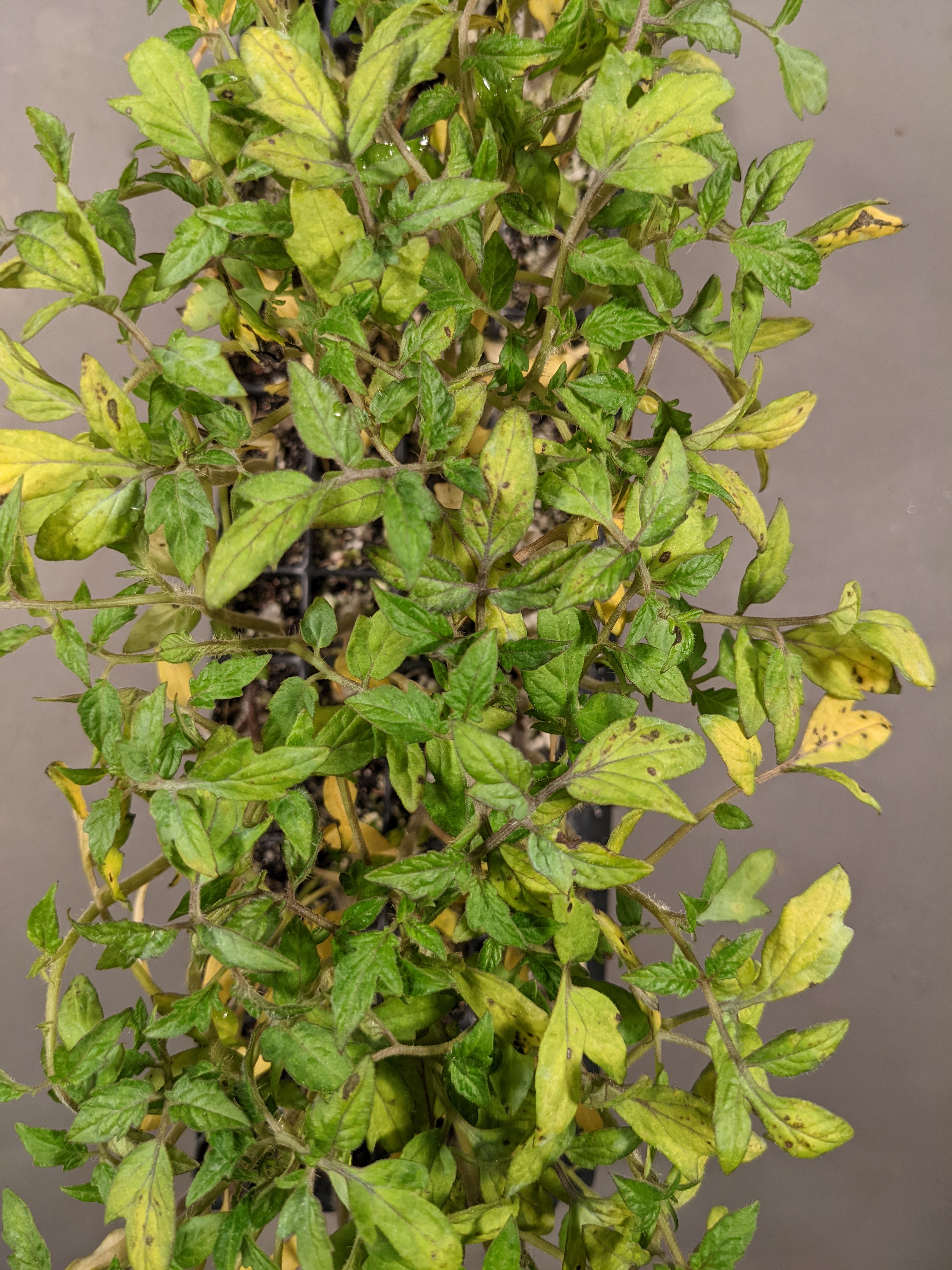 Figure 7. Bacterial spot on tomato transplants.
 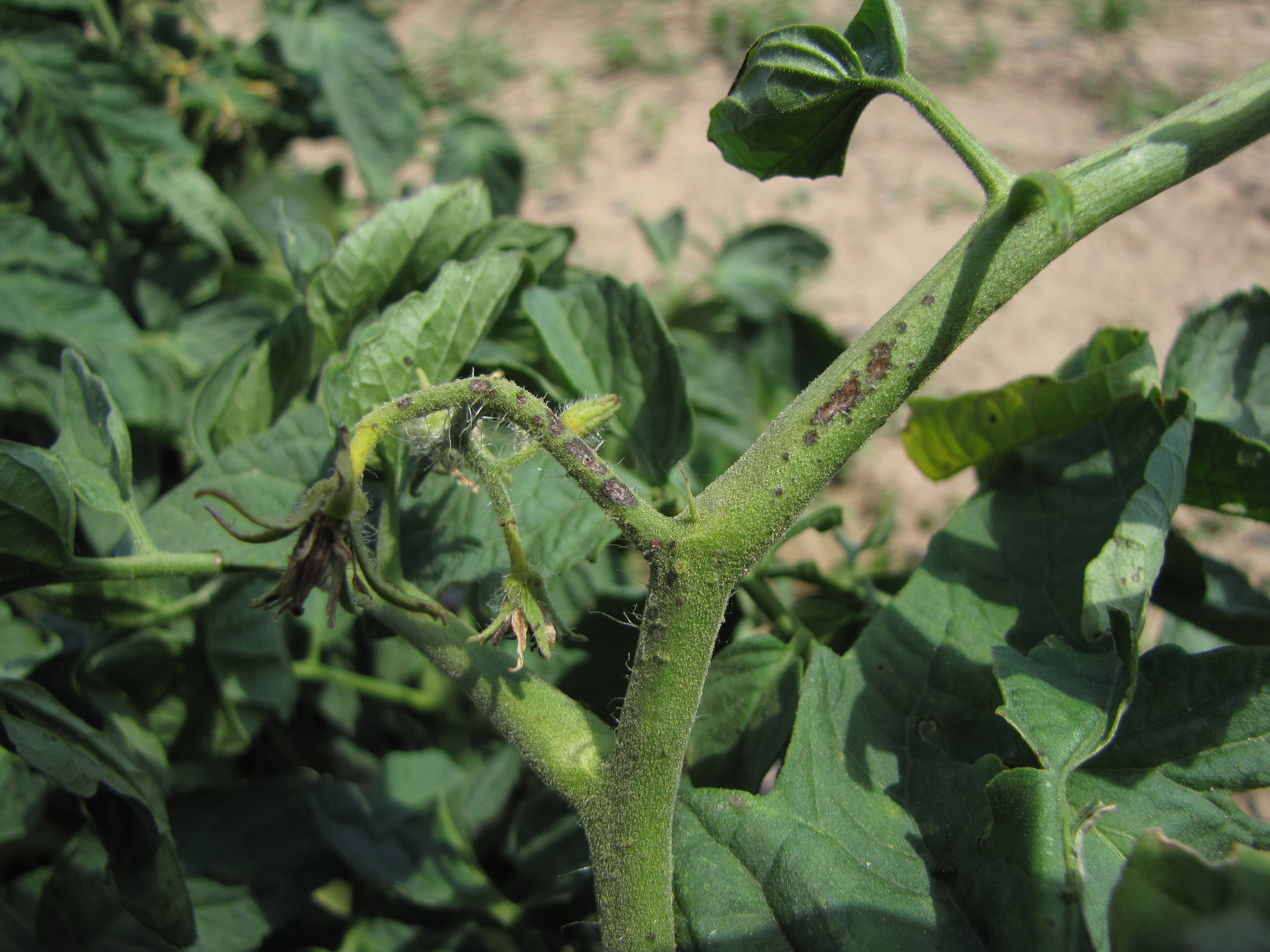 Figure 8. Bacterial spot symptoms on tomato stem.
 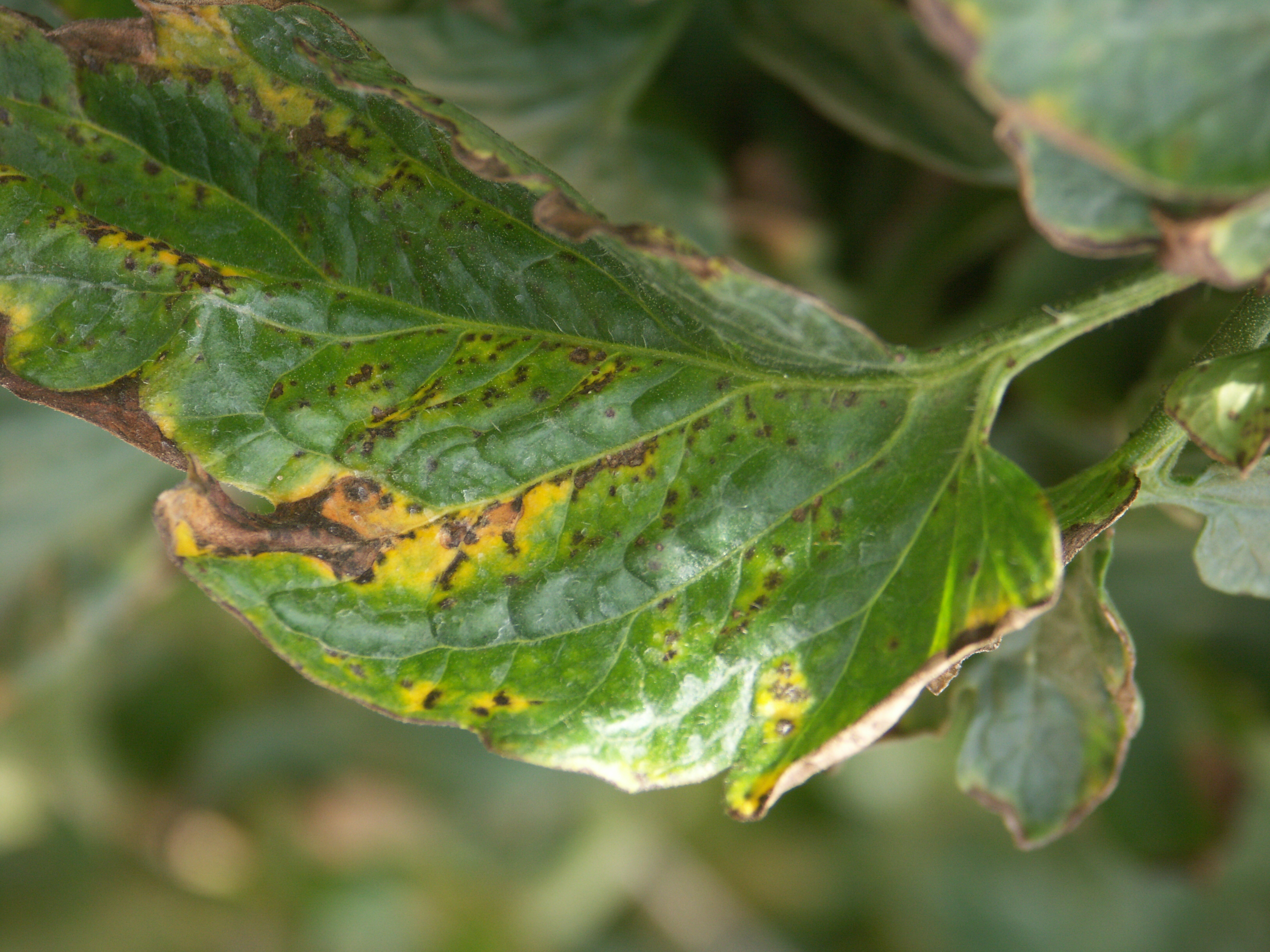 Figure 9. Lesions of bacterial spot on tomato leaf. Note that chlorotic areas often occur where several lesions occur close together.
 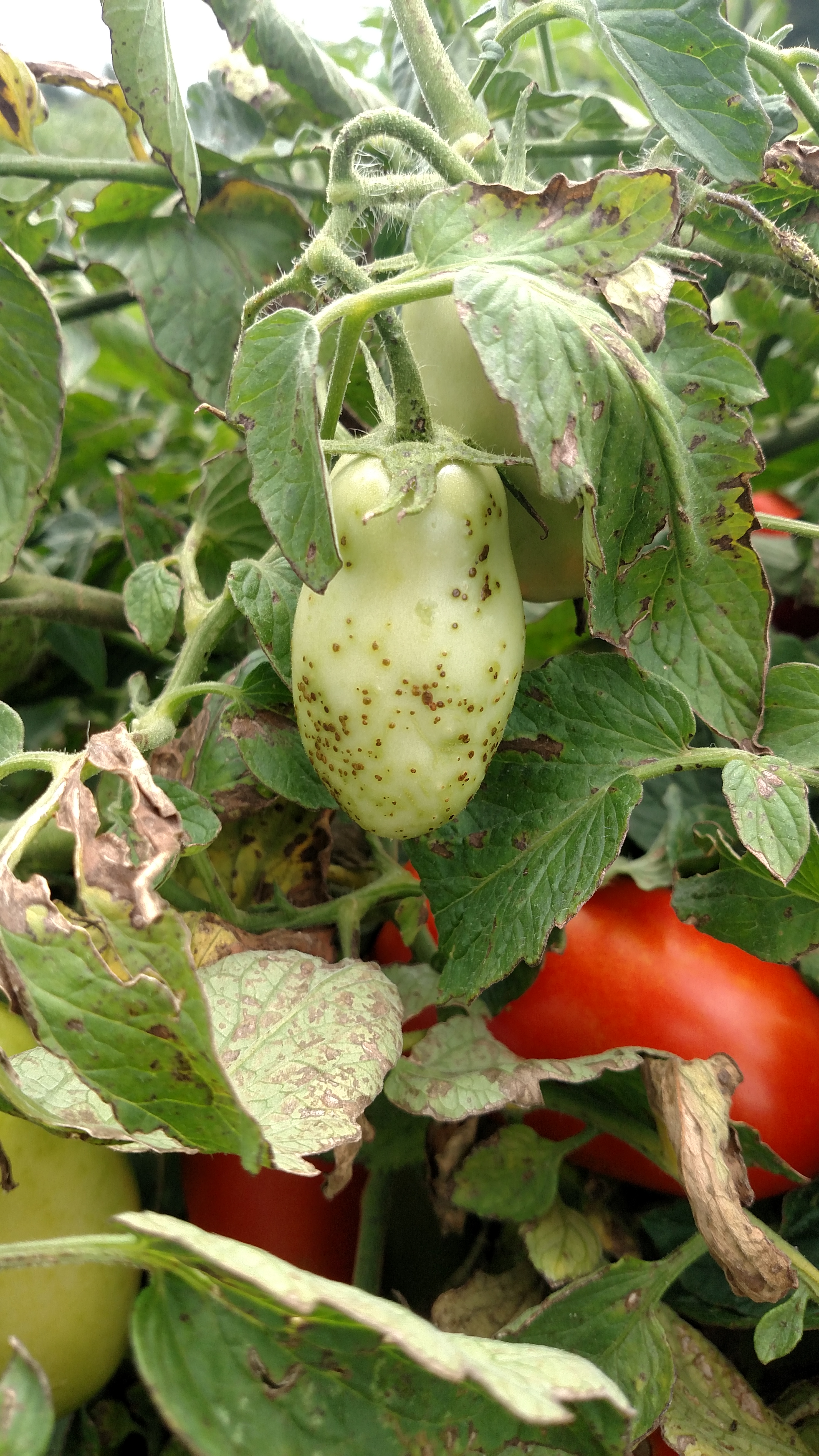 Figure 10. Tomato fruit severely affected by bacterial spot.
 Blossom end rot
Blossom-end rot-This is not an infectious disease. Nevertheless, it is important to differentiate these symptoms from infectious diseases. Note leathery-like lesions on blossom end.
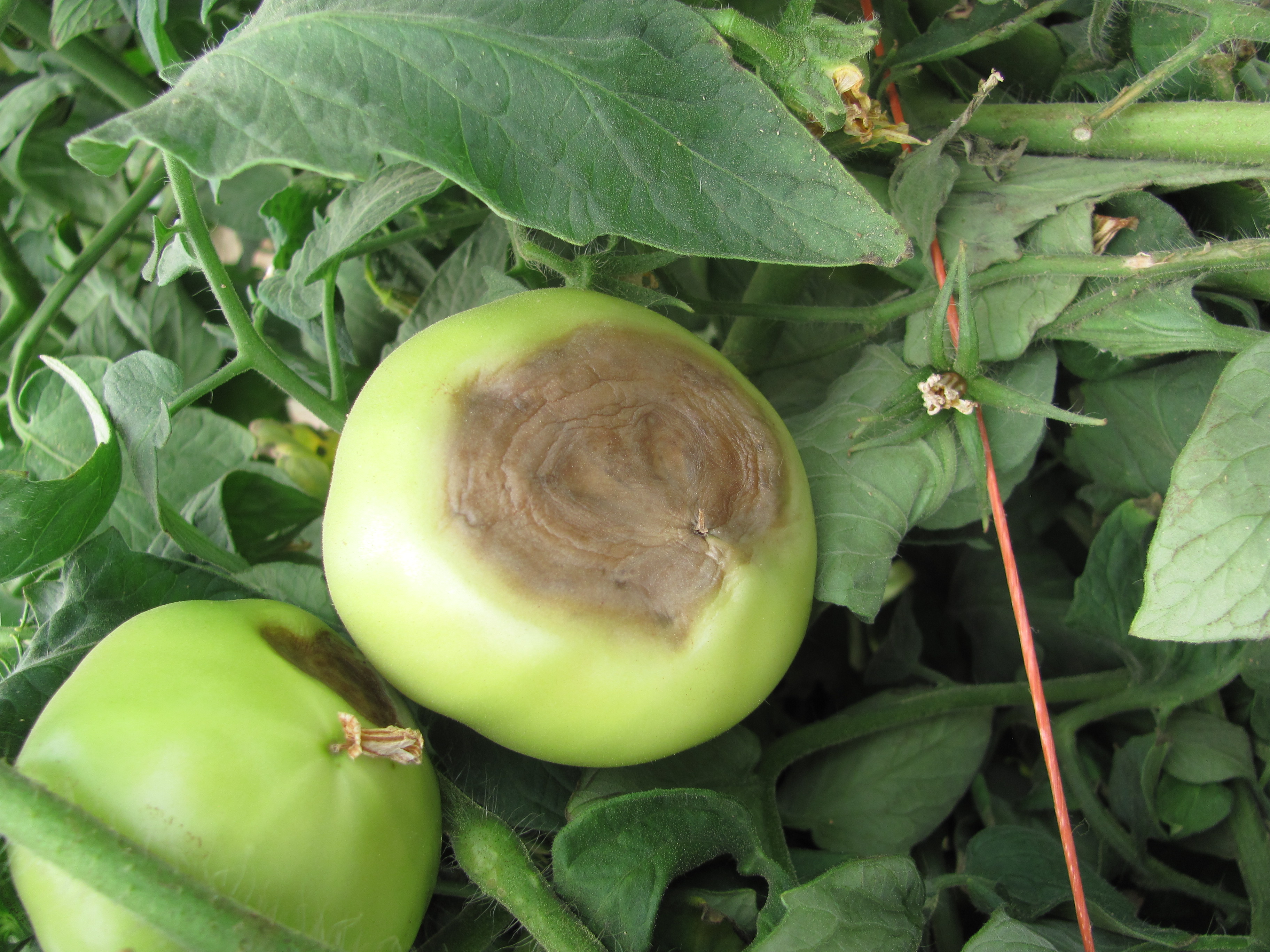 Figure 1. Blossom end rot of tomato can be recognized by a leathery brown lesion at the base of the tomato.
 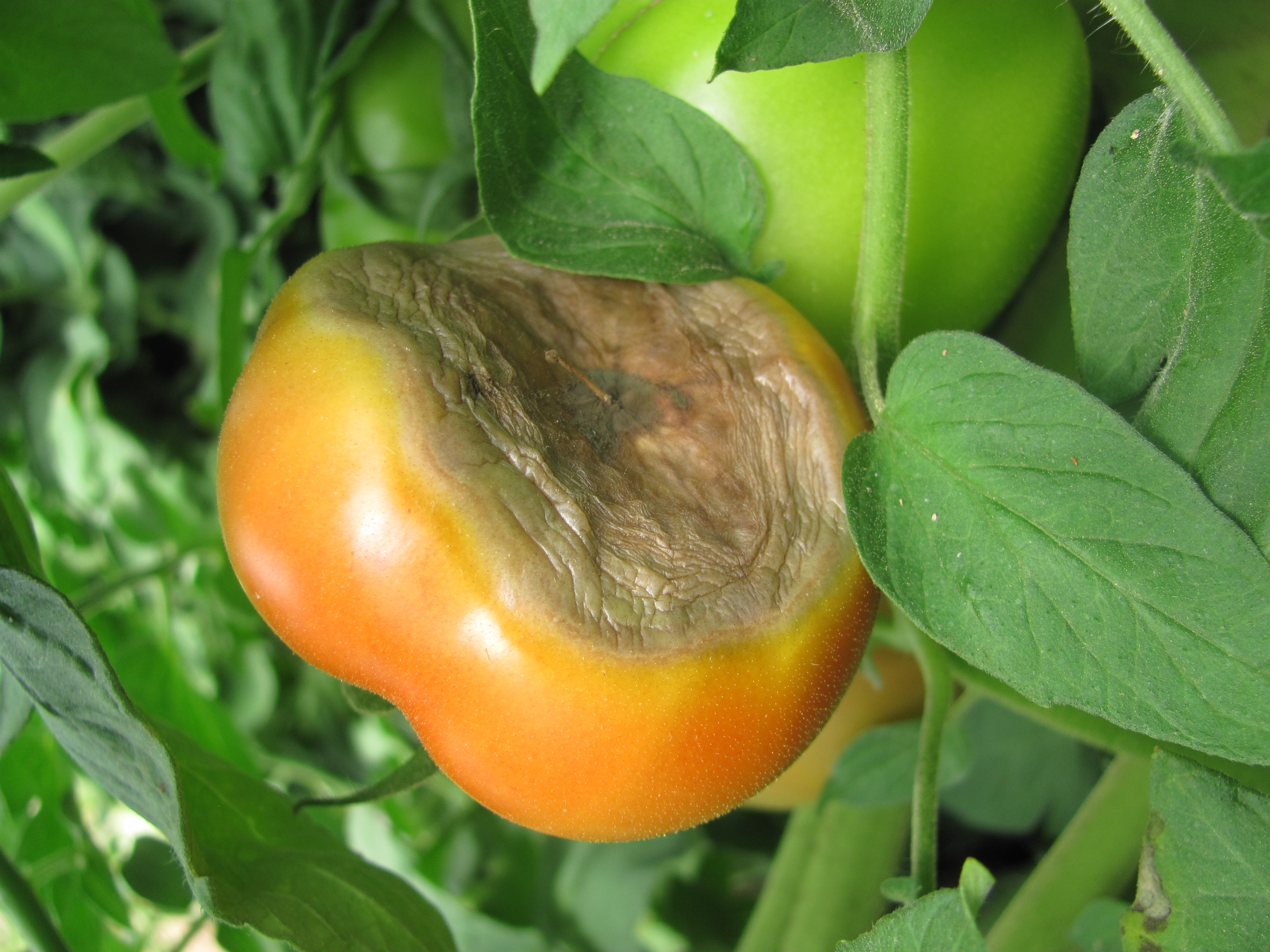 Figure 2. Blossom end rot of red tomato.
 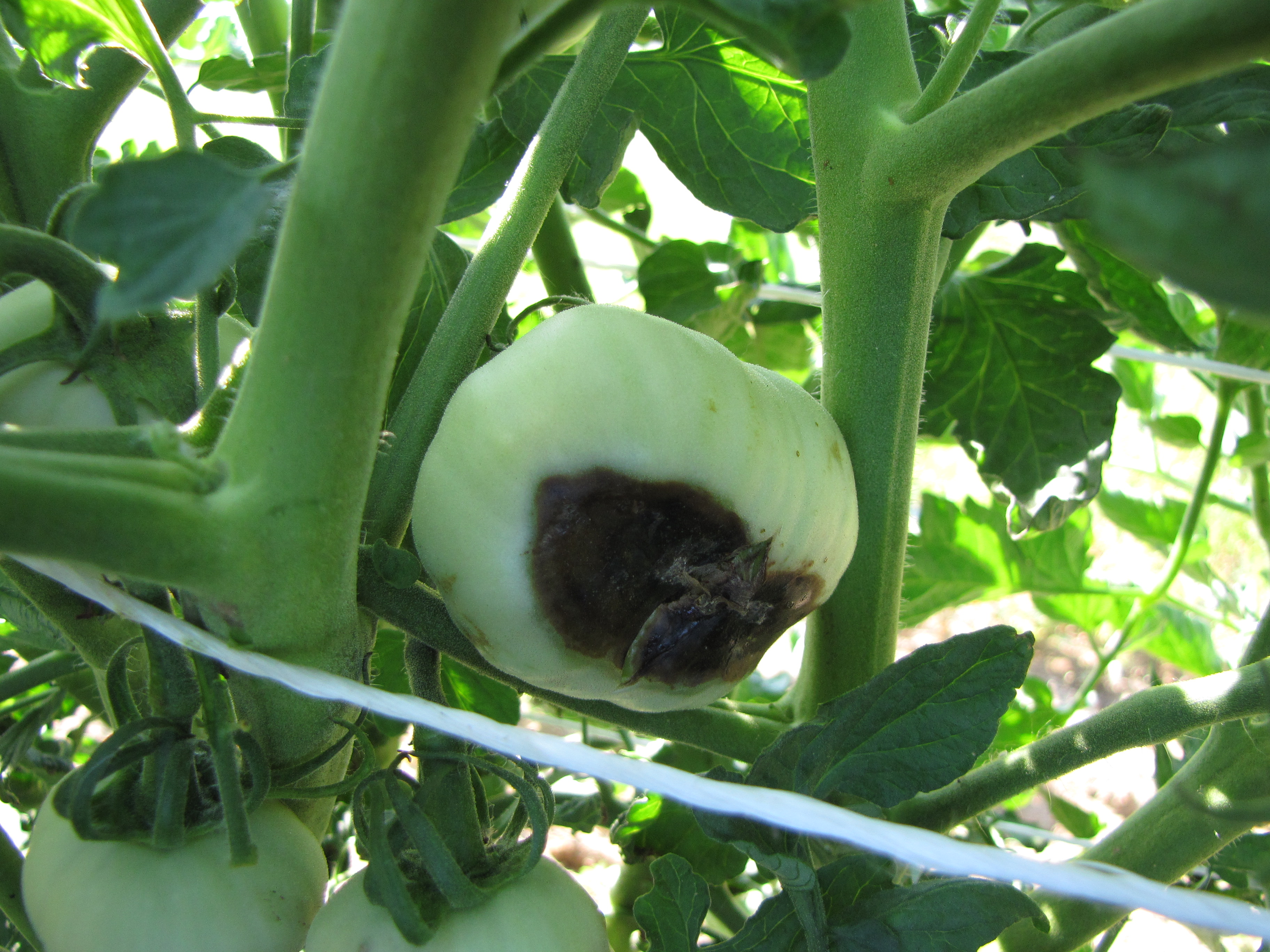 Figure 3. Blossom end rot of tomato.
 Buckeye rot
Buckeye rot-I have only observed this disease in processing fields. The characteristic symptom is the dark, concentric pattern of necrosis on tomato fruit.
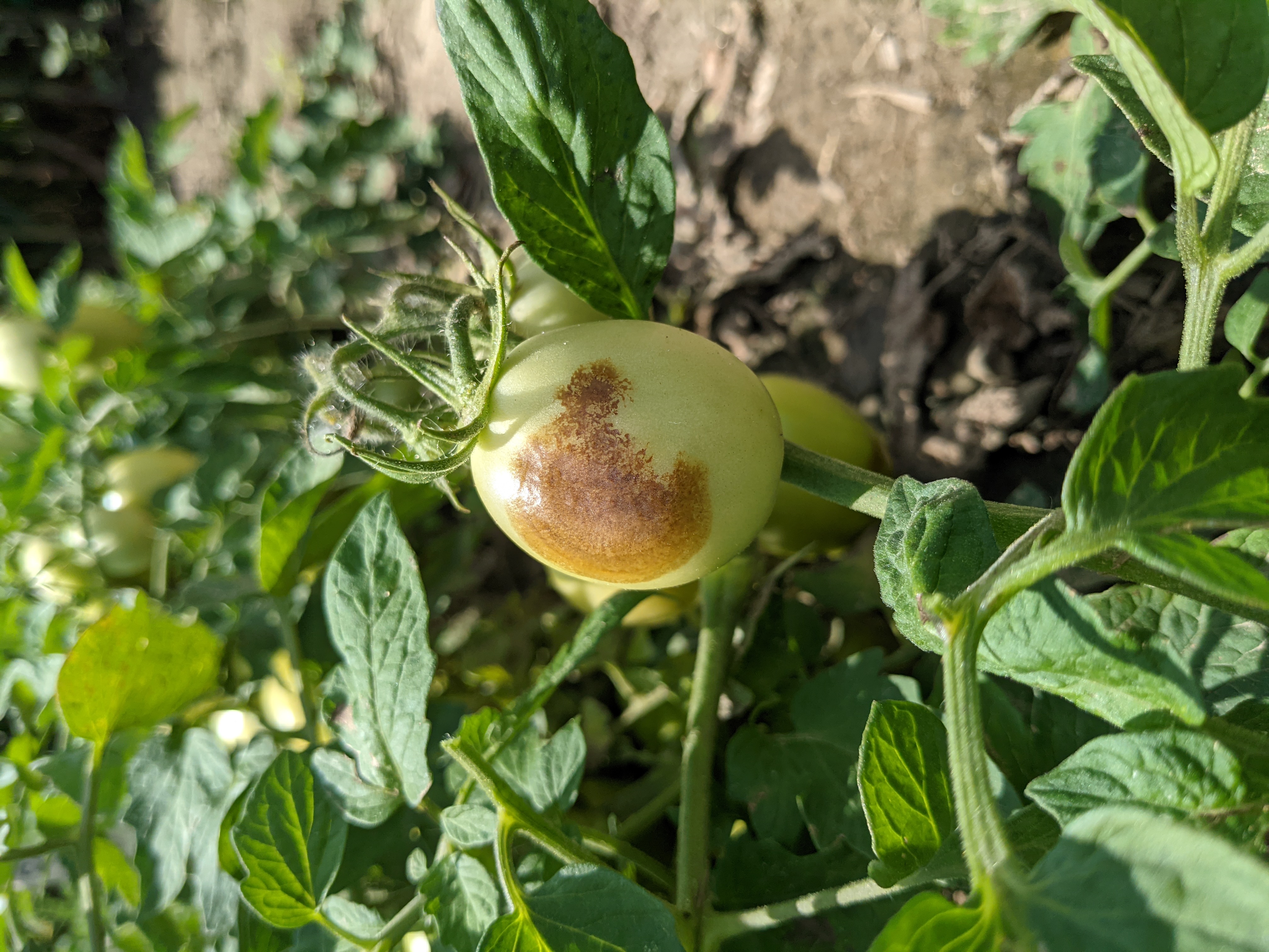 Figure 1. A processing tomato with Buckeye rot.
 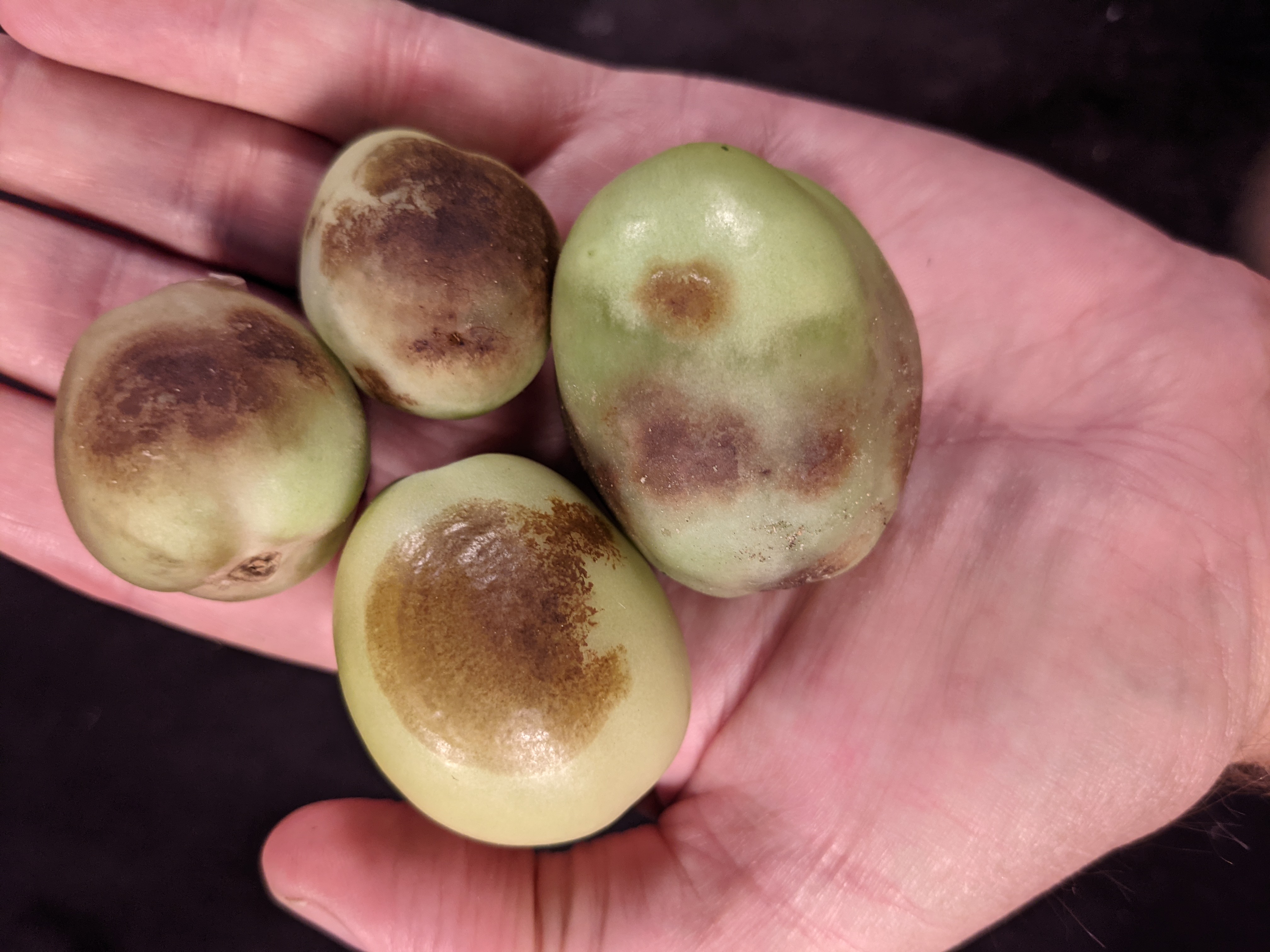 Figure 2. Four processing tomatoes with diverse symptoms of Buckeye rot.
 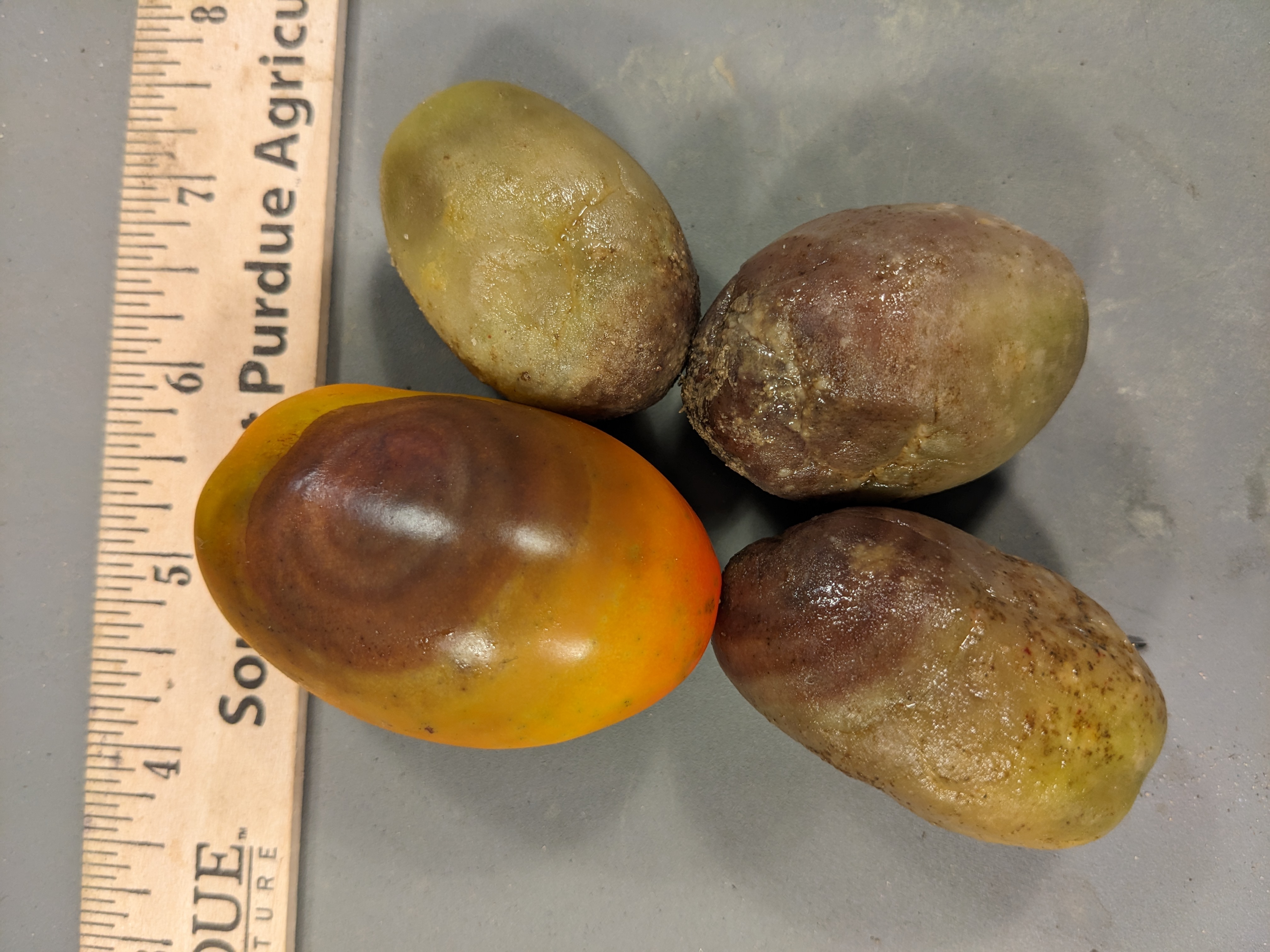 Figure 3. Four tomatoes with Buckeye rot with a range of symptoms. Concentric circles of necrosis is typical of Buckeye rot.
 Cercospora leaf mold
Cercospora leaf mold-This disease is known as a tropical or subtropical disease. In recent years, however, this disease has become more frequent in Indiana, particularly in high tunnels. Note dark or black sporulation in contrast with olive green sporulation common in leaf mold.
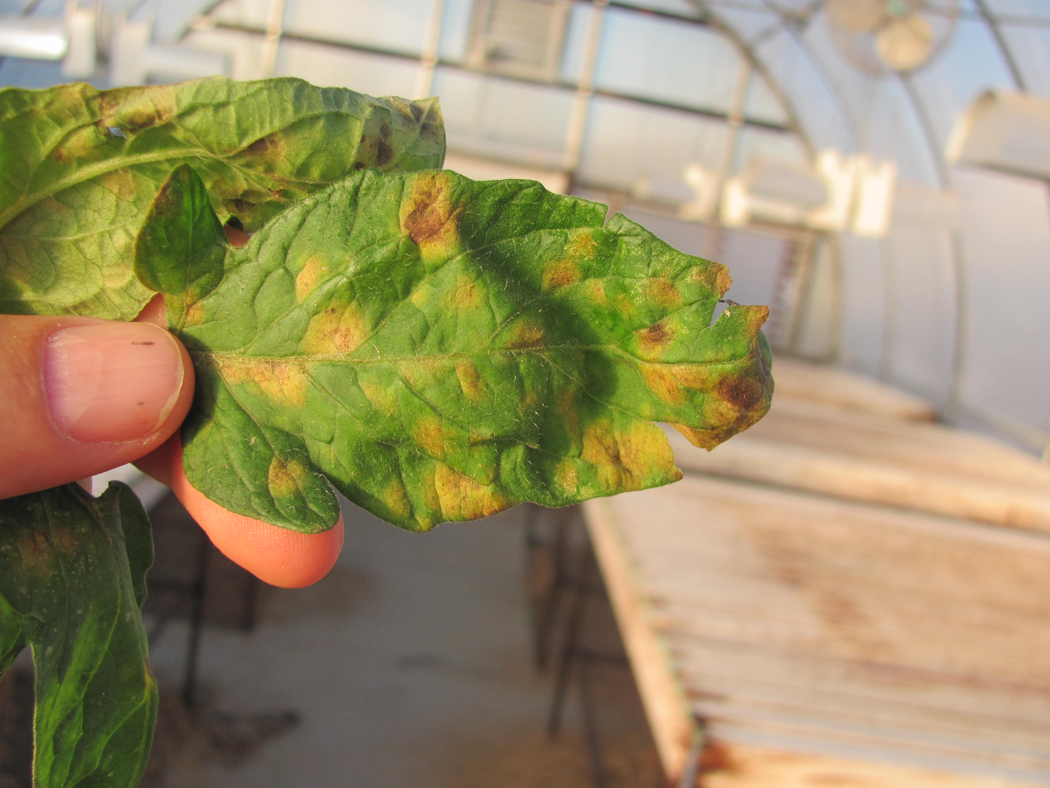 Figure 1. Cercospora leaf mold of tomato. Note relatively large lesions with diffuse chlorotic margins.
 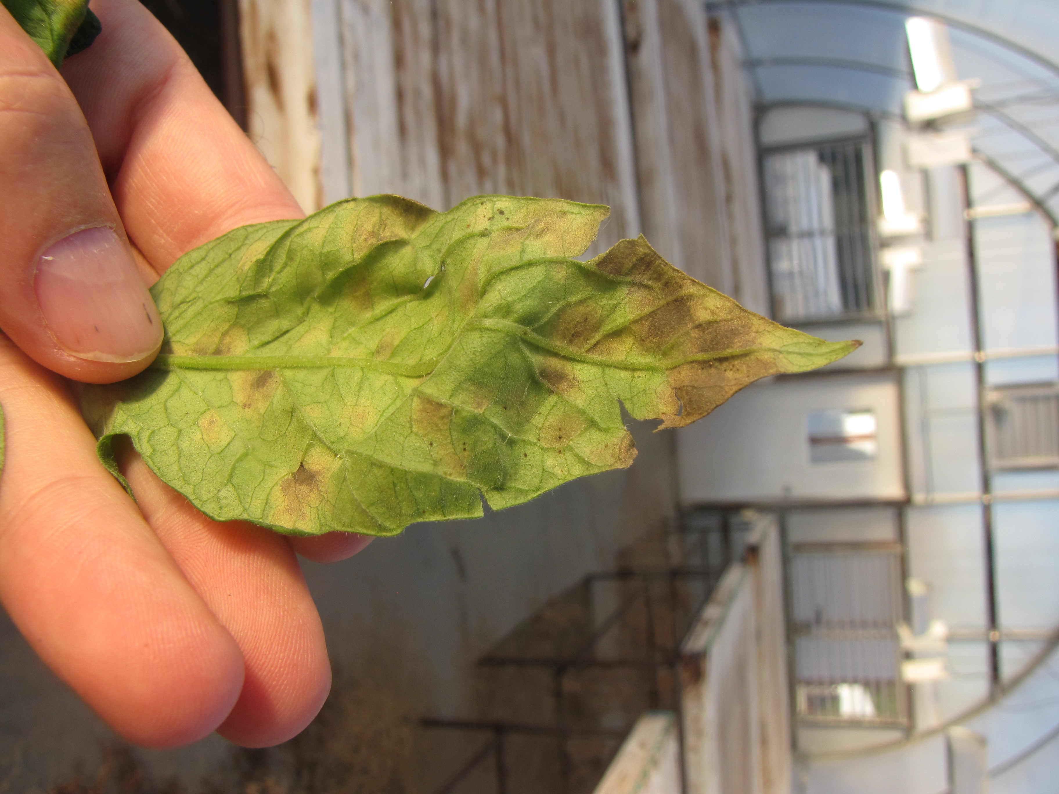 Figure 2. Underside of tomato leaf with Cercospora leaf mold. Note dark sporulation of pathogen and compare with leaf mold of tomato.
 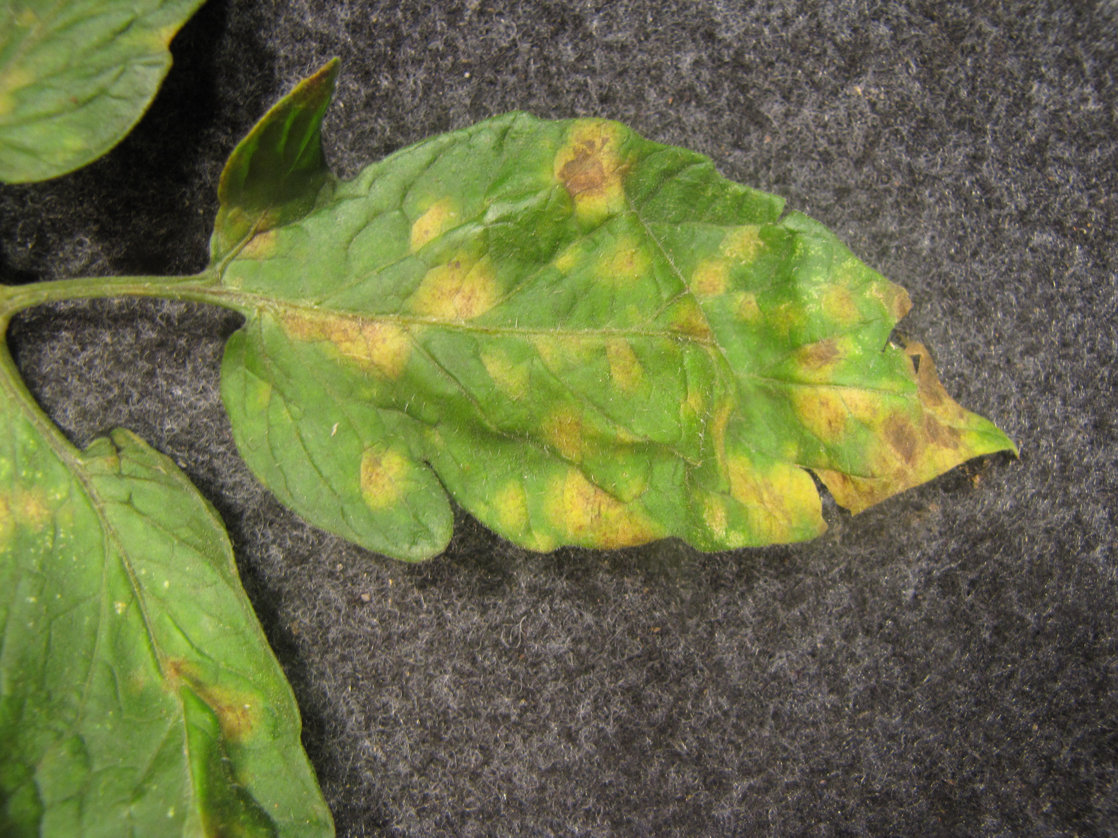 Figure 3. Cercospora leaf mold of tomato. Note that smaller lesions are chlorotic while larger lesions have become necrotic.
 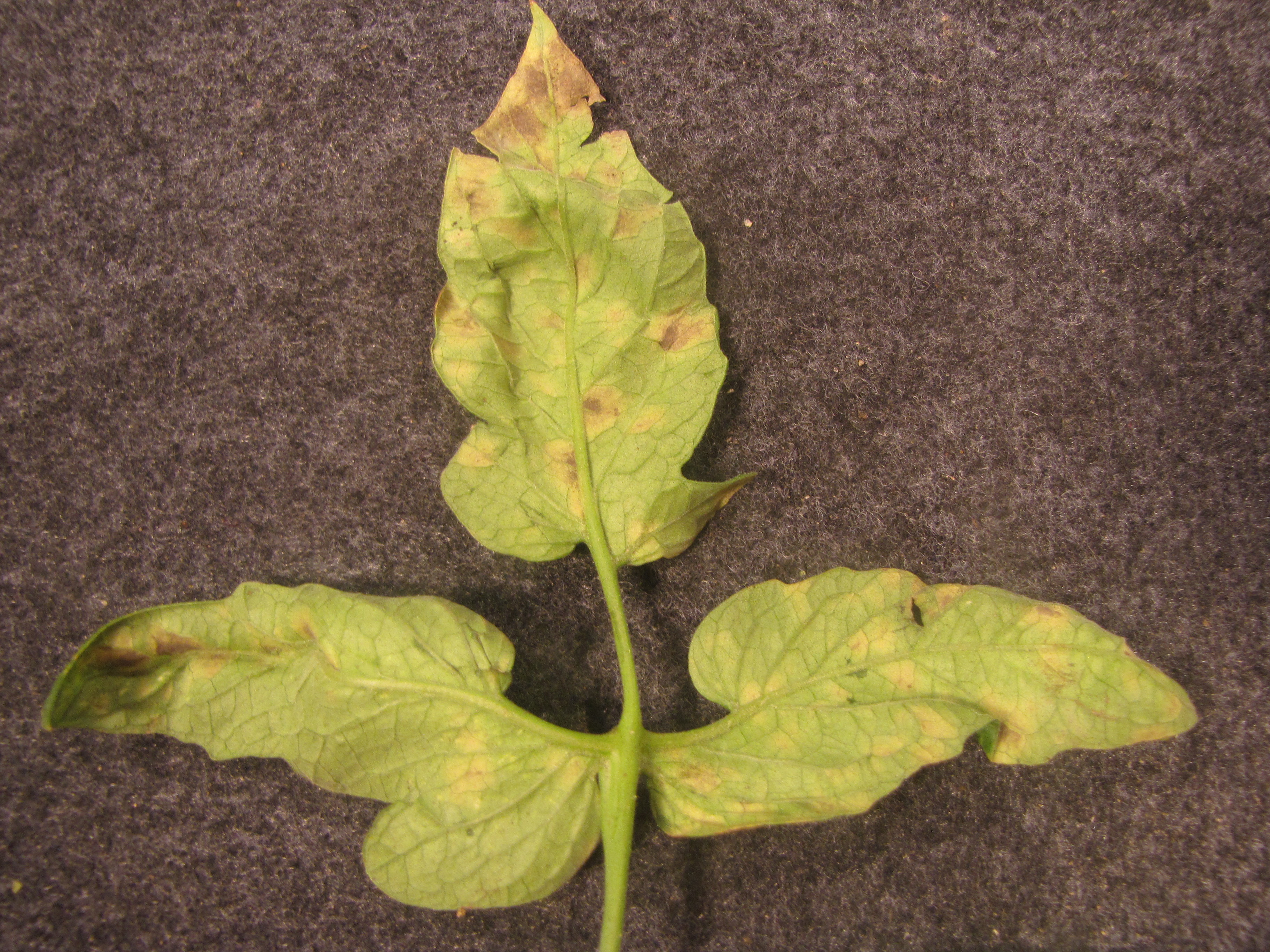 Figure 4. Another look at the underside of a tomato leaf with sporulation of Cercospora leaf mold.
 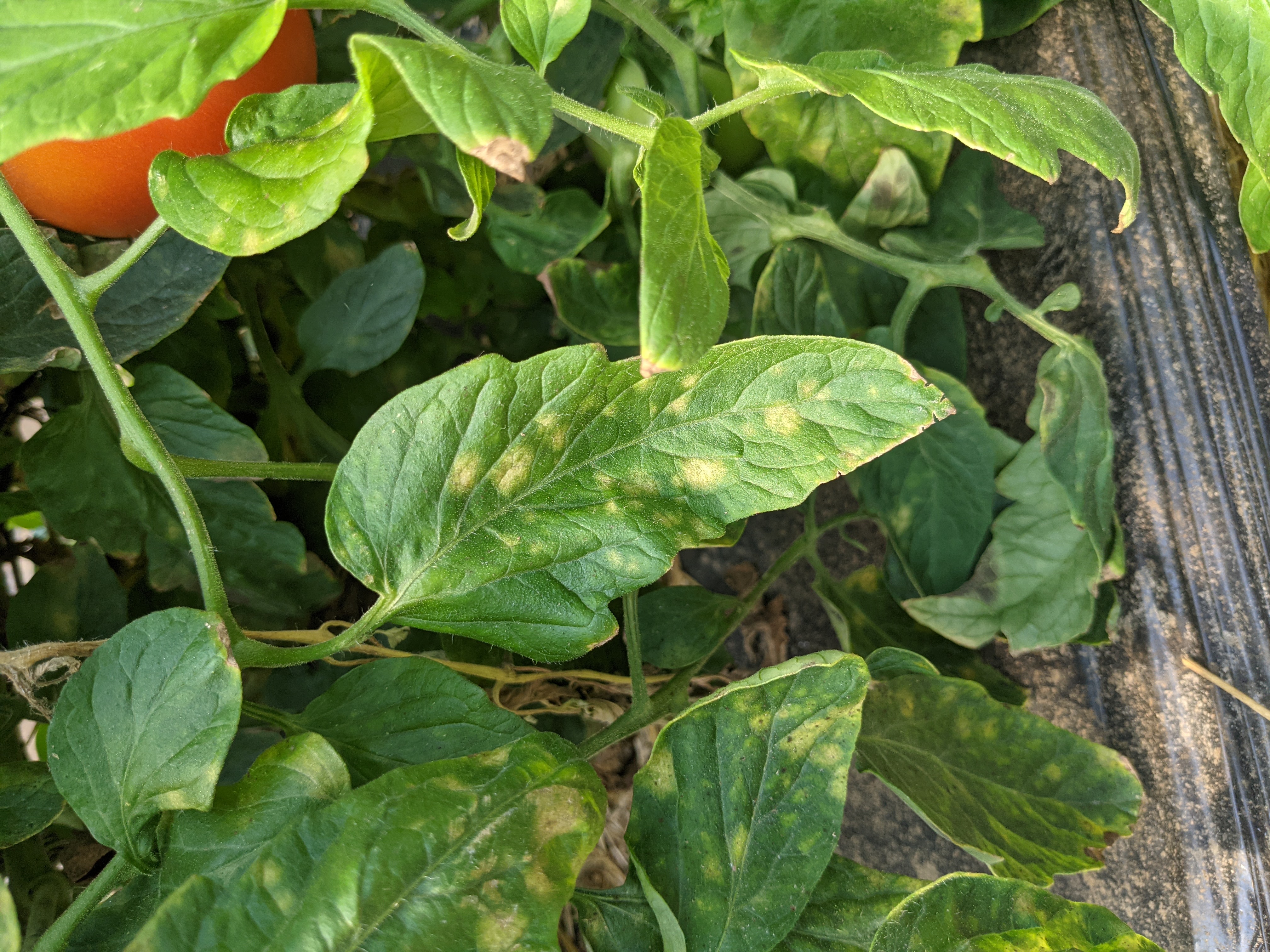 Figure 5. Cercospora leaf mold of tomato in a high tunnel.
 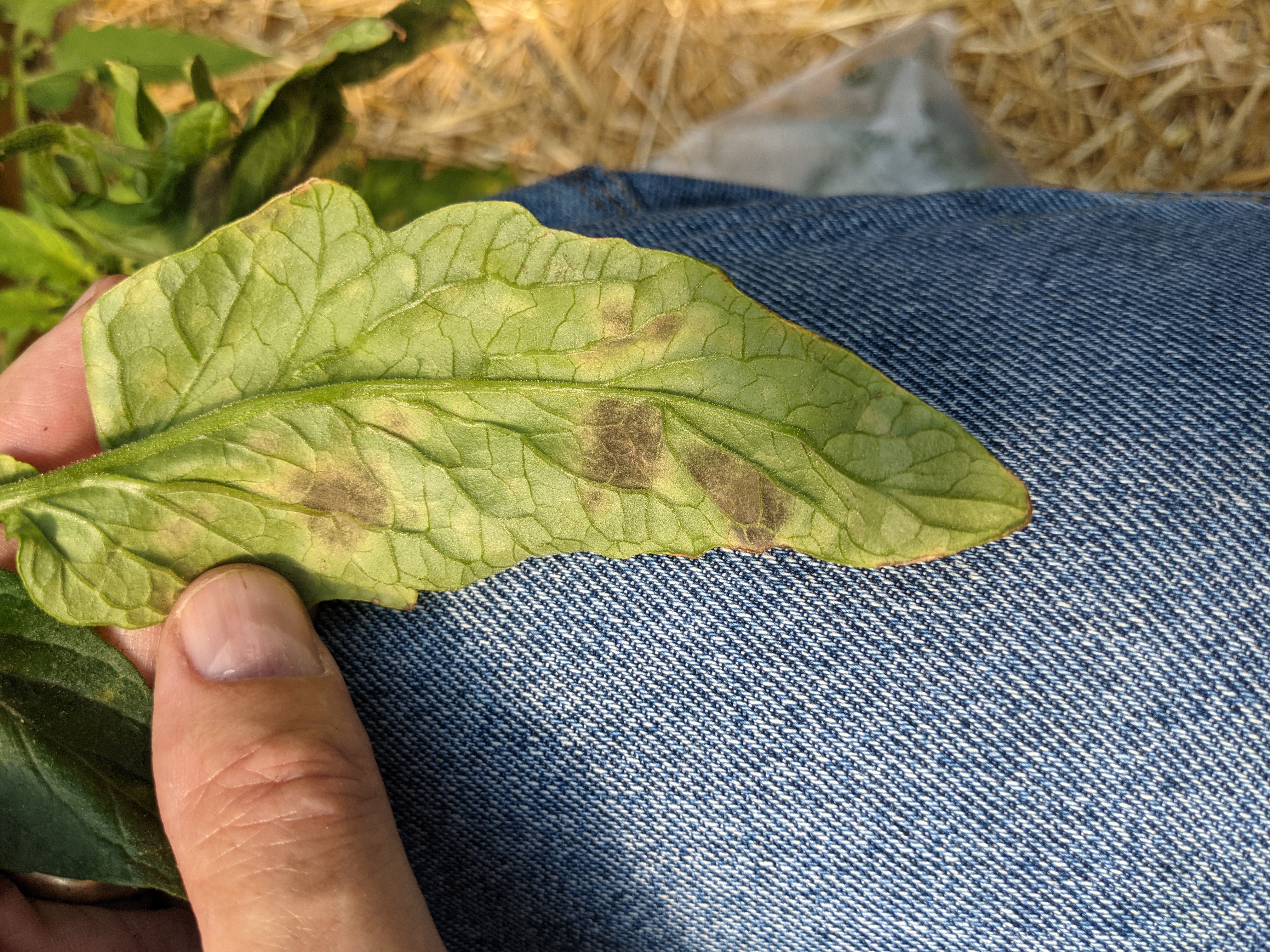 Figure 6. Another view of the dark sporulation of Cercospora leaf mold of tomato.
 Early blight
Early blight-One of the most common tomato diseases in the Midwest. Note older leaves are more severely affected. Concentric ring lesion is typical. Common in field production, but not difficult to manage. Less common in high tunnels.
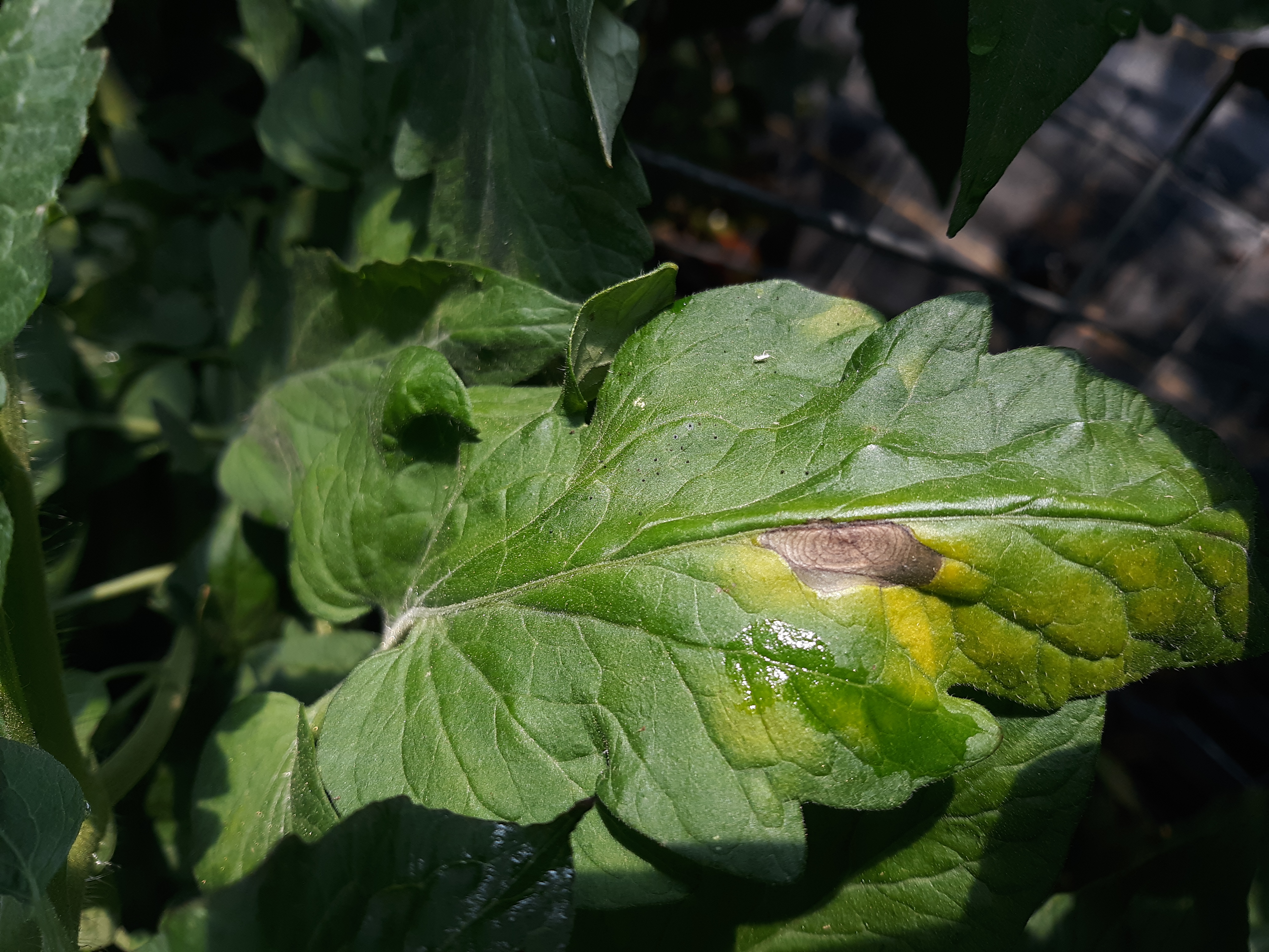 Figure 1. Early blight lesion on tomato leaf.
 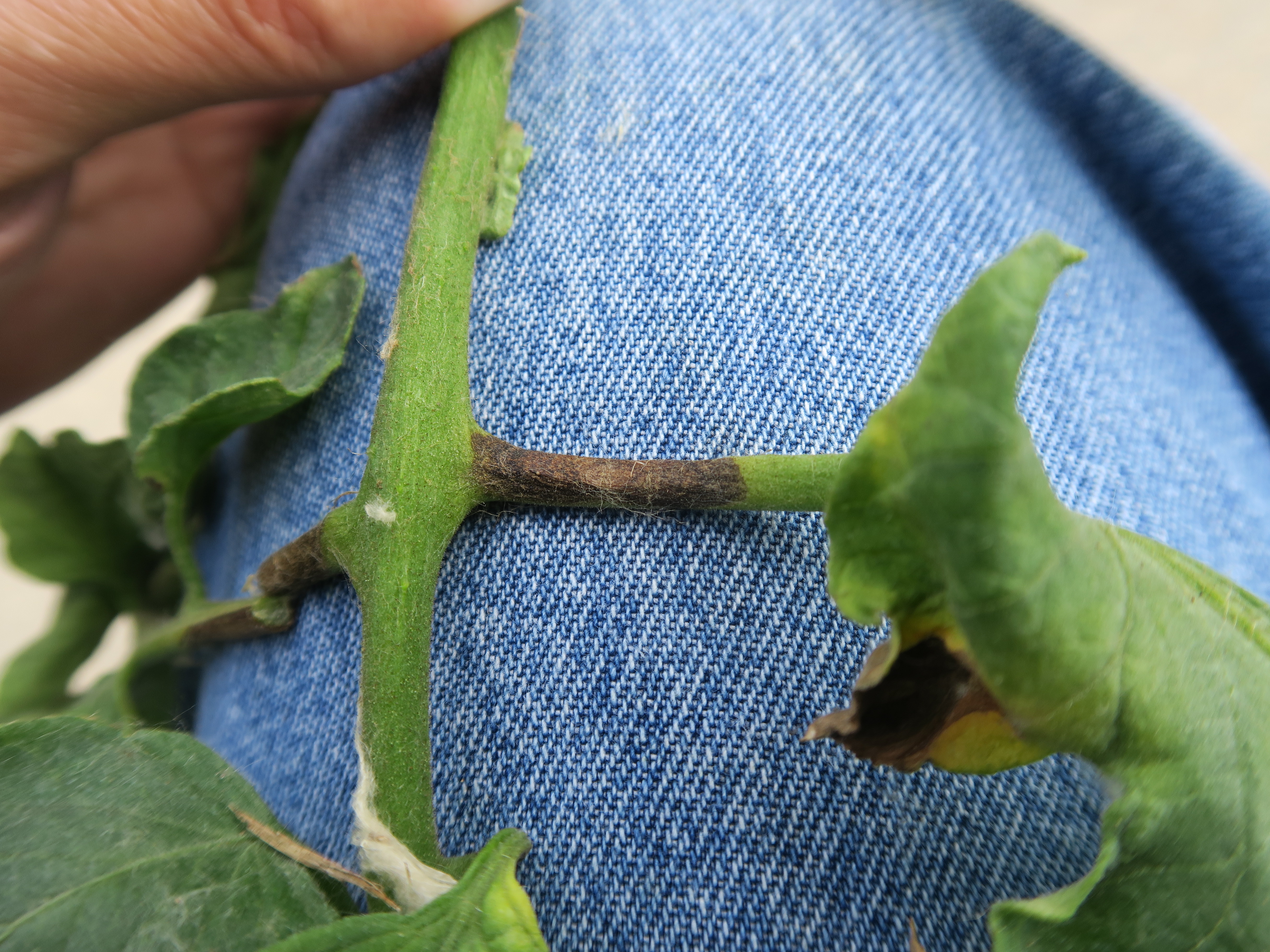 Figure 2. Petiole lesion of early blight of tomato.
 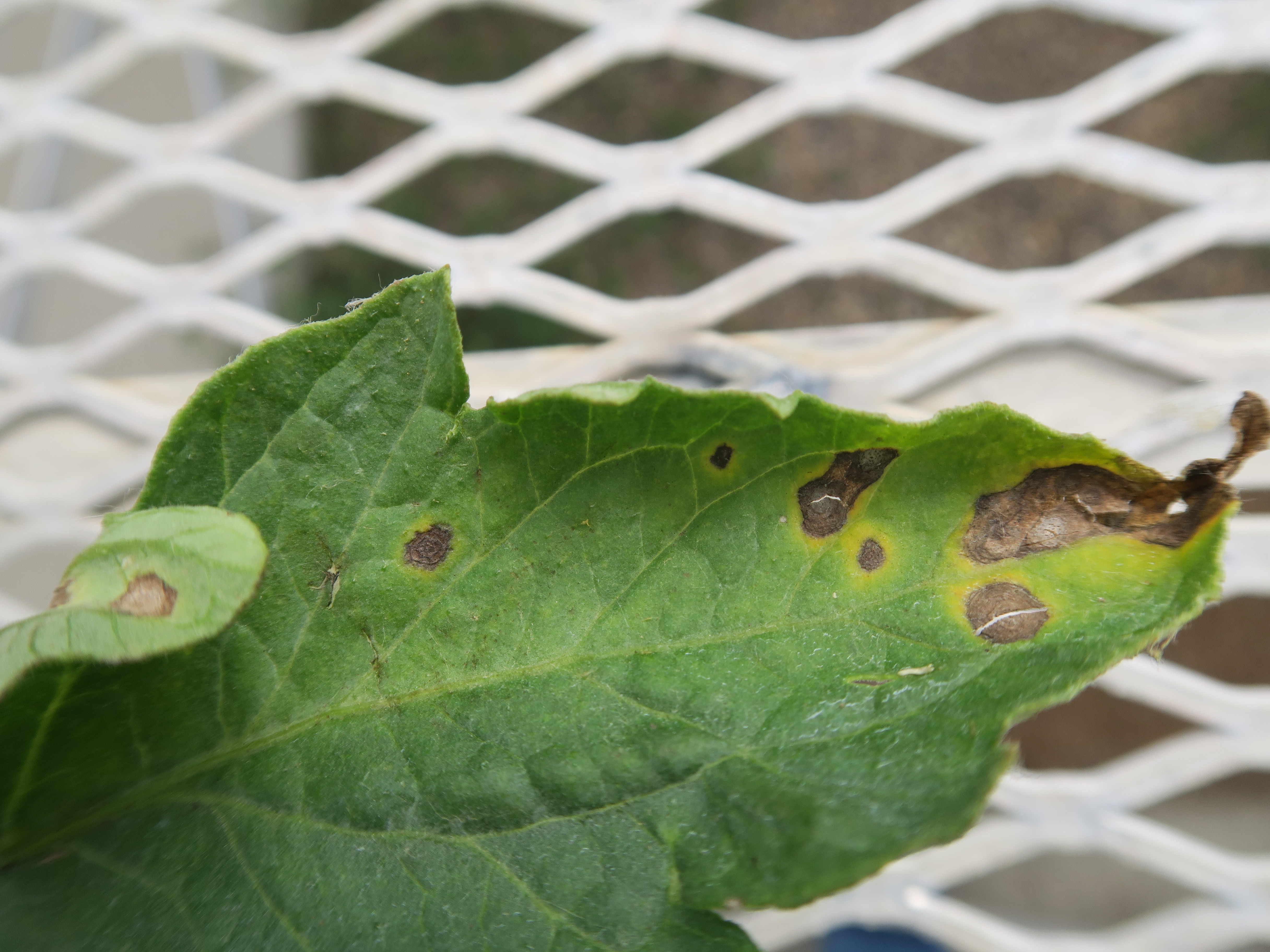 Figure 3. Early blight lesions on tomato leaf. Note cracked lesions.
 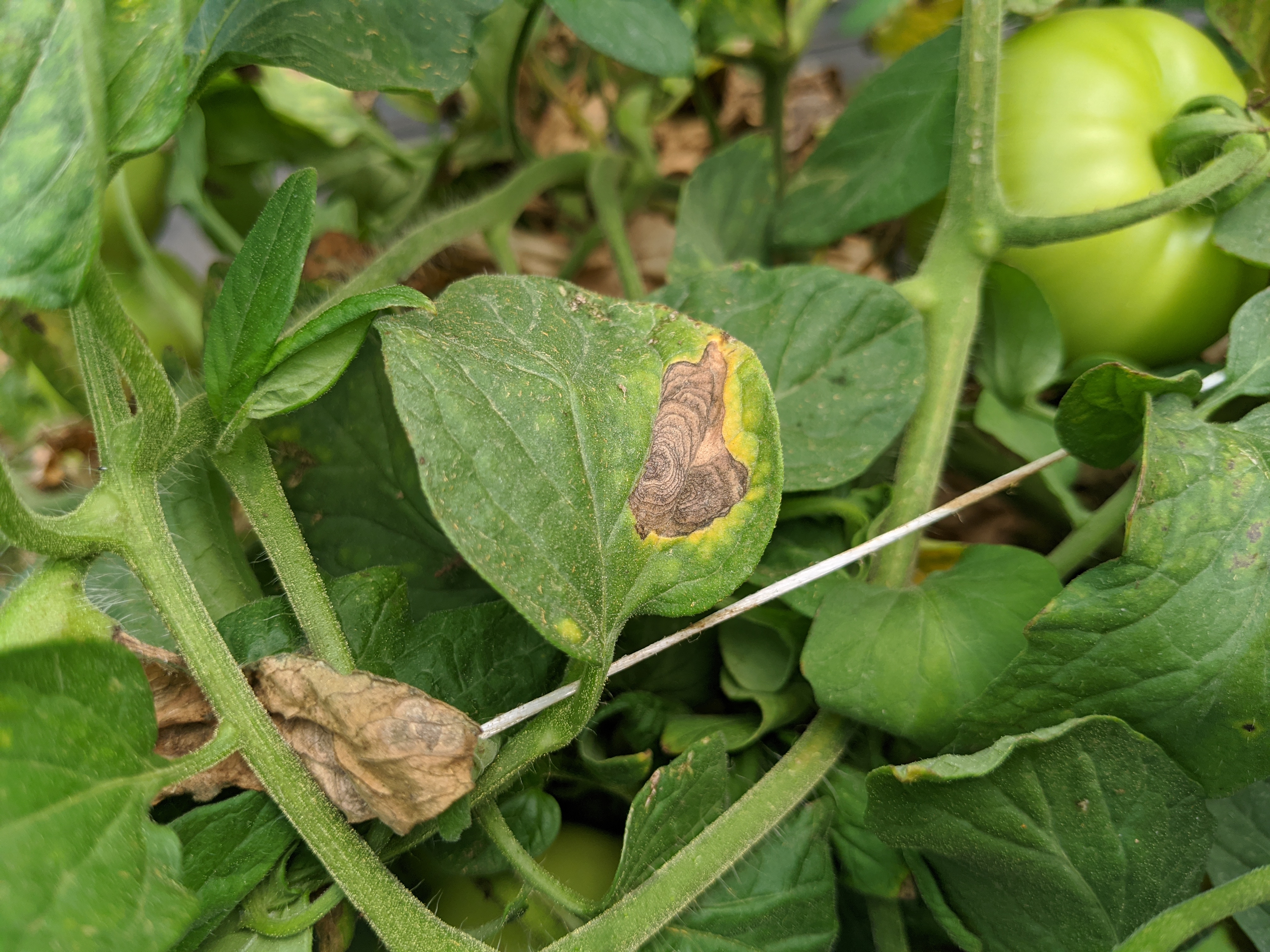 Figure 4. Early blight lesion on tomato leaf. Note lesion is restricted by vein.
 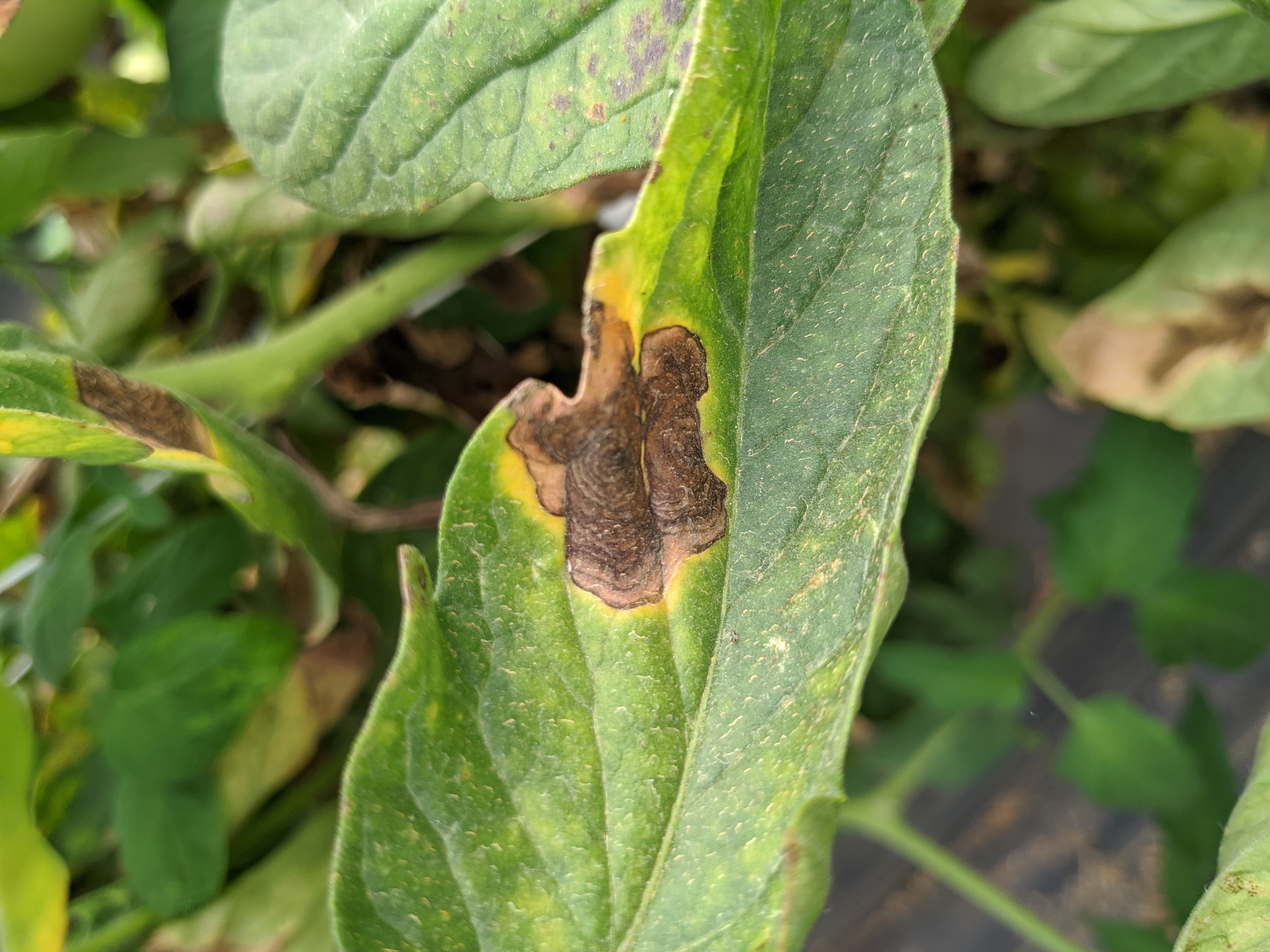 Figure 5. Lesion of early blight of tomato.
 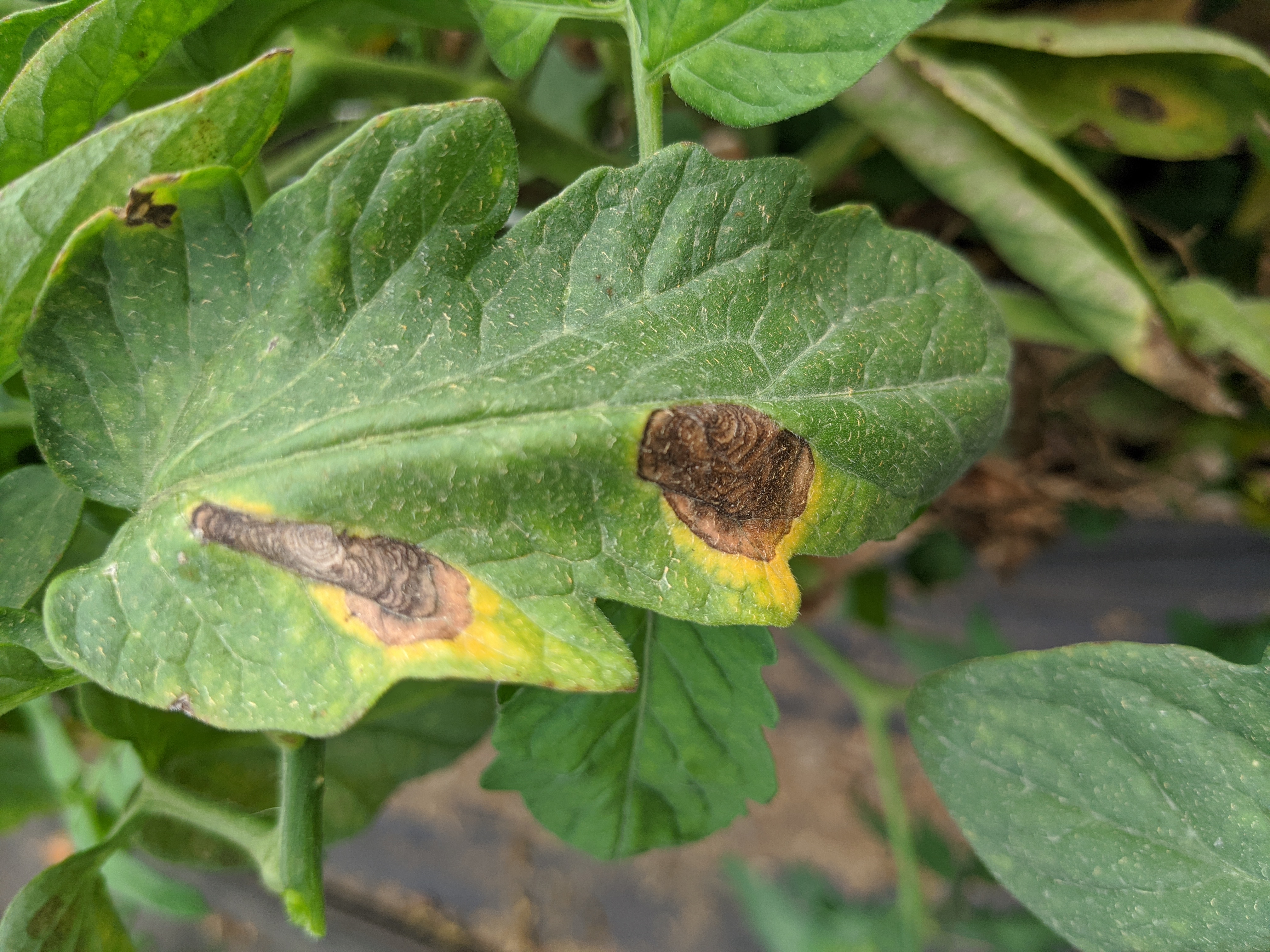 Figure 6. Early blight lesion on tomato.
 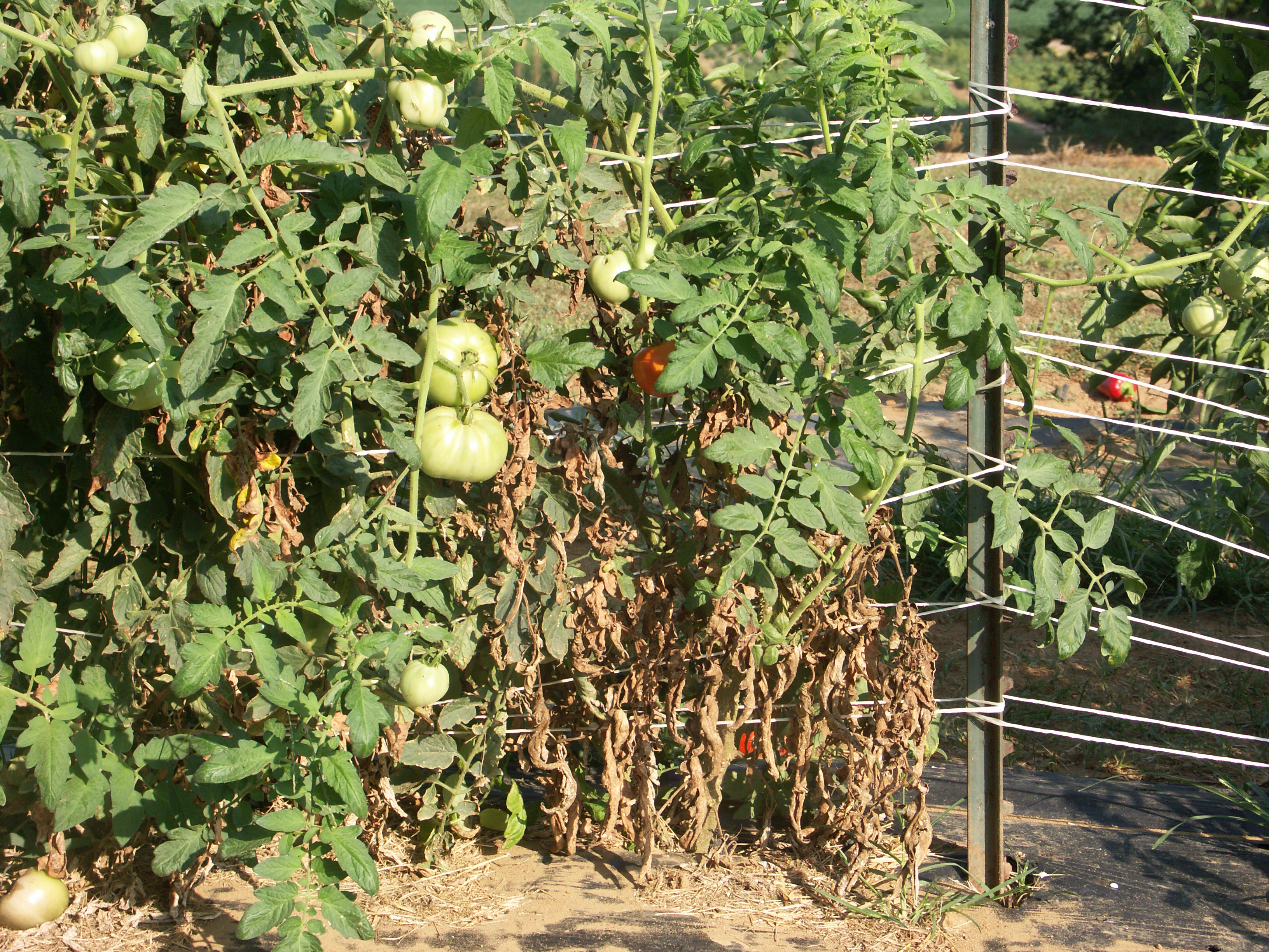 Figure 7. Older leaves are more susceptible to the early blight fungus. Therefore, tomato plants may appear to be dying from the ground up.
 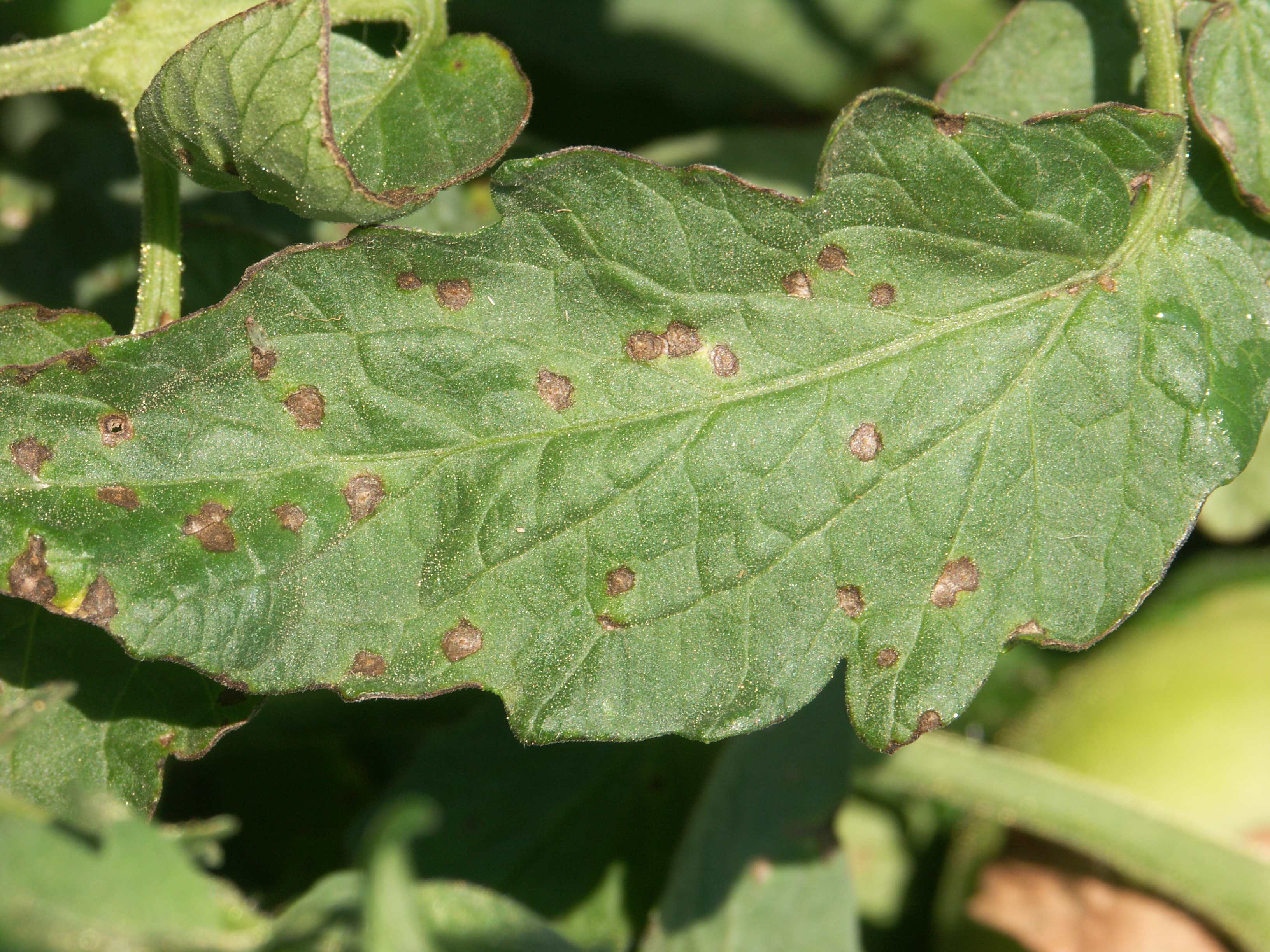 Figure 8. Note ring structure of early blight of tomato lesions.
 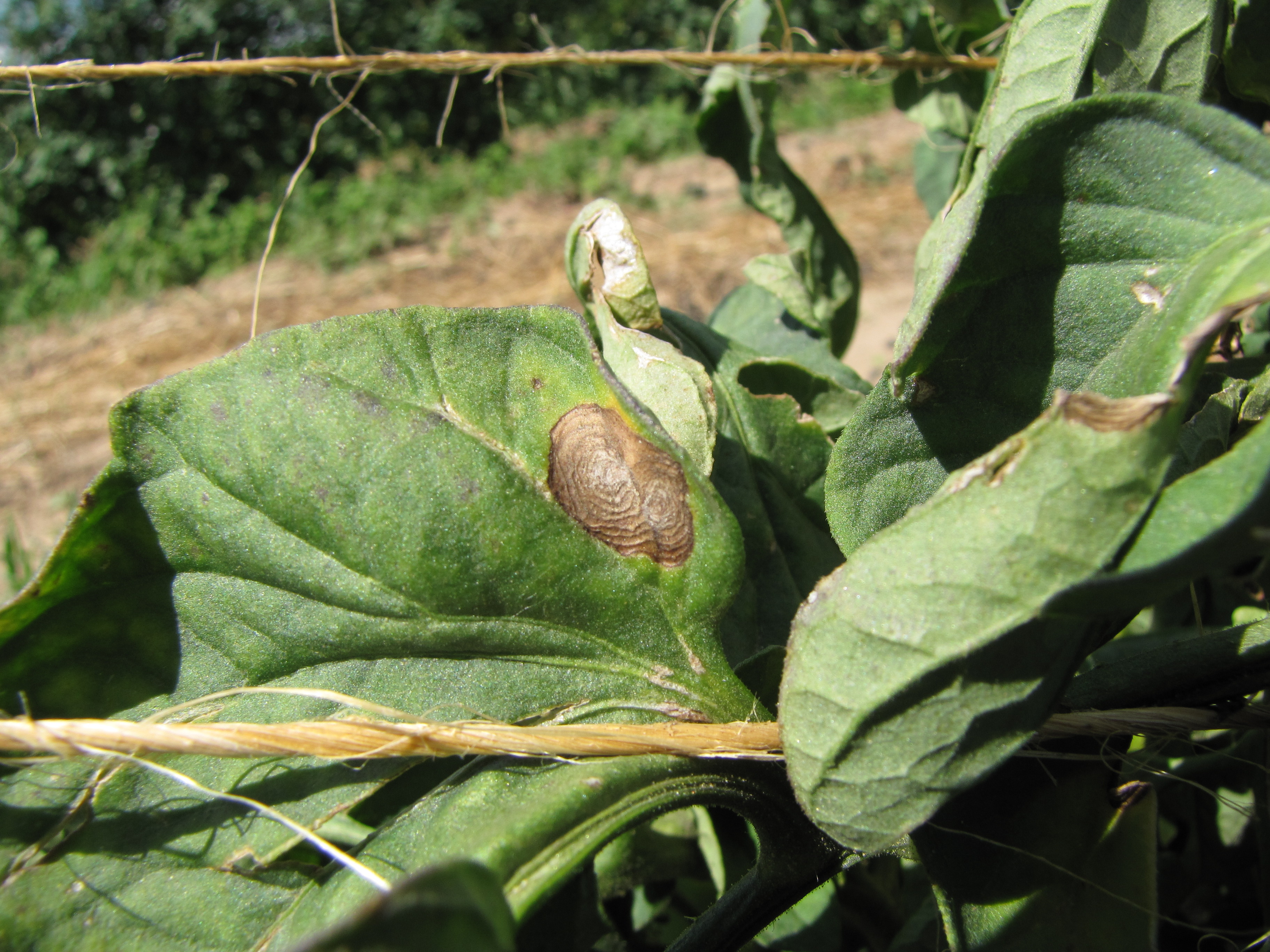 Figure 9. Clear ring structure of early blight lesion on tomato.
 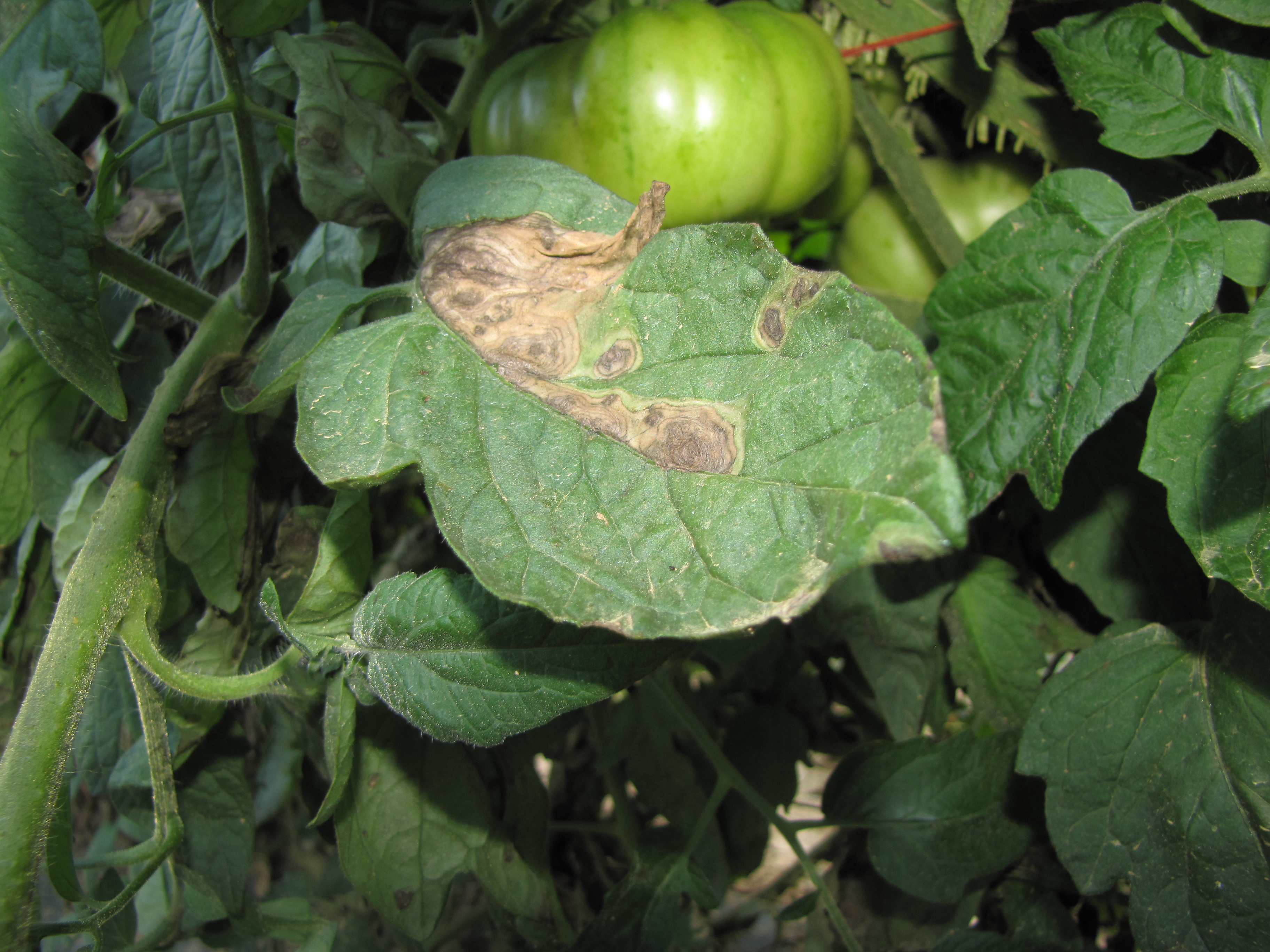 Figure 10. Early blight lesions of tomato. This is an heirloom variety and perhaps especially susceptible. Note bulls-eye lesions.
 Ethylene phytotoxicity
Ethylene phytotoxicity-This is not an infectious disease. May occur in greenhouses when there is inadequate exhaust or faulty heaters. Note epinasty.
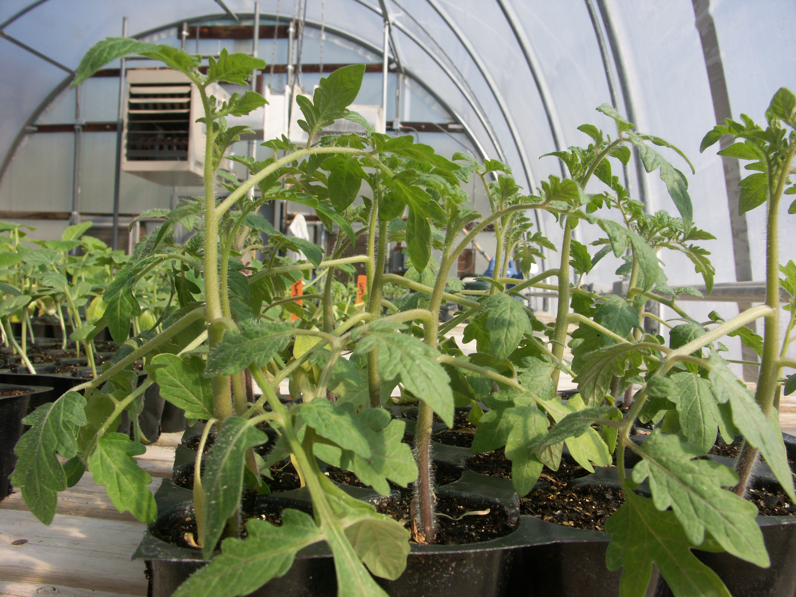 Figure 1. Ethylene damage on tomato seedlings. Ethylene has caused the branches to turn down (epinasty). Note that younger leaves are not pointed down since heaters were fixed so that ethylene was not longer a by-product.
 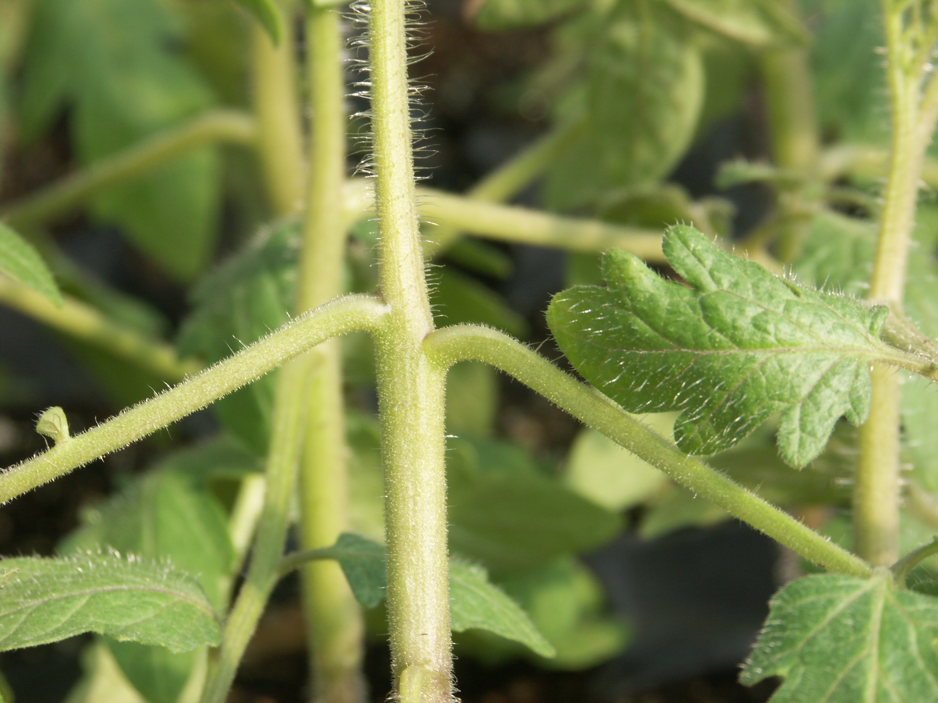 Figure 2. Close-up of branches with epinasty due to ethylene.
 Fusarium crown & root rot
Fusarium crown and root rot-Initial symptoms one might notice is wilt and decline of the plant. Lesion on base of stem is characteristic. Note vascular discoloration a few centimeters up the stem in contrast to Fusarium wilt where the discoloration continues much higher on the plant.
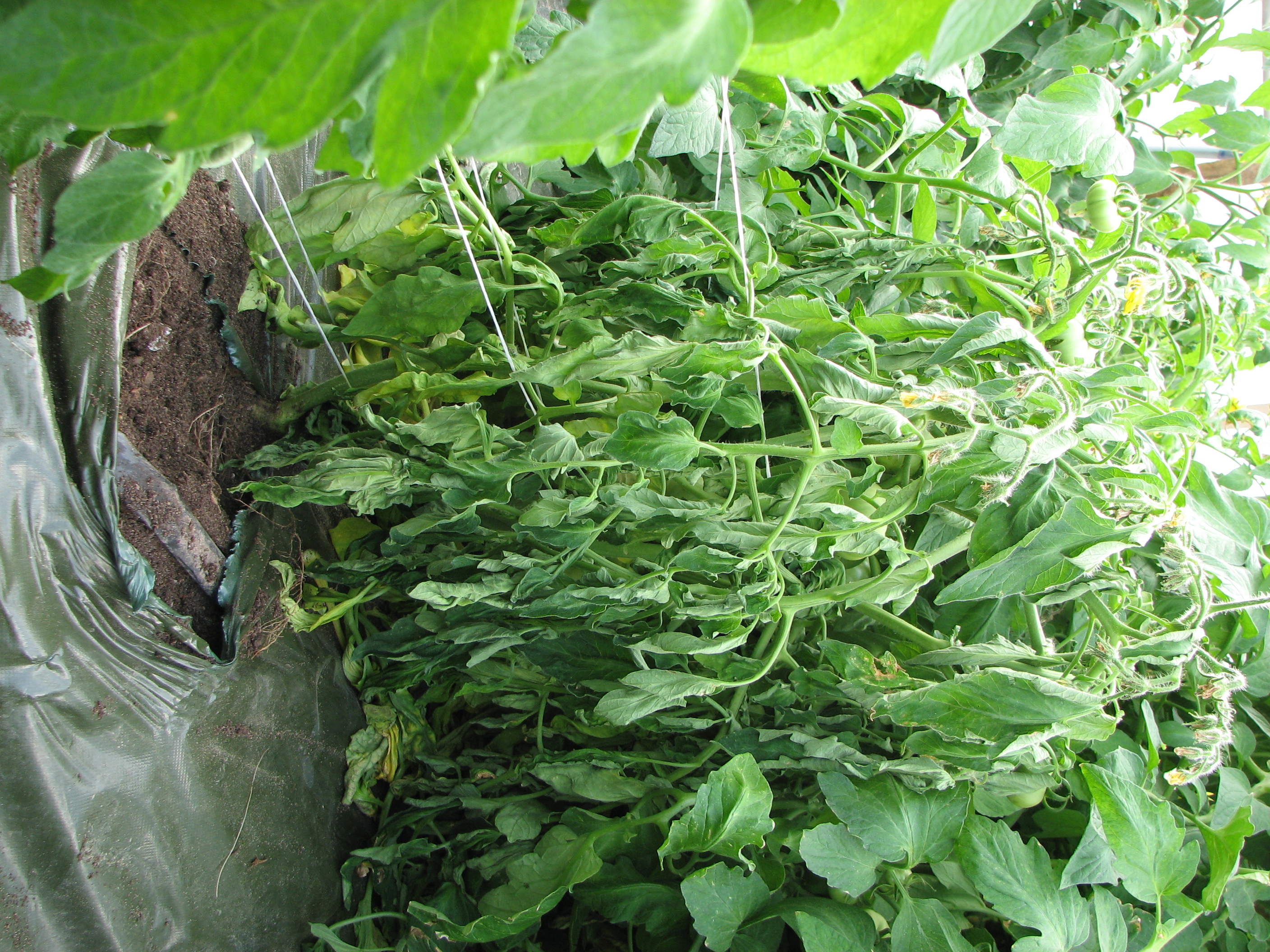 Figure 1. The first symptom of Fusarium crown rot one of tomato is likely to notice is a wilted plant.
 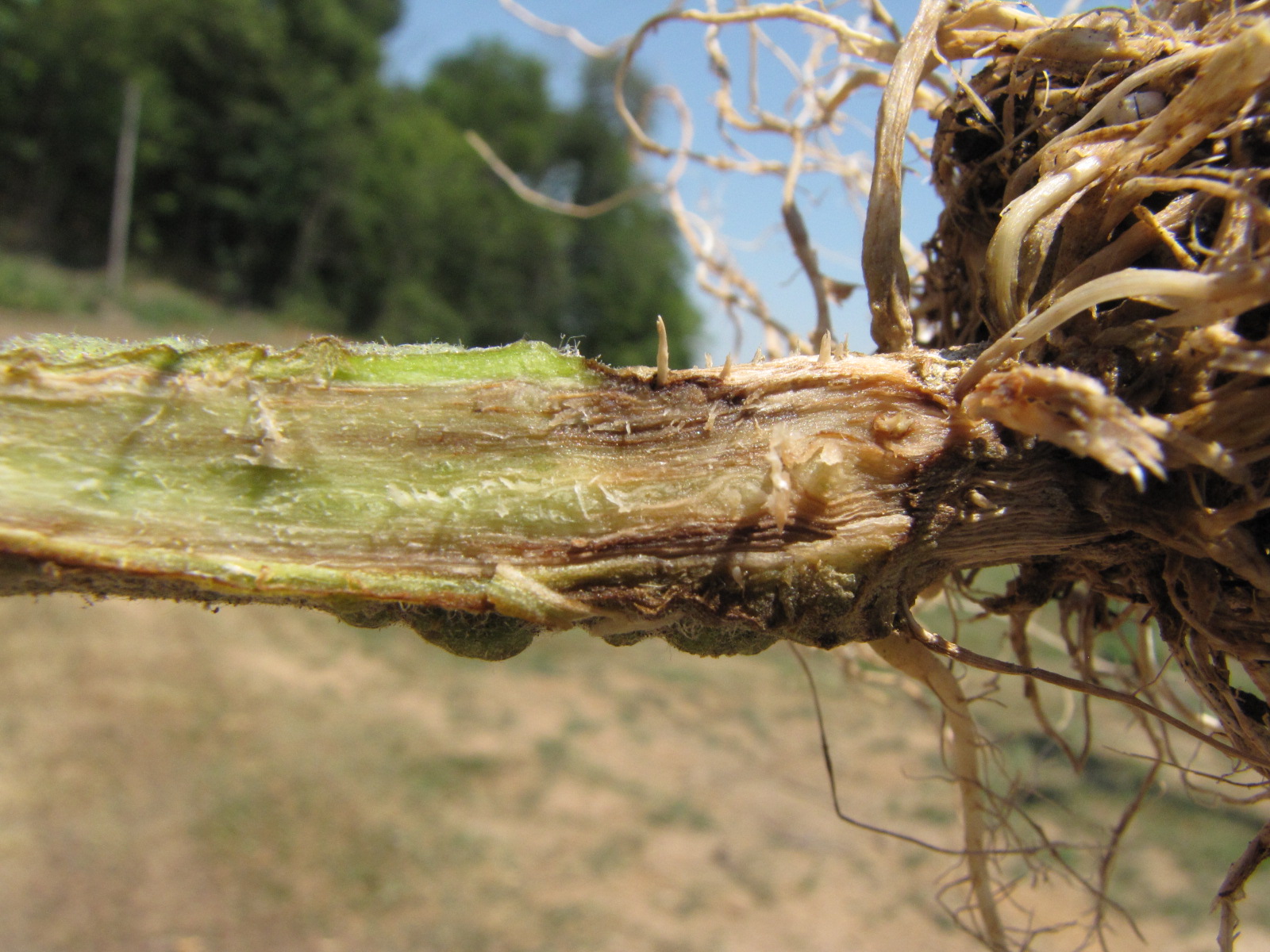 Figure 2. Dark vascular discoloration is typical of crown rot of tomato. Whereas vascular discoloration of crown rot typically extends an inch or two up the stem, discoloration due to Fusarium wilt will extend for several inches. Note also that wilt due to Fusarium wilt tends to be one-sided whereas wilt due to crown rot is likely to be the entire plant.
 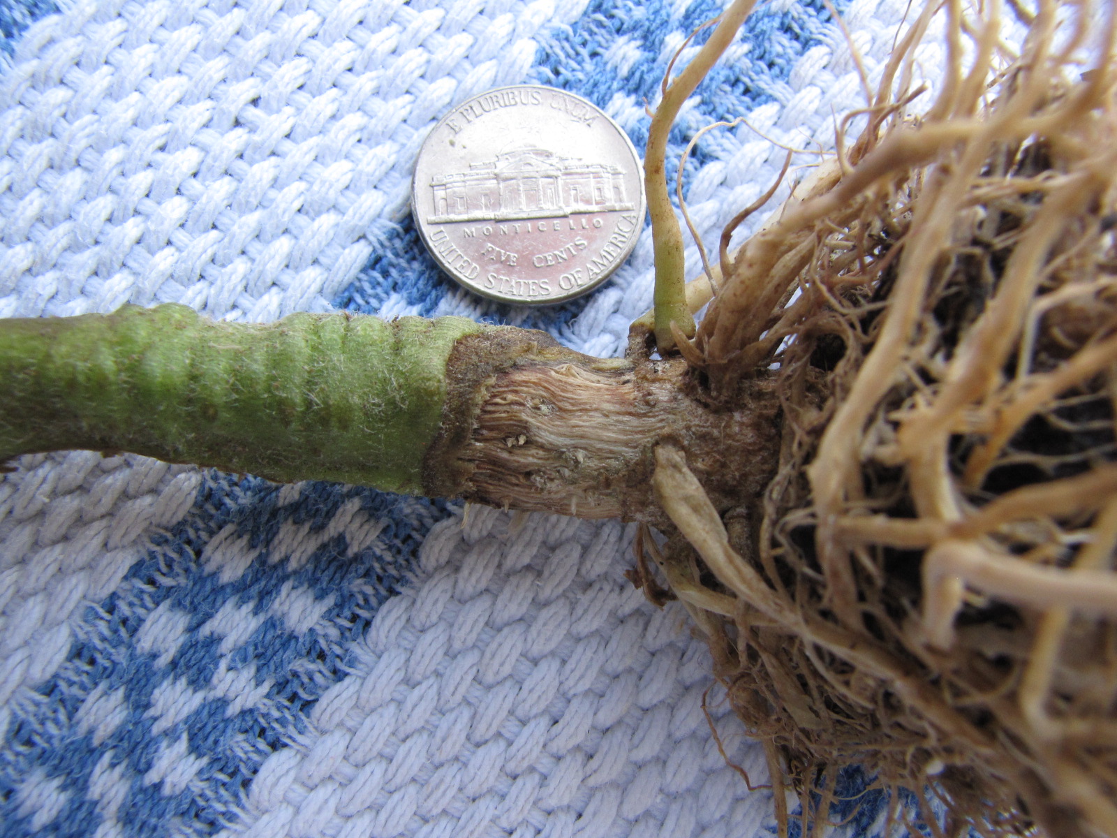 Figure 3. Canker on outside of stem due to crown rot of tomato.
 Fusarium wilt
Fusarium wilt-One sided wilt is a diagnostic symptom of Fusarium wilt of tomato. An additional symptom is the vascular discoloration that extends many centimeters up the stem. This disease is not common perhaps due to good resistance in modern hybrids.
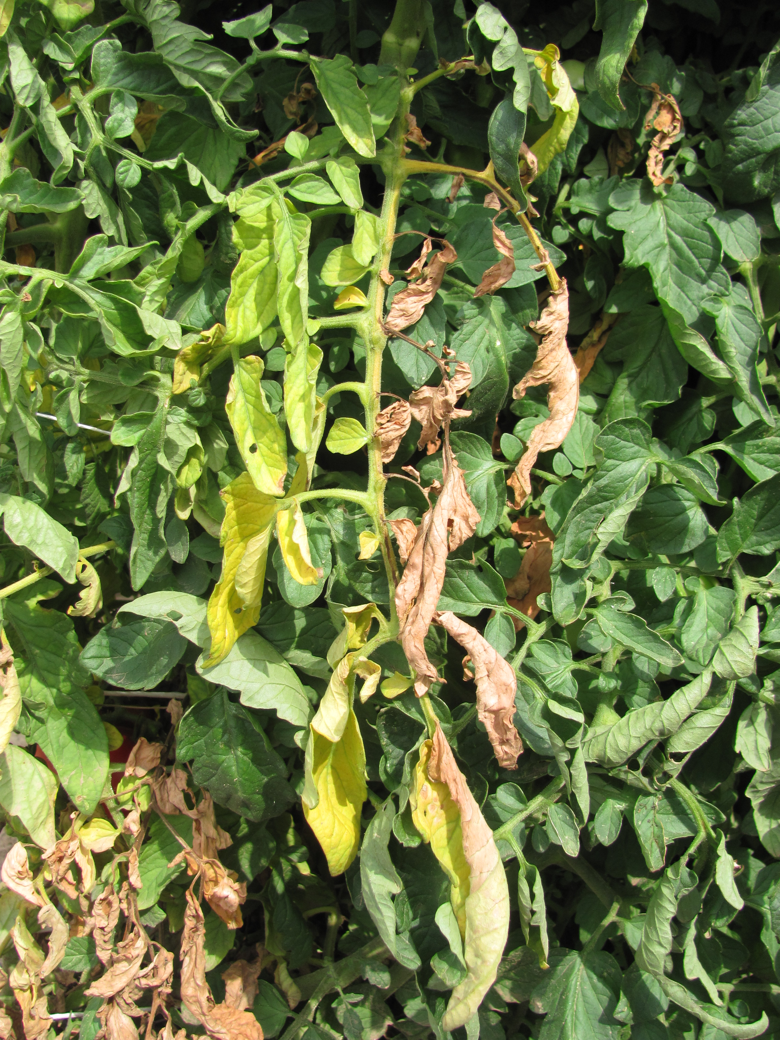 Figure 1. Fusarium wilt of tomato, as in many other vascular wilts, can cause a one-sided wilt.
 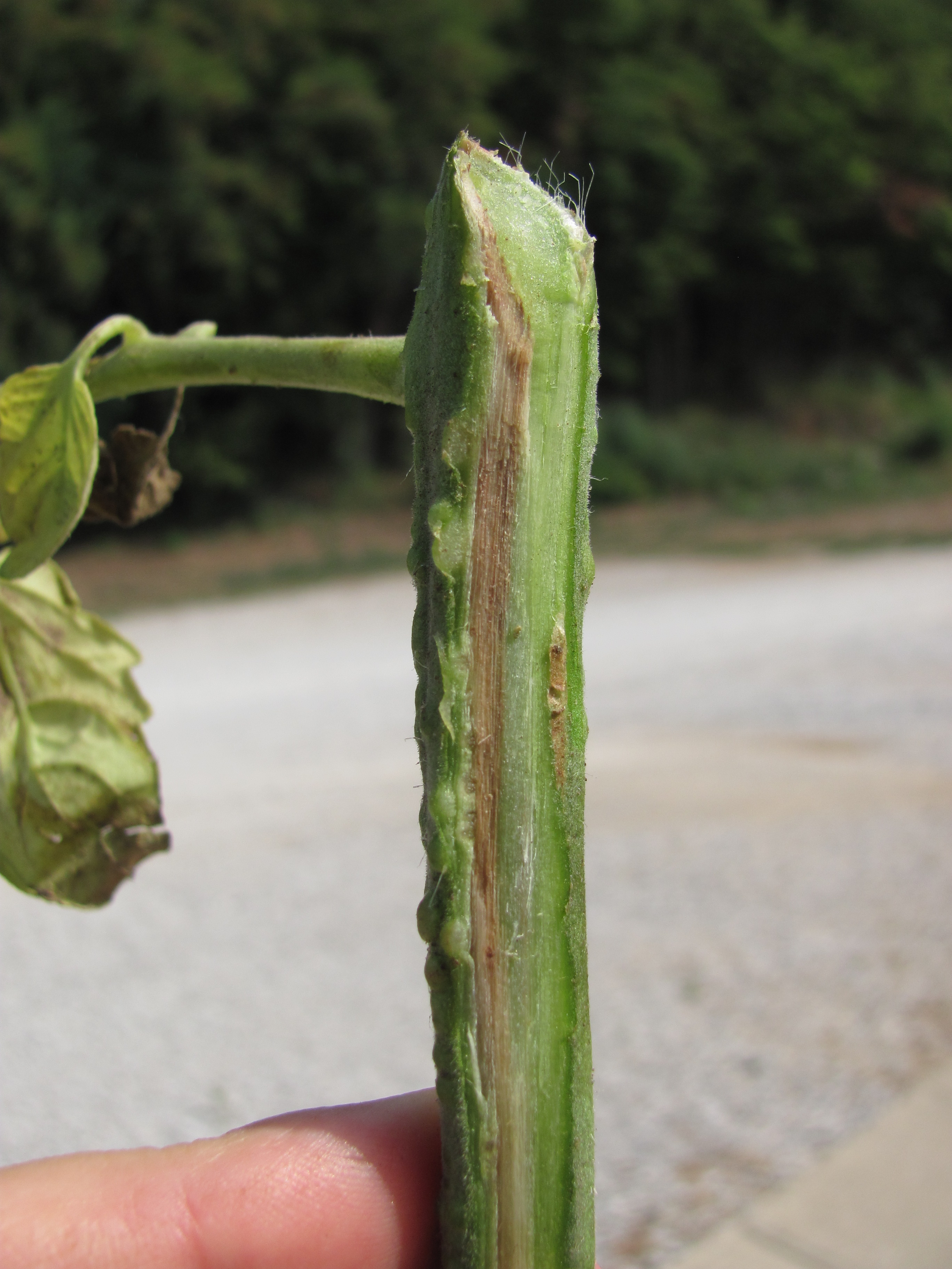 Figure 2. Fusarium wilt of tomato can cause a vascular discoloration on one side of the stem. The one-sided vascular discoloration typically goes along with the one-sided wilt.
 Gray mold
Gray mold-Symptoms occur on leaves, stems and fruit. Leaf lesions tend to be light brown. Stem lesions may be a darker brown. Fruit may appear soft. All plant parts may be covered with a gray mold.
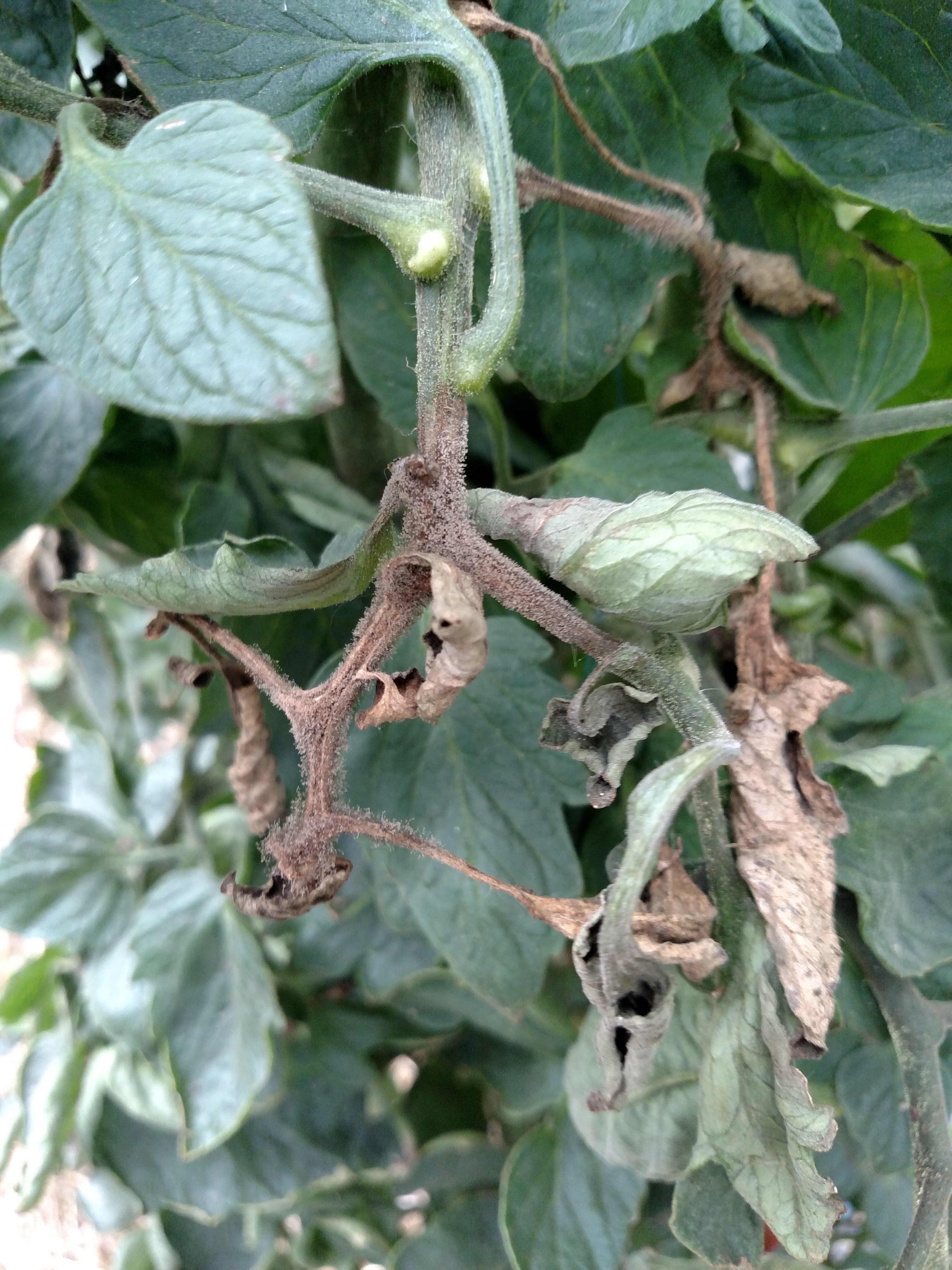 Figure 1. The fungus that causes gray mold often sporulates on infected tomato stems.
 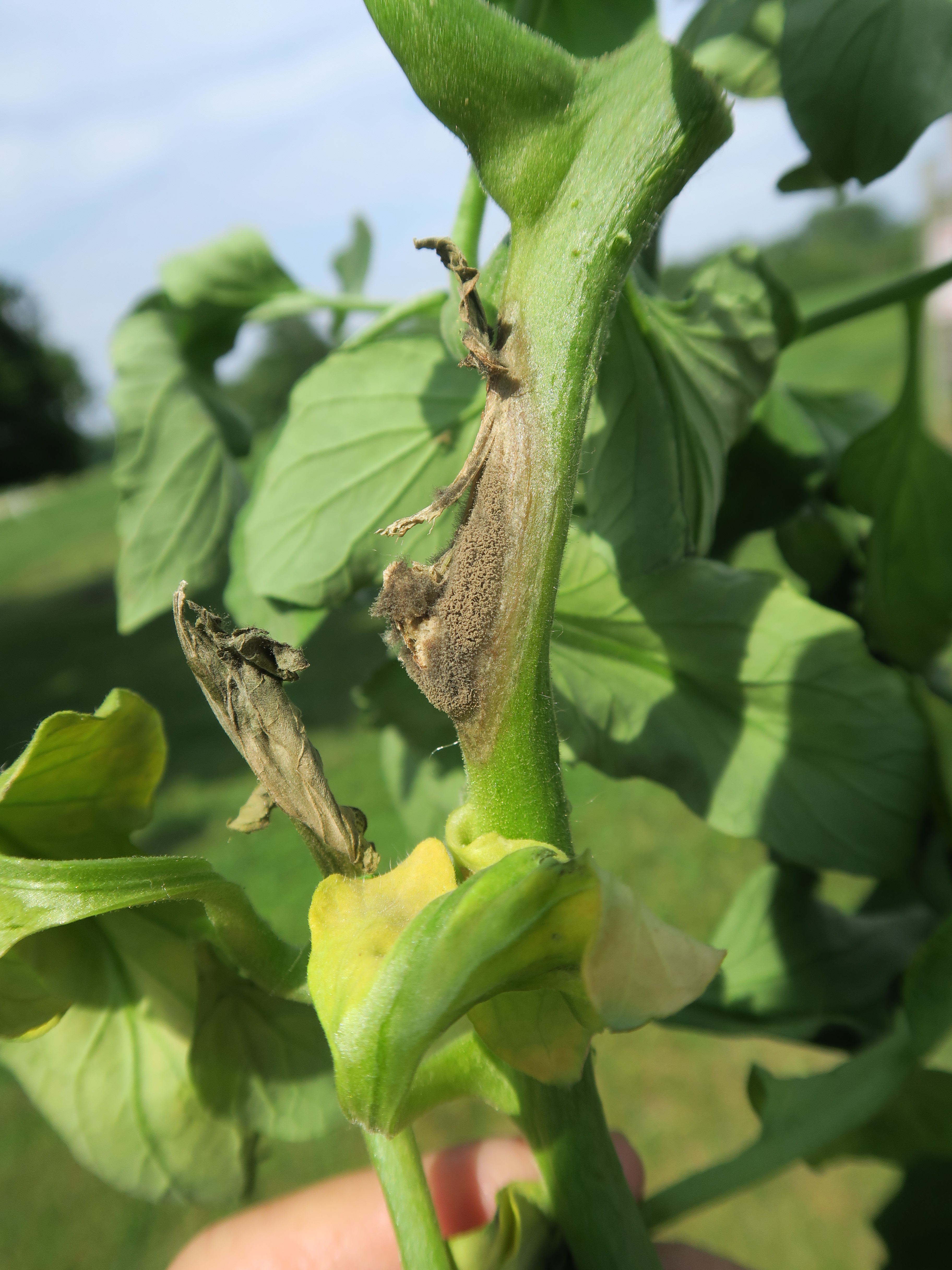 Figure 2. Gray mold on infected tomato stem.
 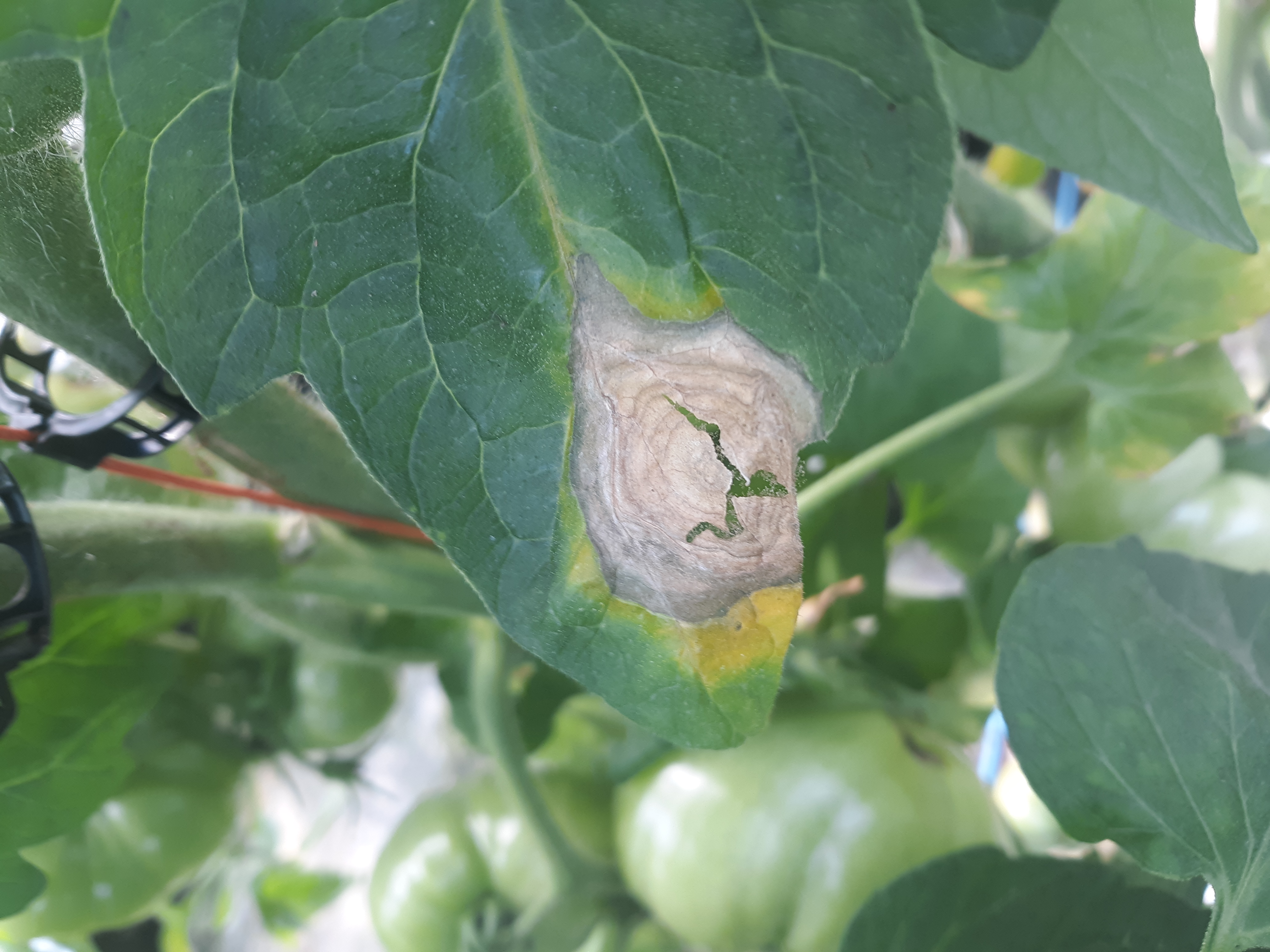 Figure 3. Gray mold lesions on leaves are often light brown or gray, often on the edge of the leaf and may show a ring-structure. Note sporulation of fungus observed in the crack of the lesion.
 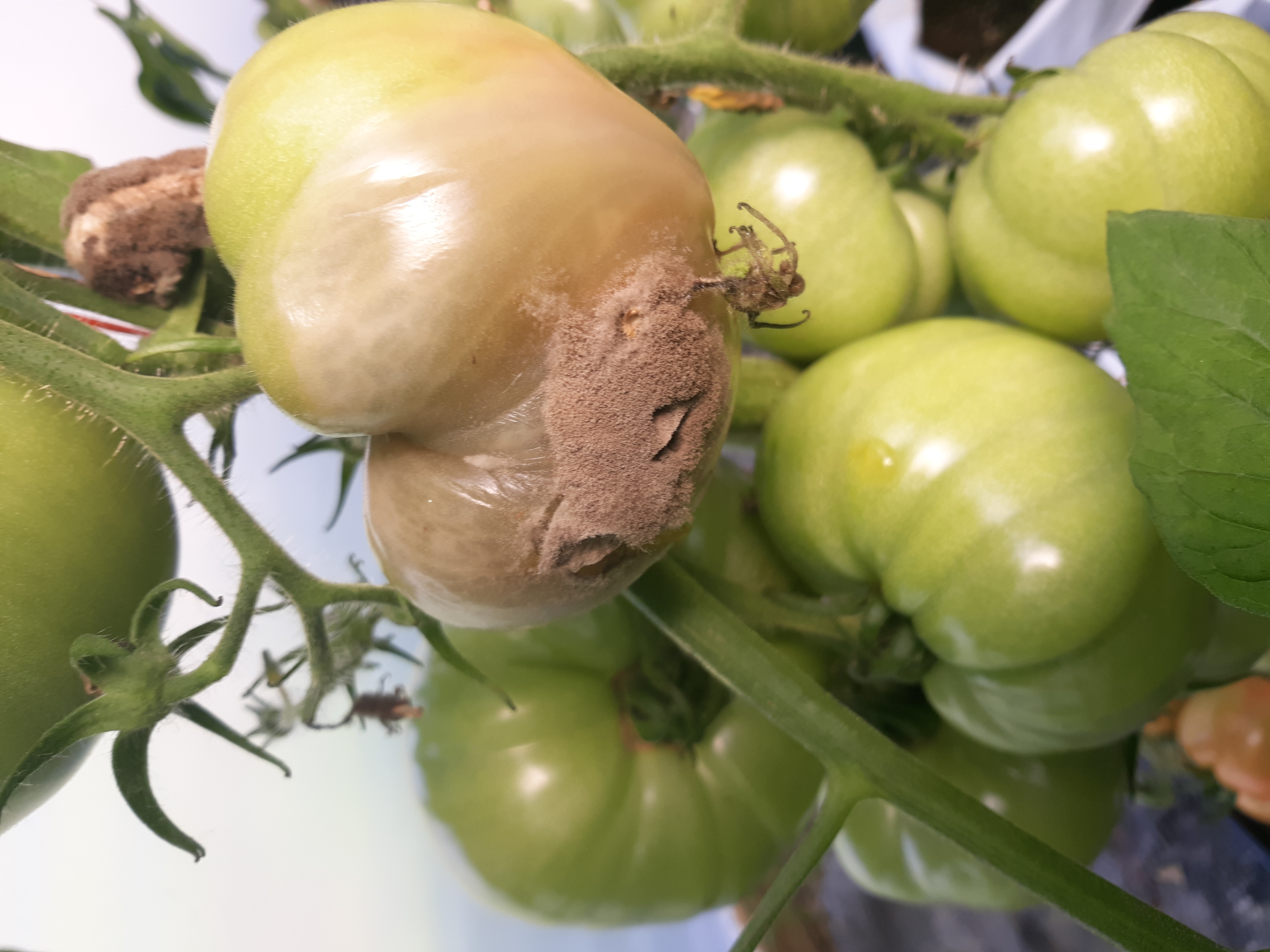 Figure 4. Gray mold of tomato fruit. Note sporulation of fungus.
 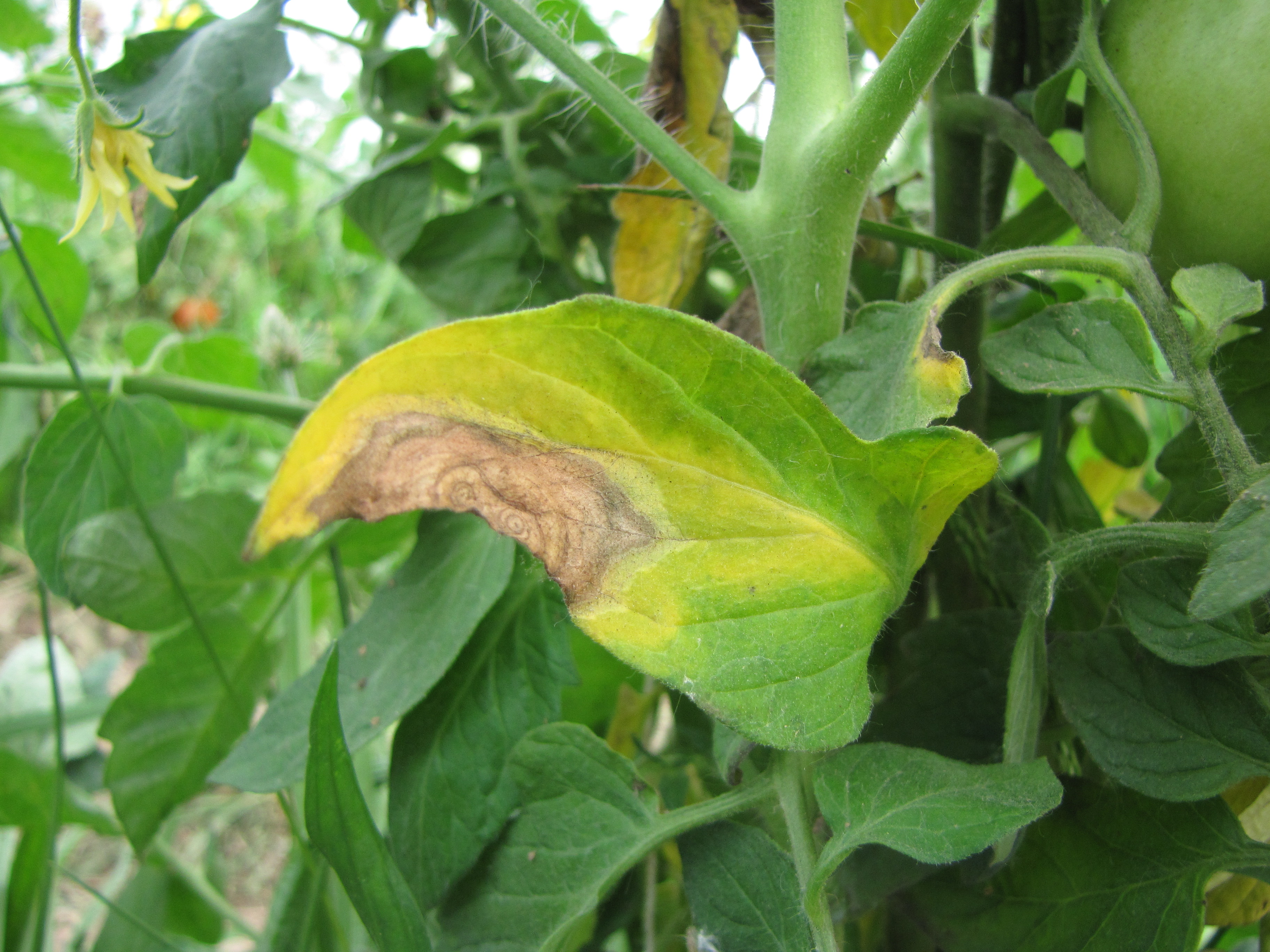 Figure 5. Gray mold lesion on tomato leaf.
  Figure 6. Gray mold on tomato fruit.
 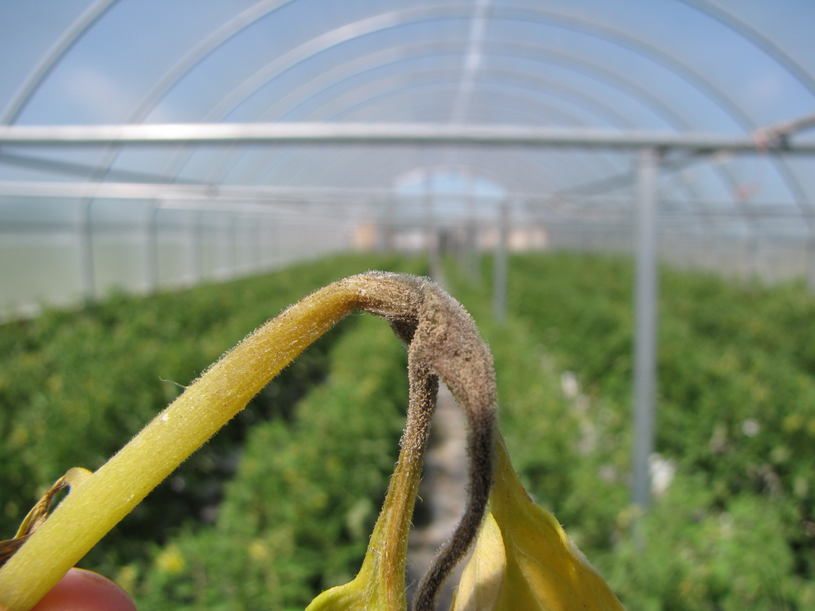 Figure 7. Gray mold on tomato leaf petiole.
 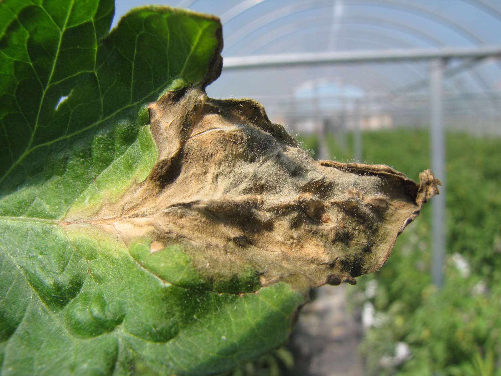 Figure 8. Gray mold on leaf lesion on margin of tomato leaf. Note sporulation.
 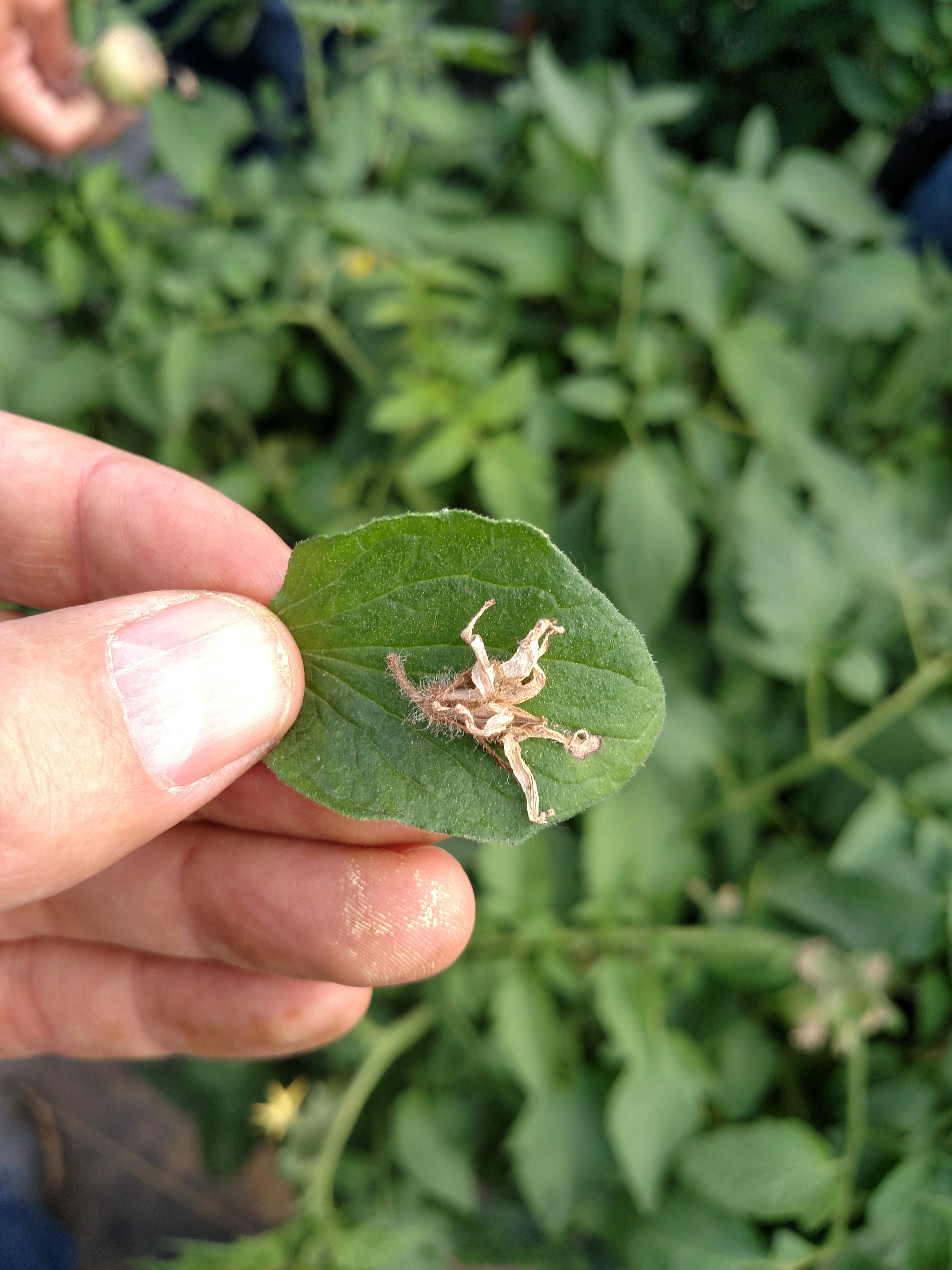 Figure 9. Tomato flower blossom with gray mold sporulation appears to have fallen on leaf where a new lesion has started.
 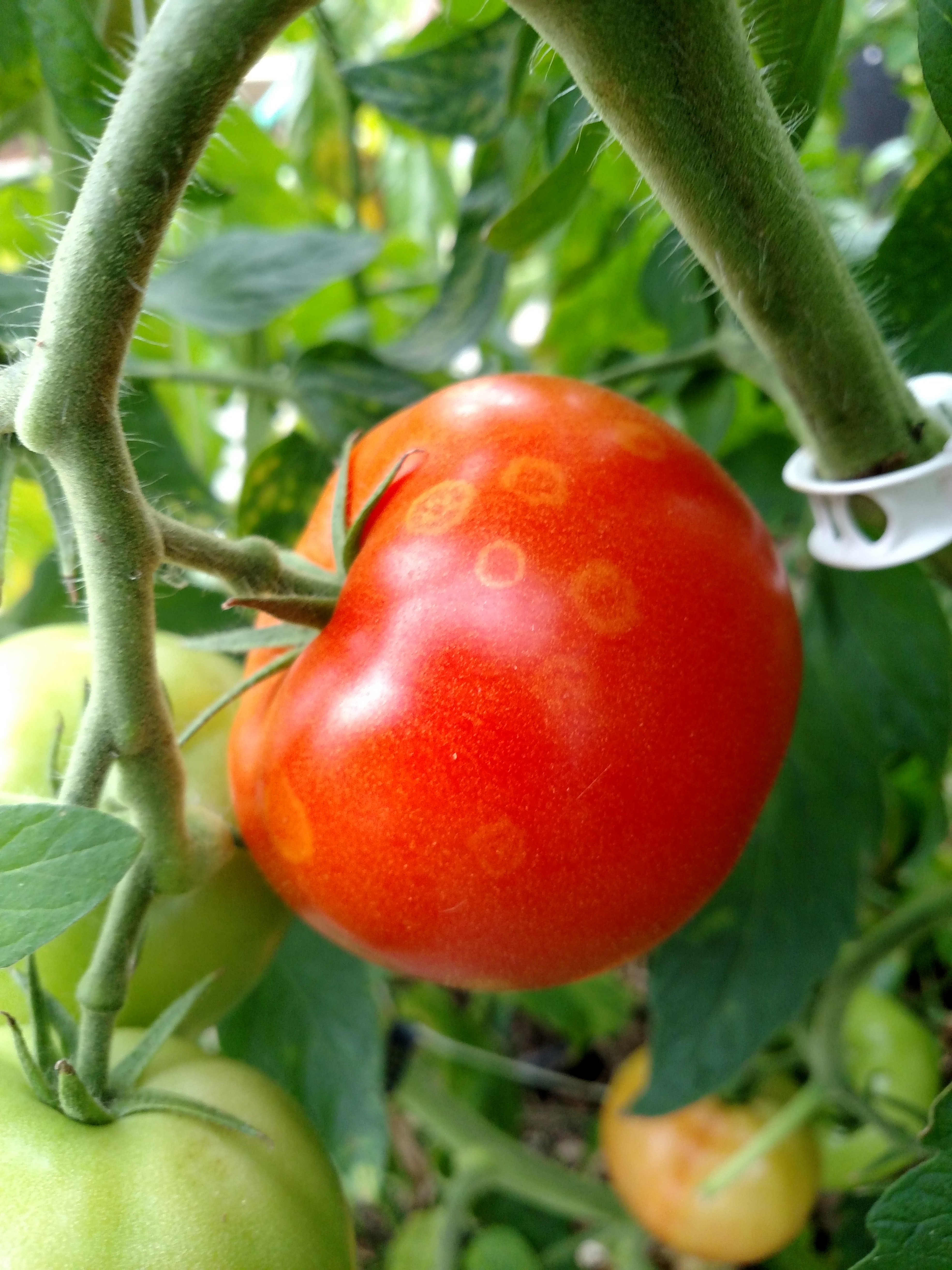 Figure 10. Conidia of the gray mold fungus fall onto tomato fruit where they may induce the reaction shown here resulting in a ‘ghost lesion’. While the lesions are unlikely to expand further, the appearance may reduce marketability.
 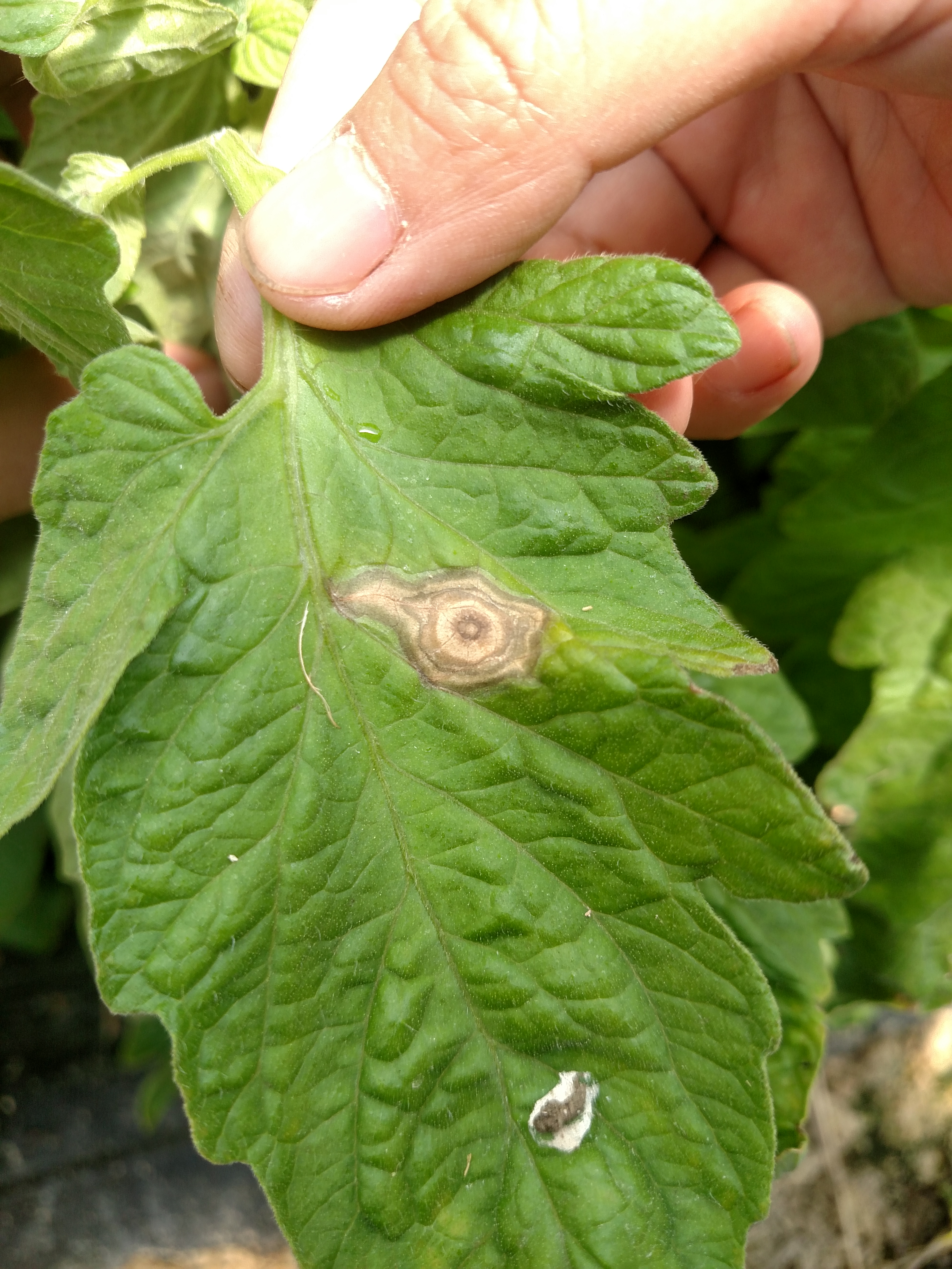 Figure 11. Lesion of gray mold on tomato leaf. Note ring structure.
 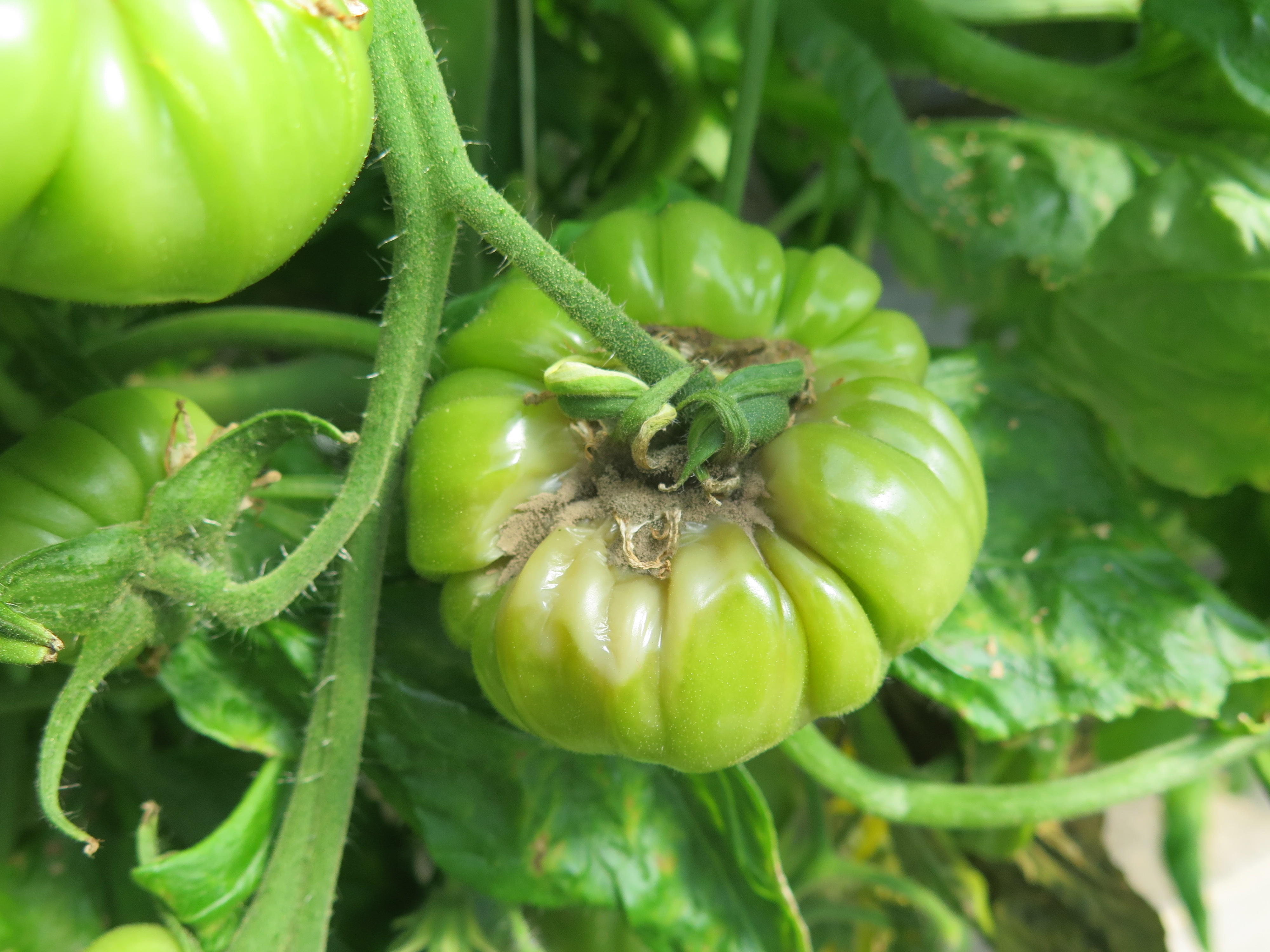 Figure 12. Gray mold on green tomato fruit.
 Intumescence
Intumescence-Not an infectious disease. Symptoms occur only on plants in greenhouse or growth chamber and may consist of bumpy growth. Not an important or common problem.
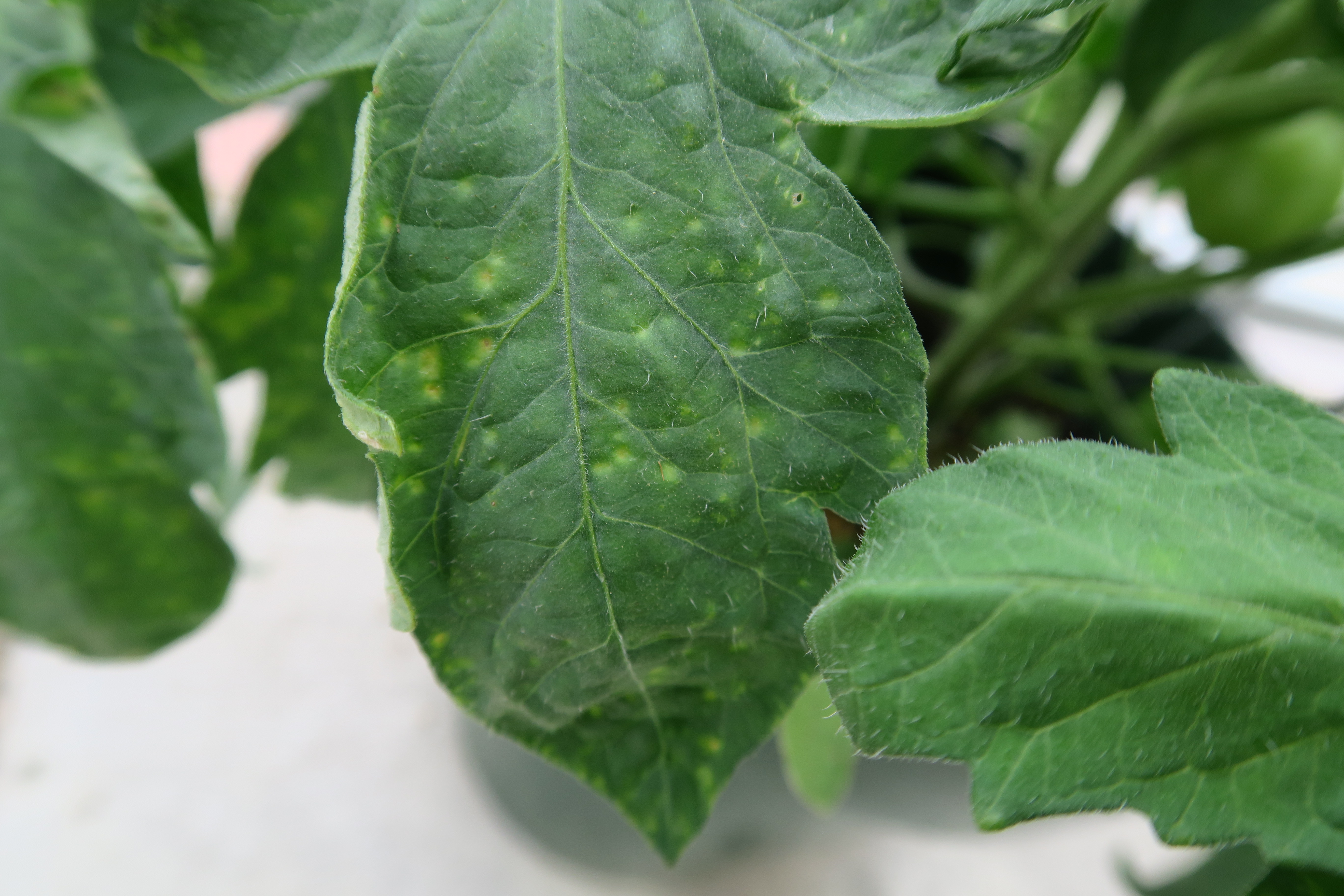 Figure 1. Yellow lesions due to intumescence.
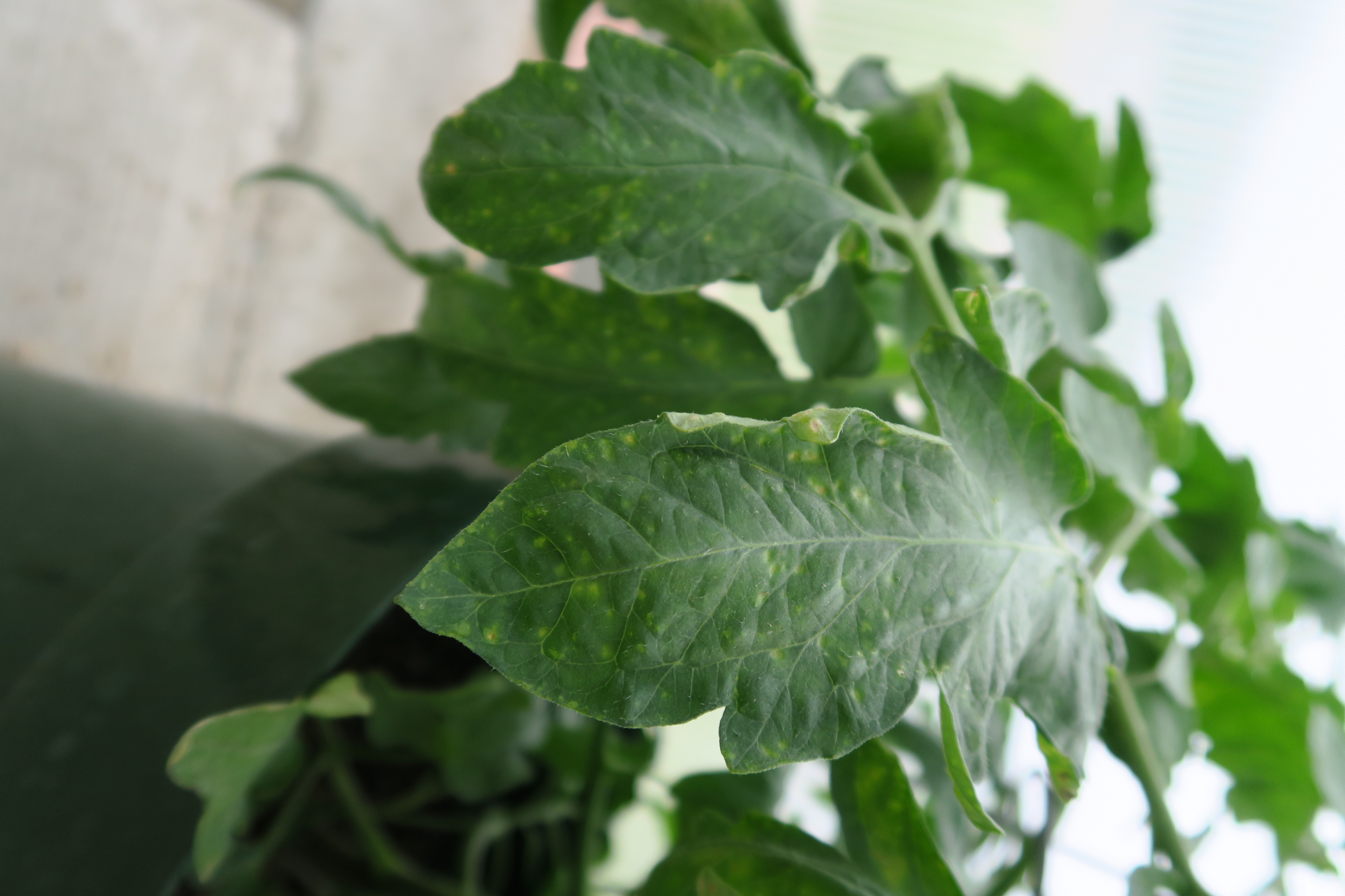 Figure 2. Intumescence of tomato leaves.
 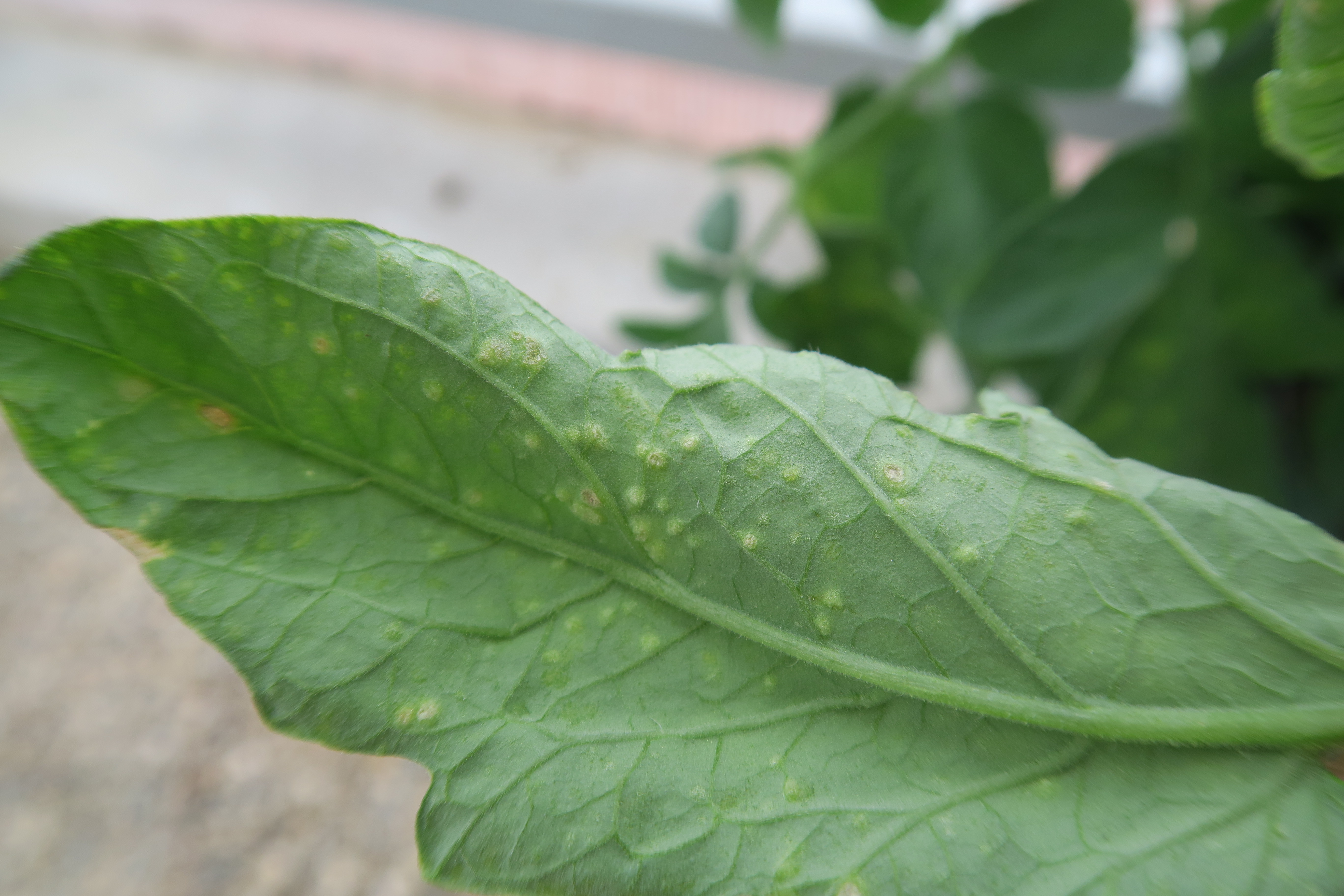 Figure 3. Intumescence on underside of tomato leaves.
 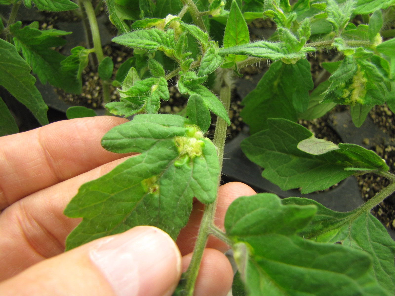 Figure 4. Intumescence on tomato leaves in a growth chamber.
 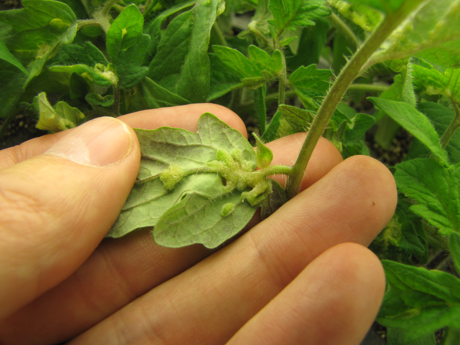 Figure 5. Intumescence of tomato leaf.
 Late blight
Late blight-Not a common problem in Indiana since the causal fungus-like organisms doesn’t usually overwinter in Indiana. However, when this disease does occur quick action and a specific set of fungicides will be required. Necrotic leaf lesions may be ringed with the white sporulation of the causal fungus. Fruit lesions appear to have a soft brown area.
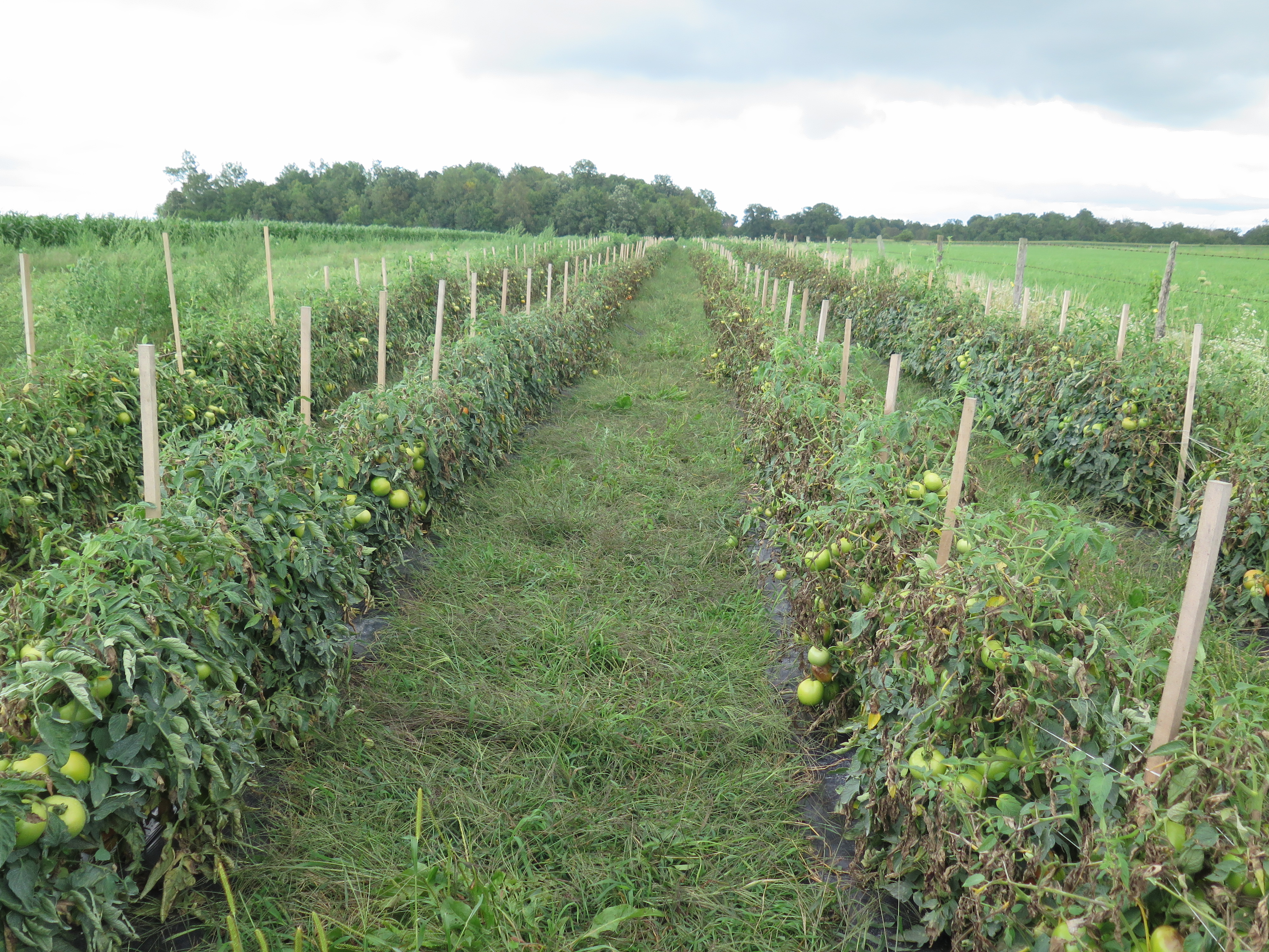 Figure 1. Although late blight is not common in Indiana, when it occurs, it can spread rapidly in a field such as this one.
 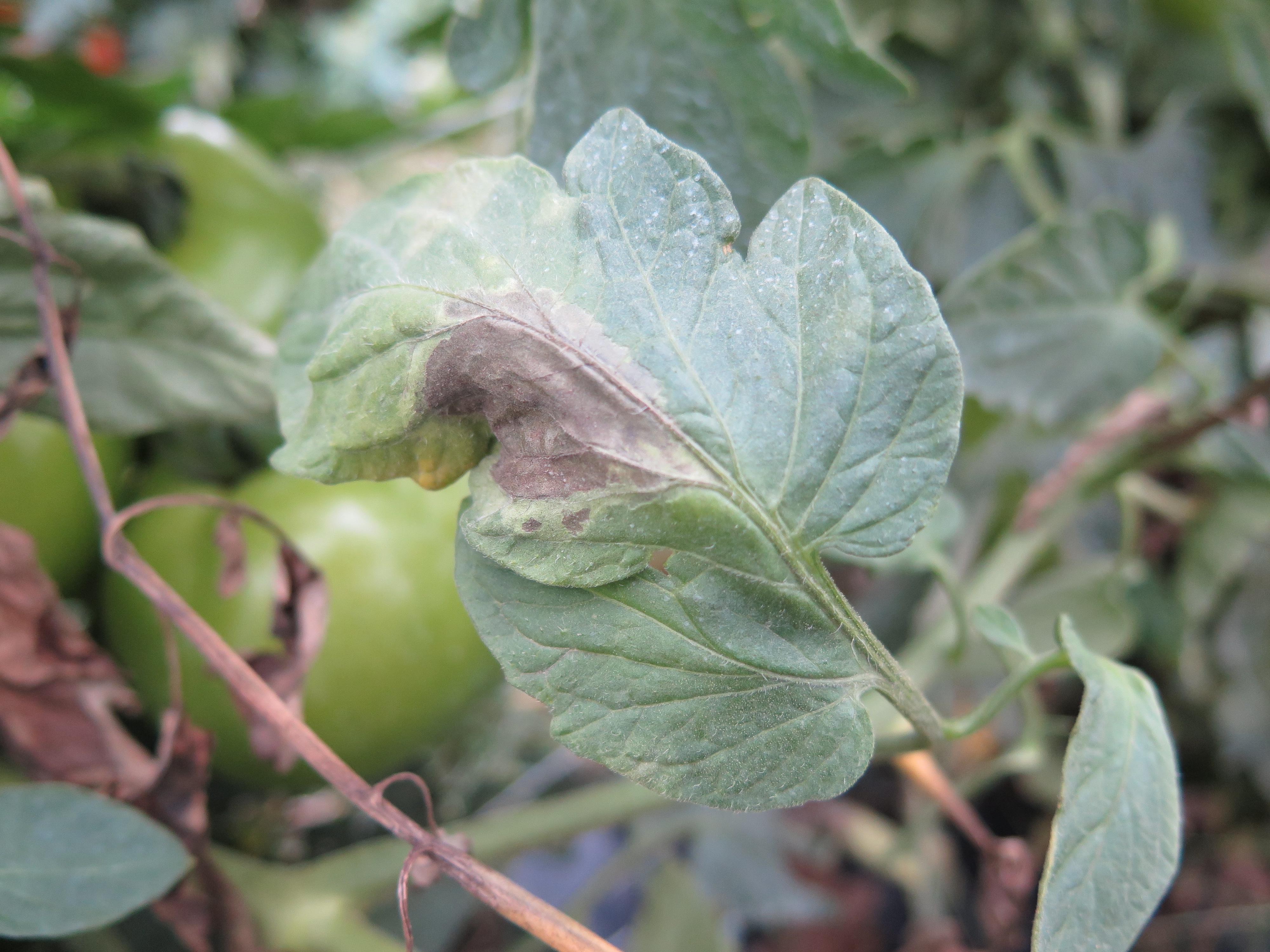 Figure 2. Leaf lesion of late blight of tomato.
 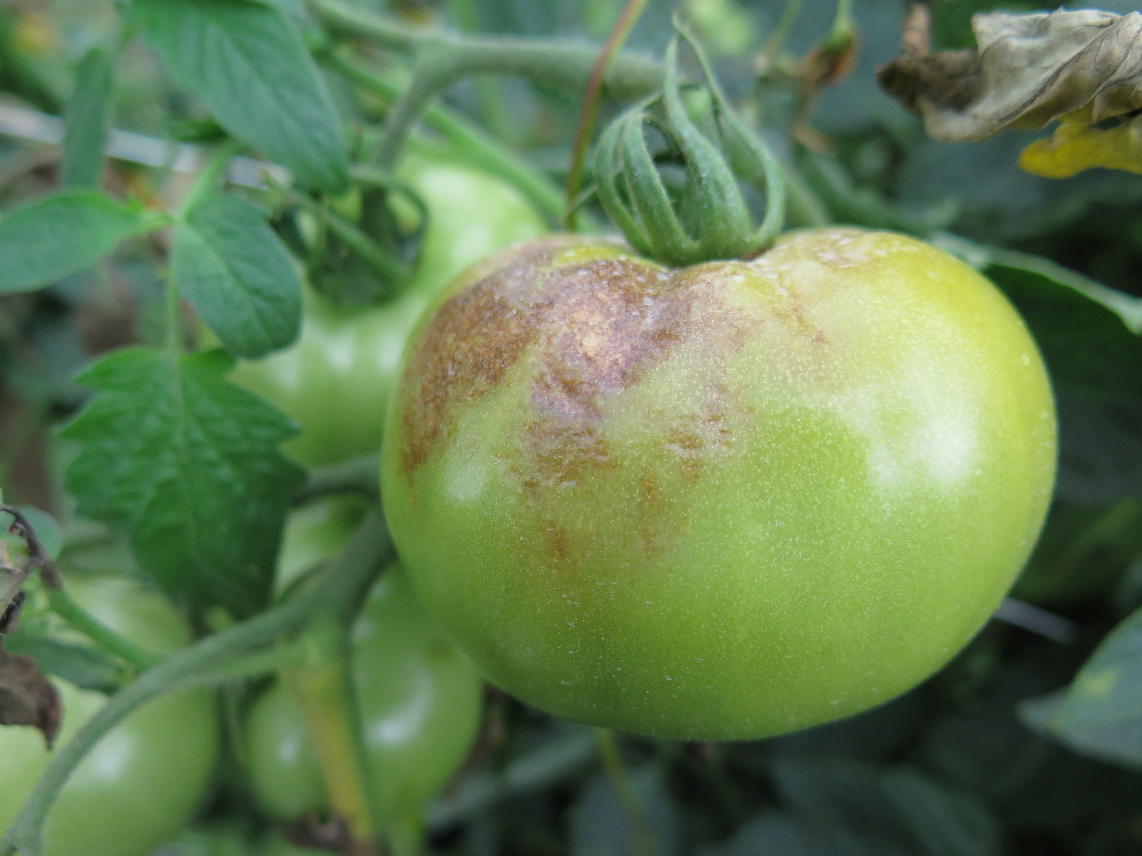 Figure 3. Late blight symptoms on tomato fruit
 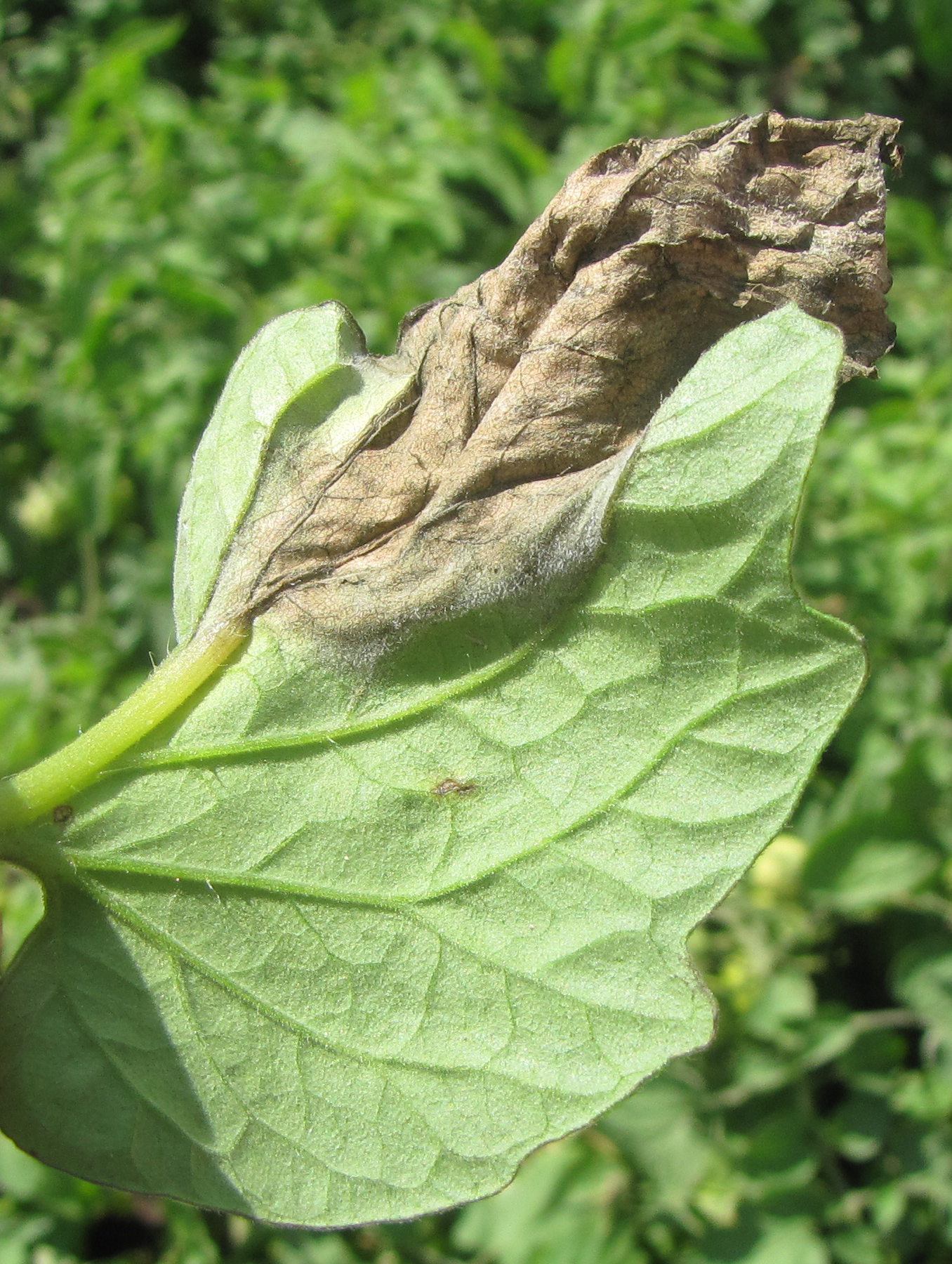 Figure 4. The white cast on this tomato leaf with late blight indicates sporulation of the causal organism.
 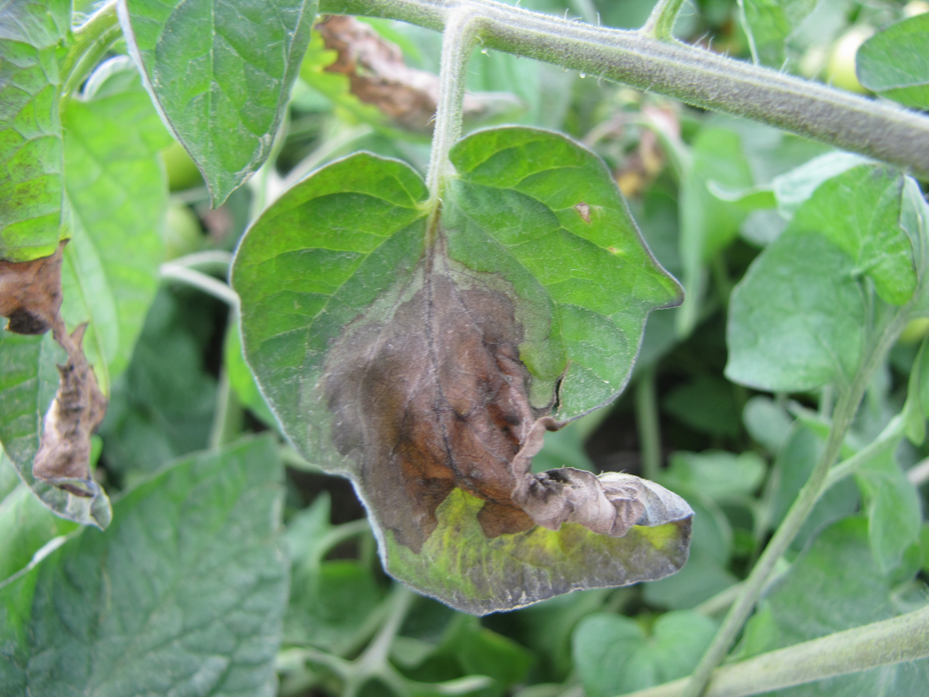 Figure 5. Lesion of late blight of tomato on leaf. Note margin between necrotic portion of leaf and healthy leaf.
 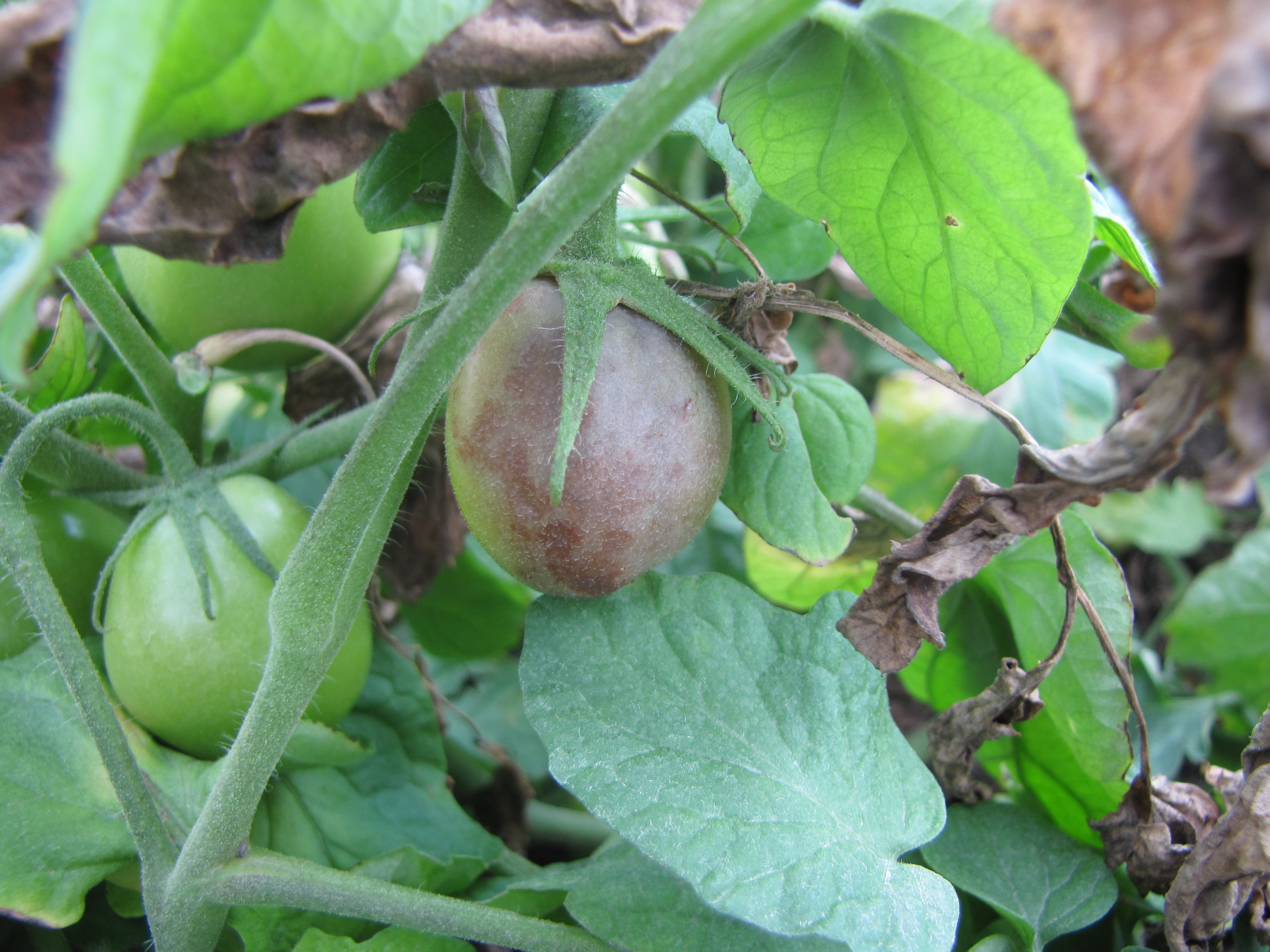 Figure 6. Late blight of tomato symptoms on fruit.
 Leaf mold
Leaf mold-Lesions of leaf mold are often chlorotic with indistinct borders. On the underside of leaves, the olive-green fungus may be sporulating. For the most part, this is a greenhouse or high tunnel disease.
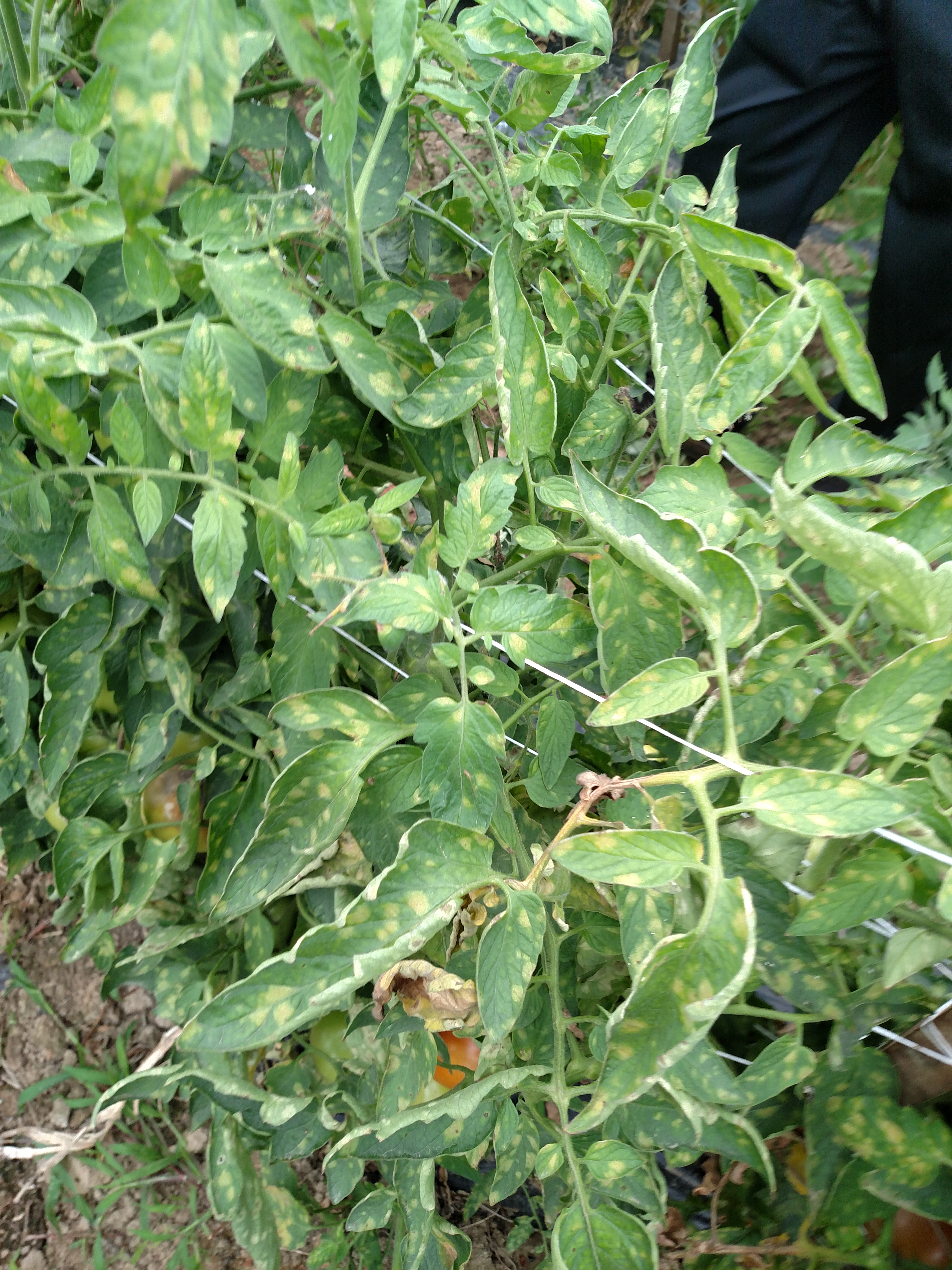 Figure 1. Chlorotic lesions on leaves caused by leaf mold of tomato.
 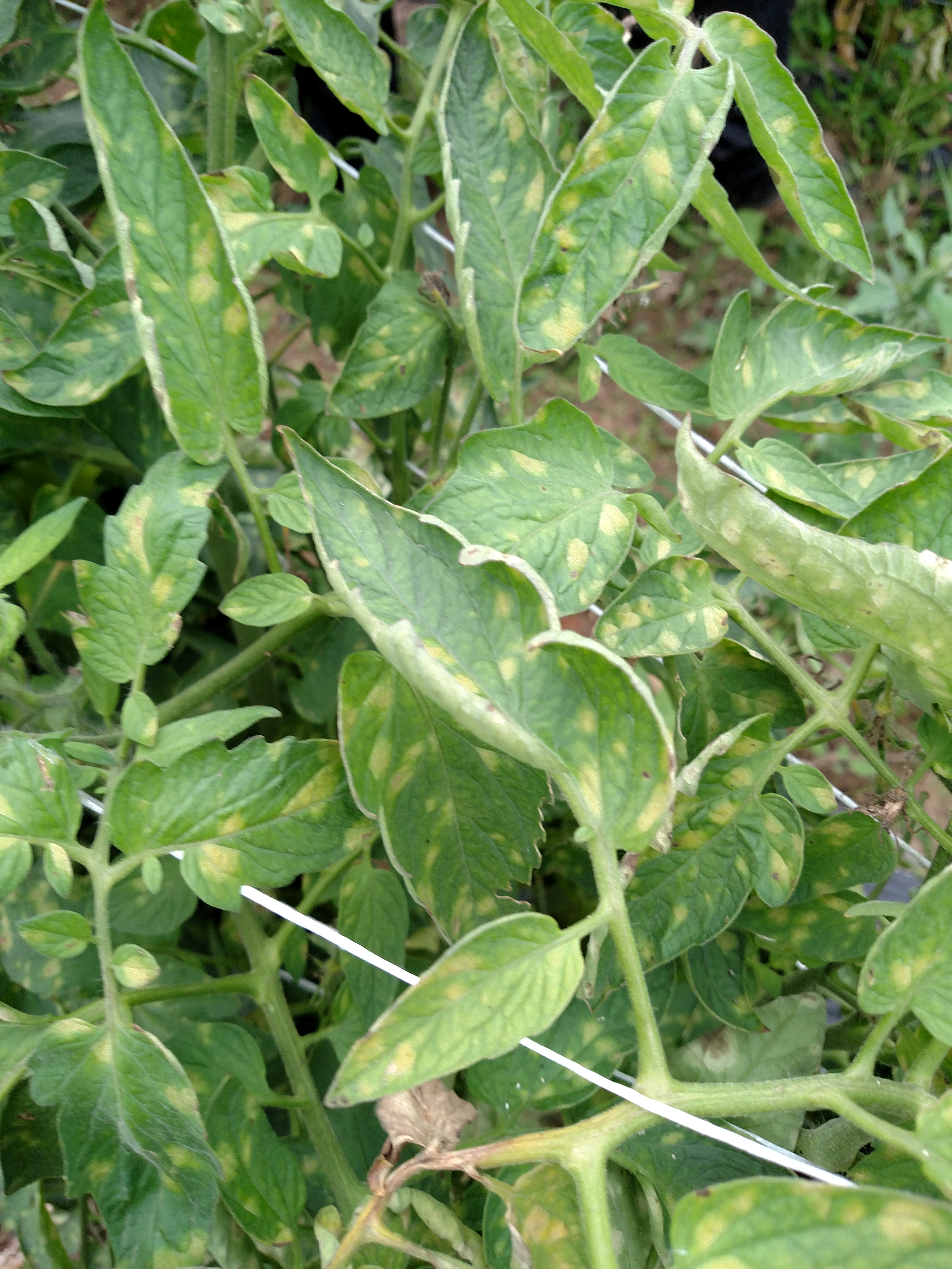 Figure 2. Leaf mold of tomato.
 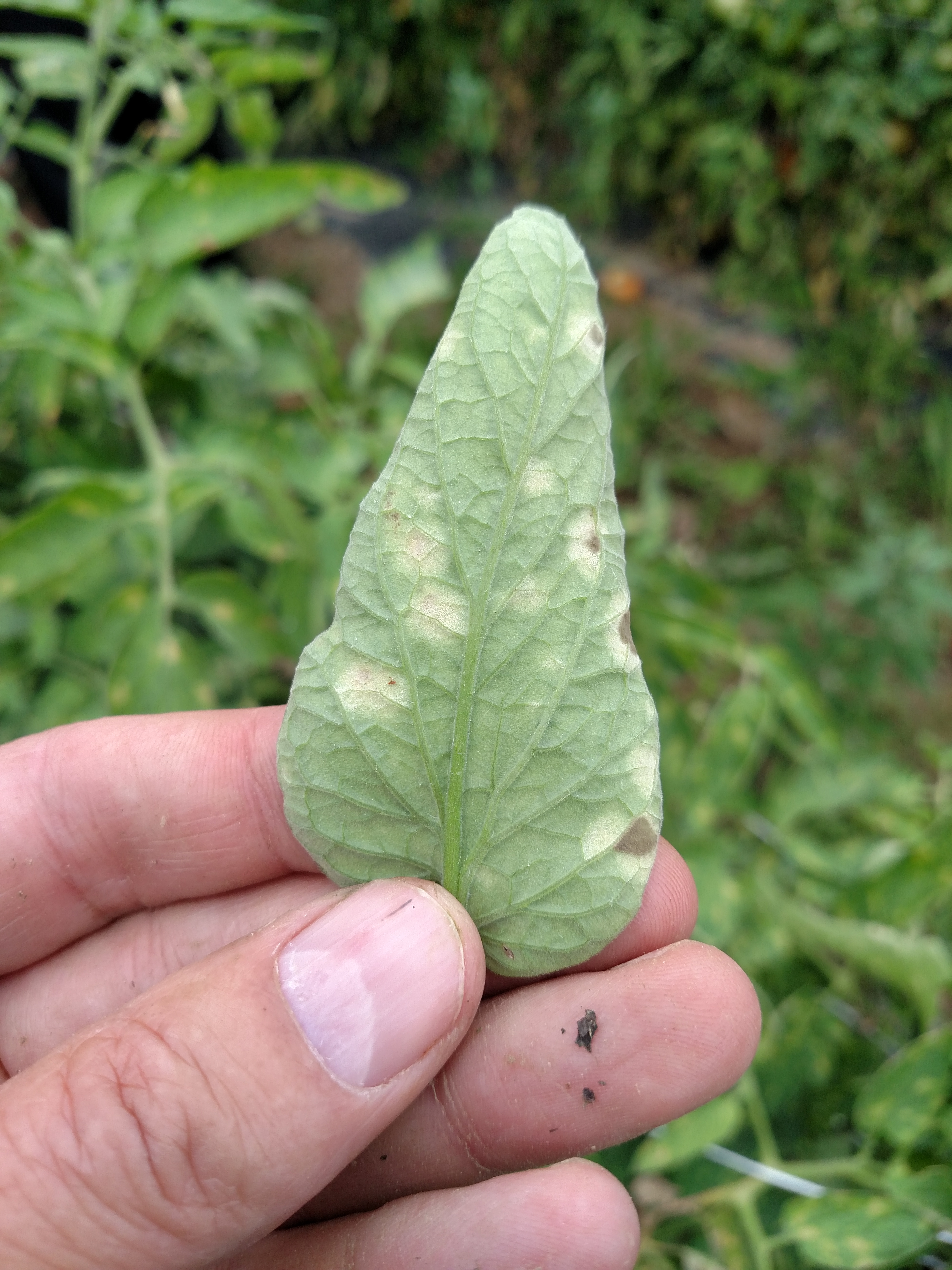 Figure 3. Underside of tomato leaf with leaf mold showing sporulation of causal fungus just starting.
 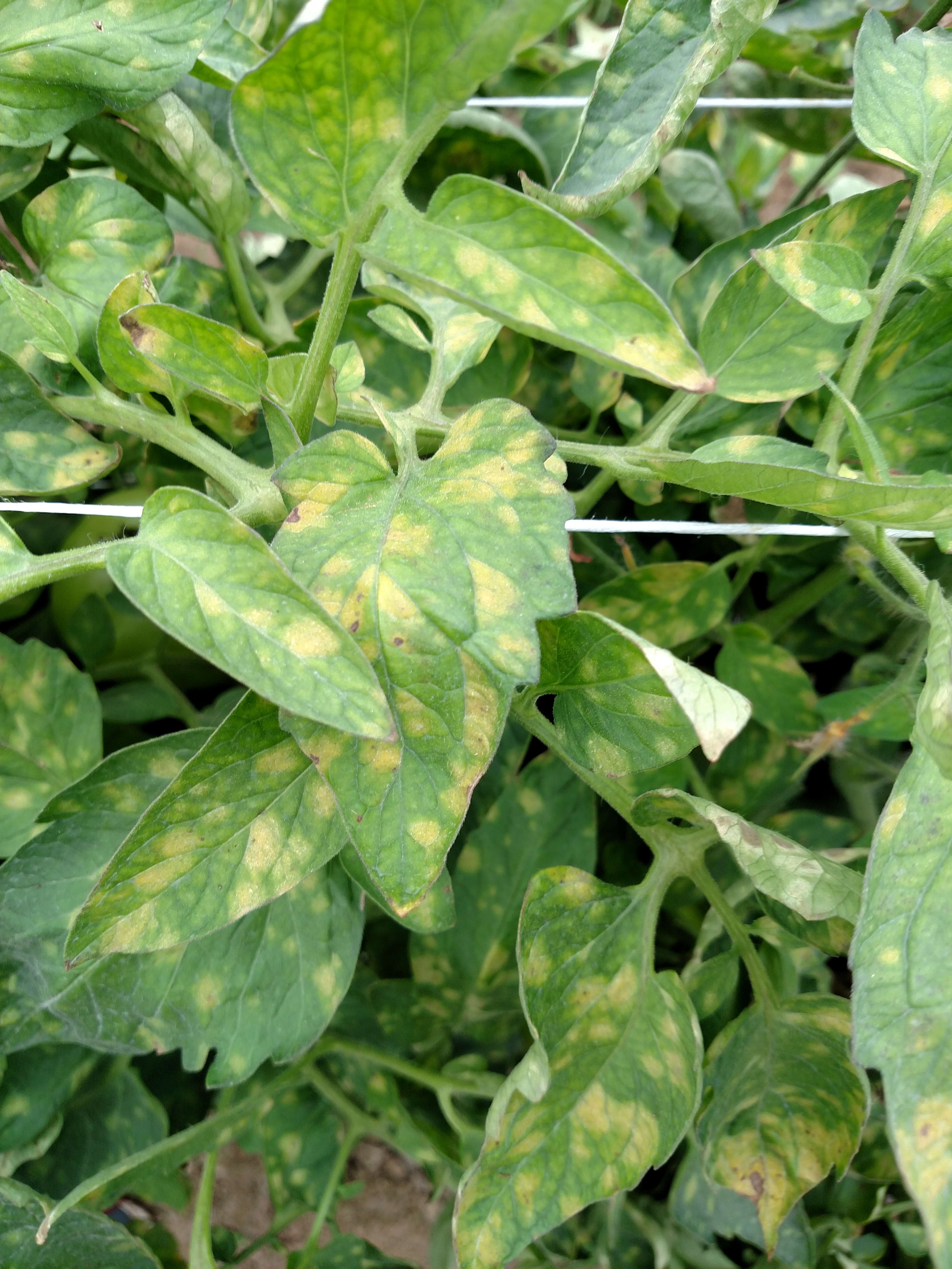 Figure 4. Leaf mold of tomato.
 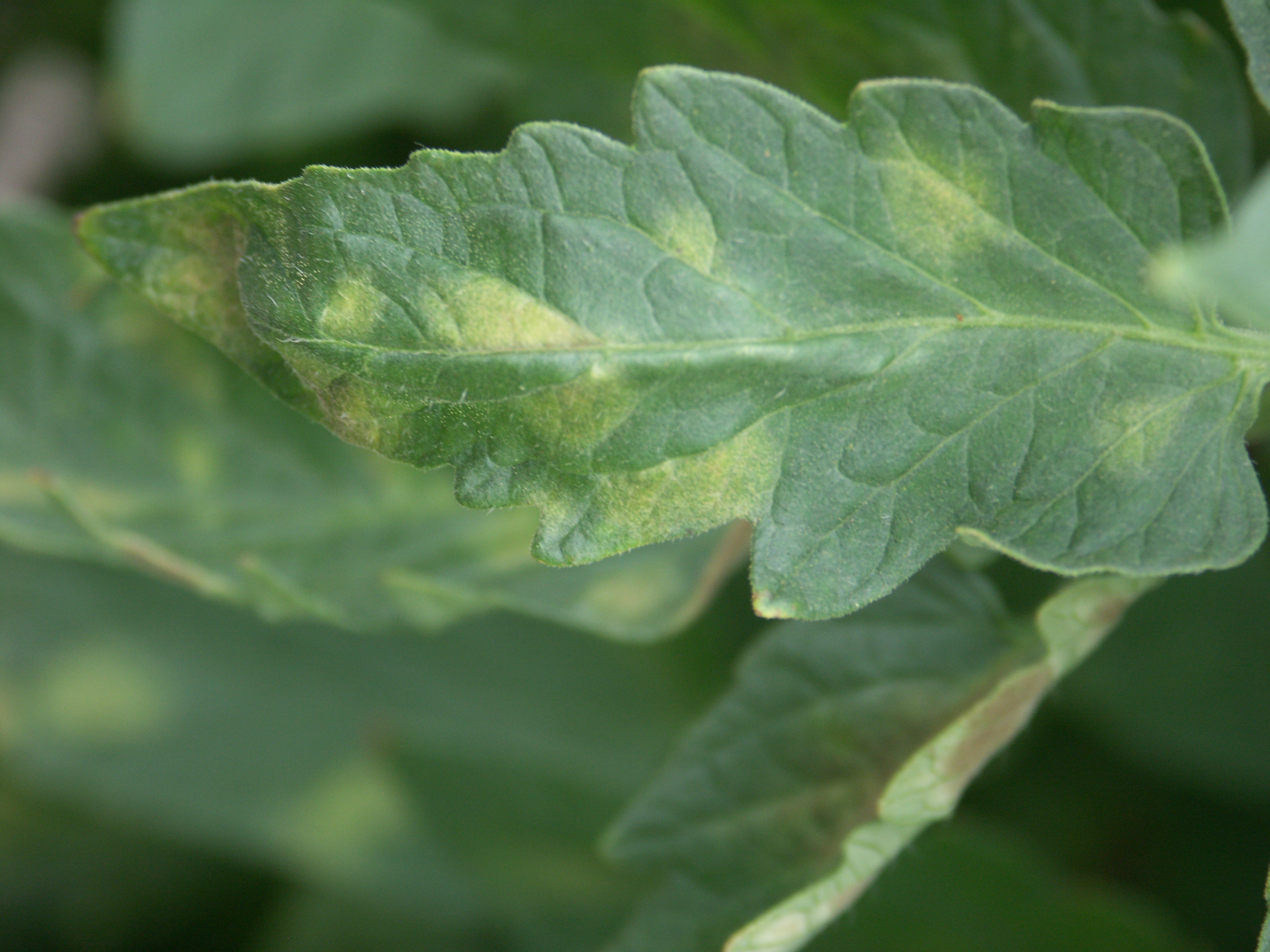 Figure 5. Close up of leaf with tomato leaf mold. Note chlorotic lesions with diffuse margins.
 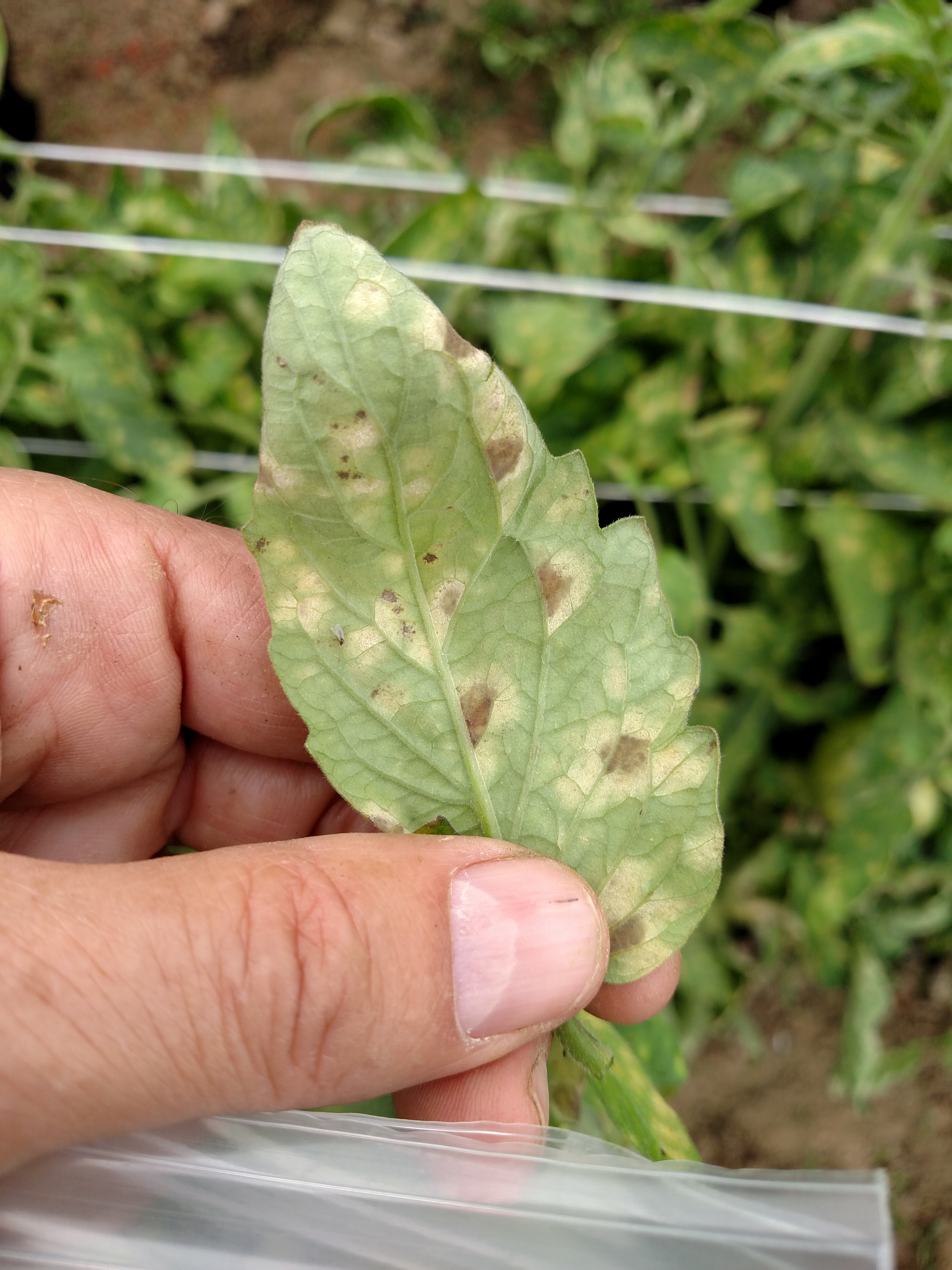 Figure 6. Underside of a leaf with tomato leaf mold.
 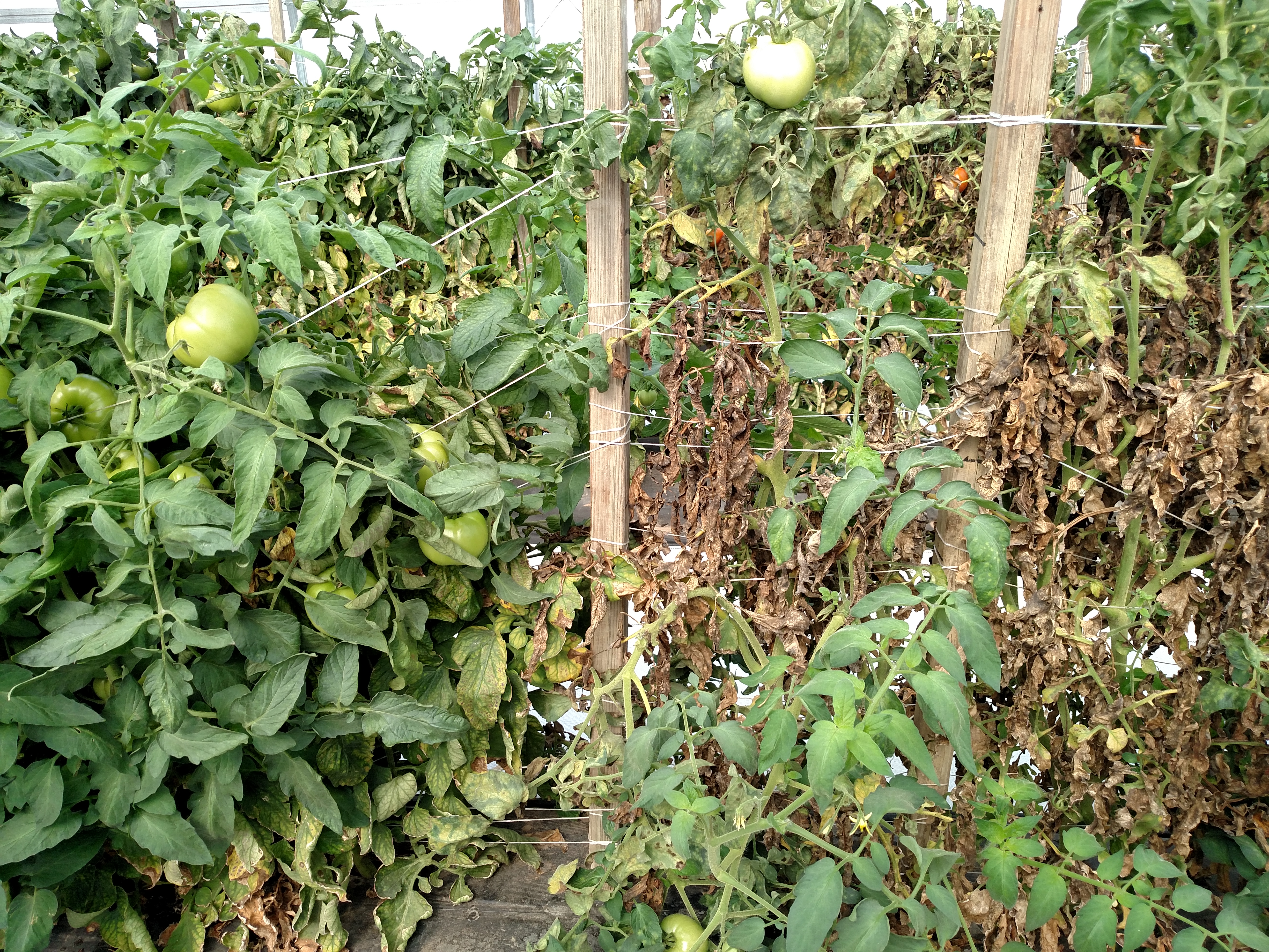 Figure 7. Leaf mold of tomato. The variety on the left is partially resistant, while the variety on the right is susceptible.
  Figure 8. Underside of leaf with sporulation of causal fungus visible for tomato leaf mold.
 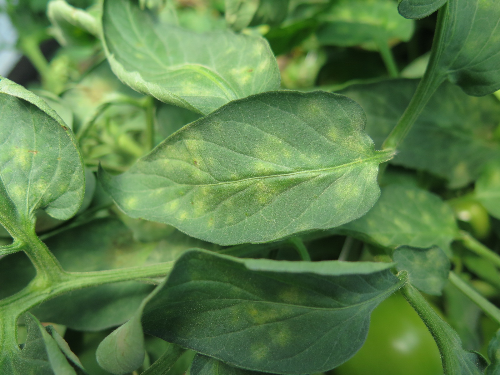 Figure 9. Leaf mold of tomato.
 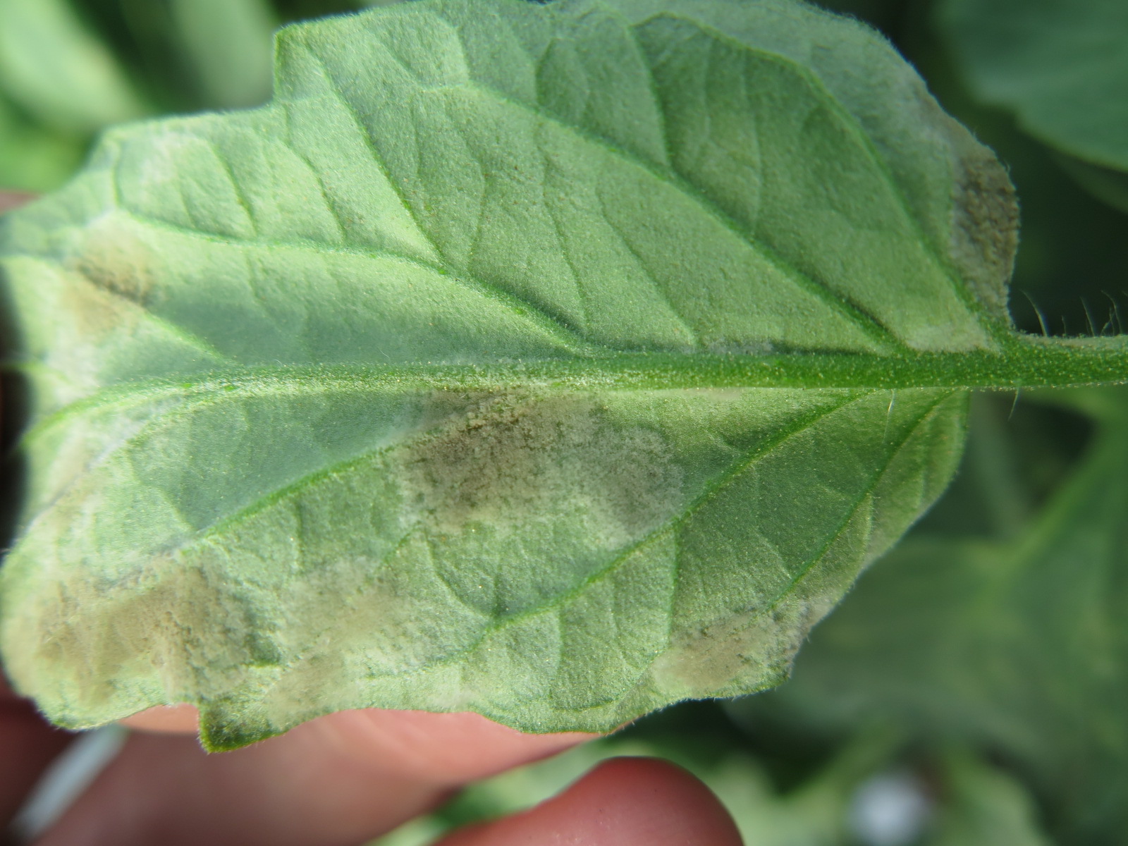 Figure 10. Underside of tomato leaf with leaf mold.
 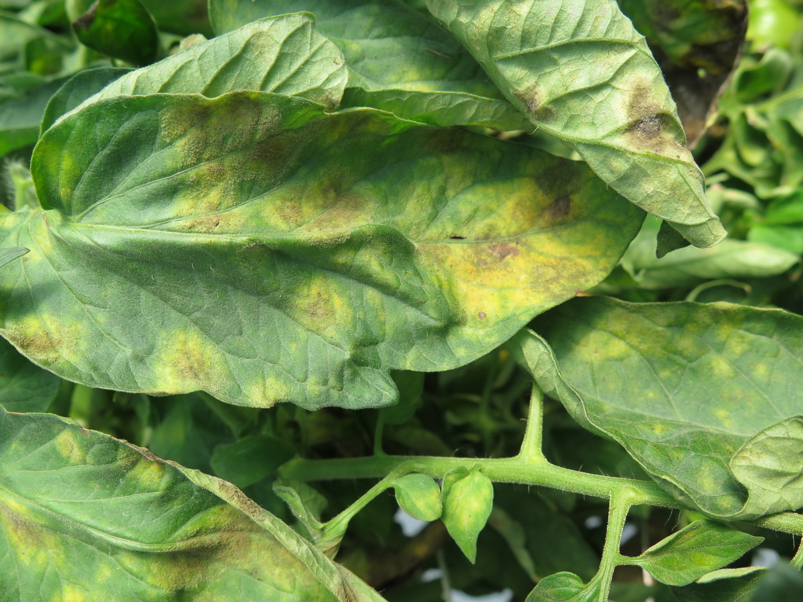 Figure 11. Leaf mold of tomato. While the sporulation of the causal fungus can often be observed on the bottom of the leaf, occasionally sporulation can be seen on the top of the leaf.
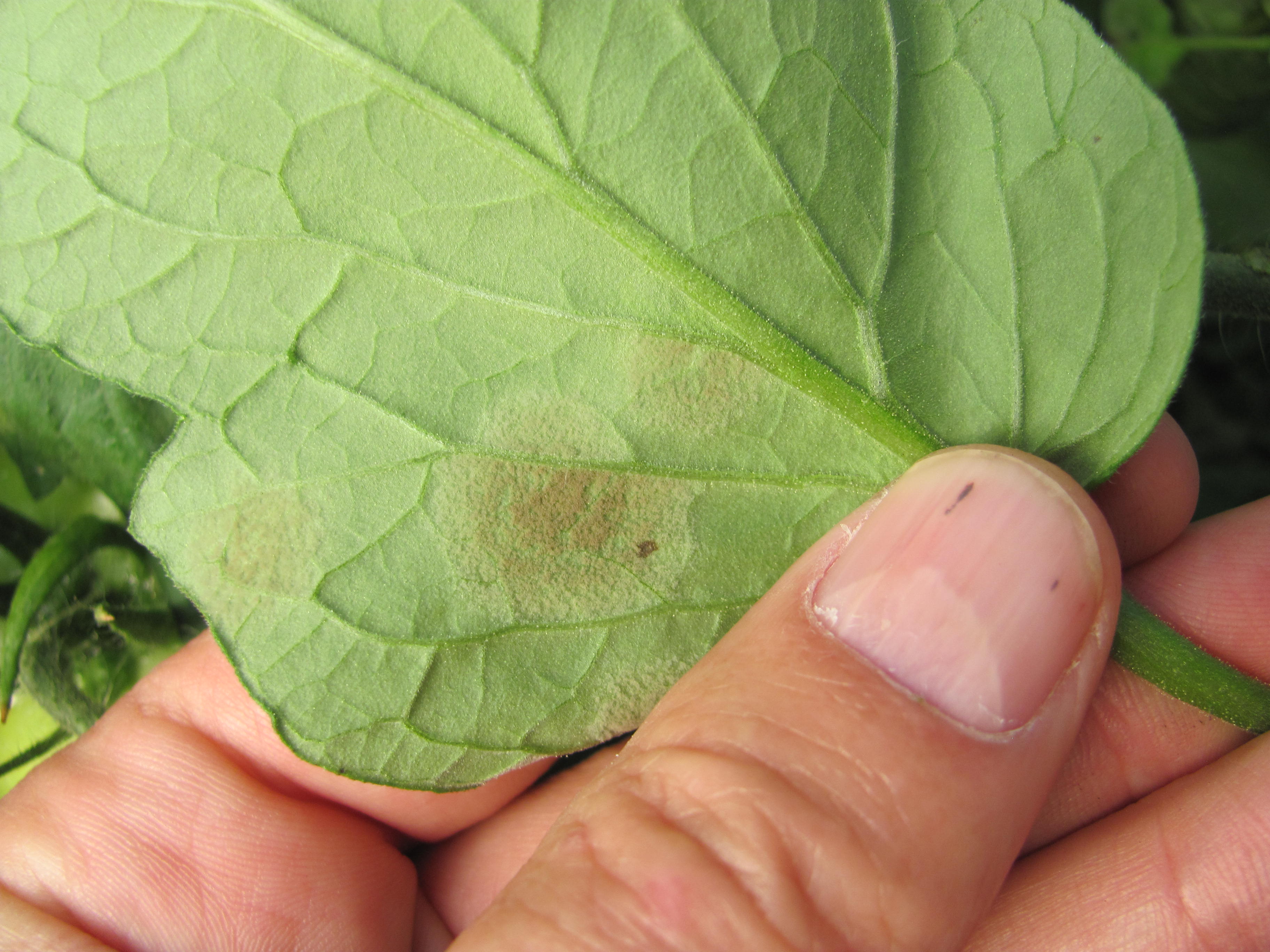 Figure 12. Close up of sporulation on underside of tomato leaf with leaf mold.
 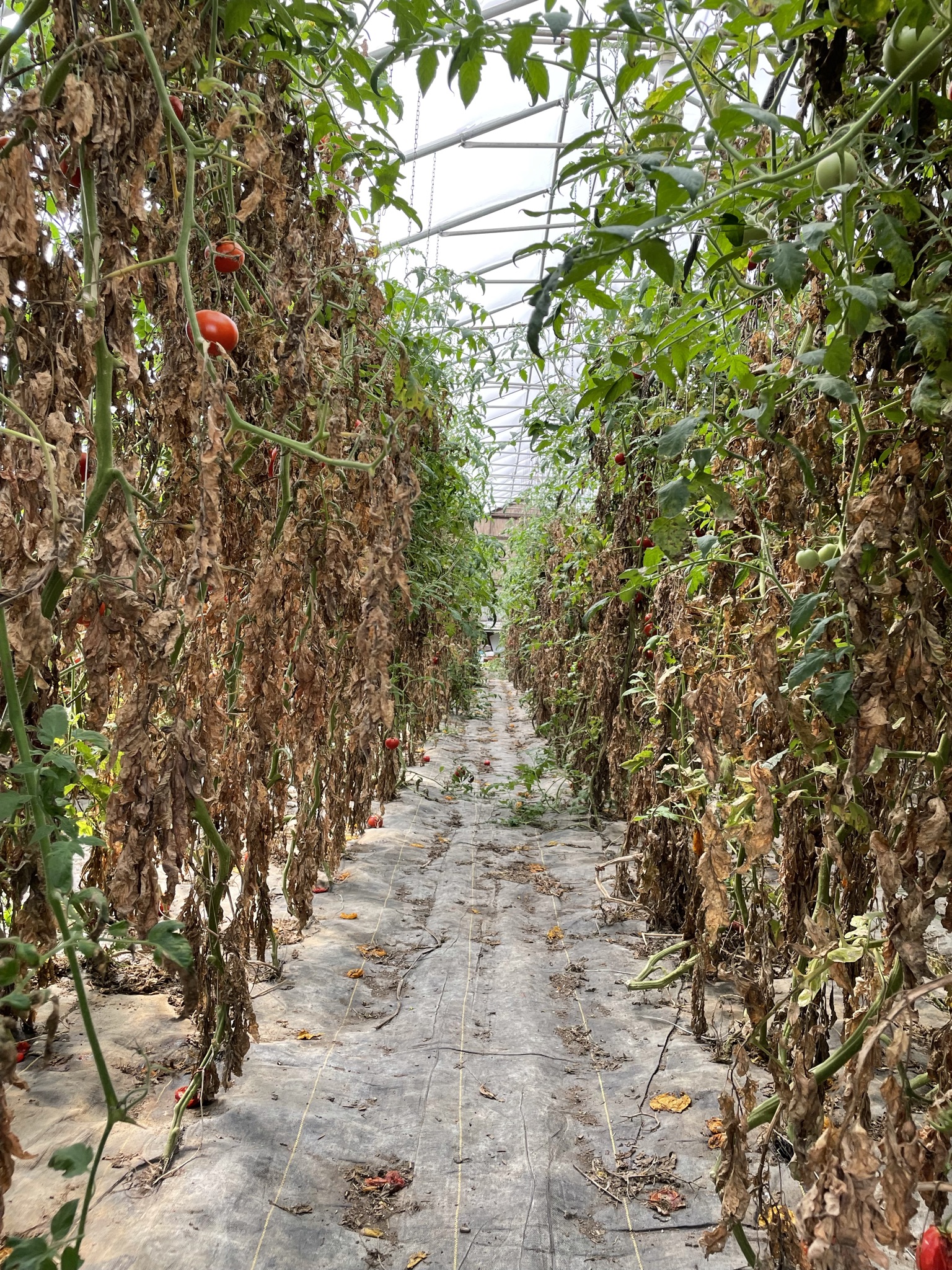 Figure 13. Severe leaf mold of tomato.
 Leaf roll
Leaf roll-this symptom is not necessarily due to an infectious disease. Leaf roll may occur due to any type of plant stress. In addition, some tomato varieties are more likely to exhibit leaf roll than others. Leaf roll does not necessarily represent a problem.
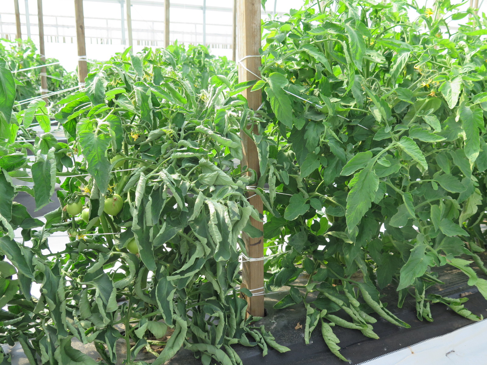 Figure 1. Although leaf roll of tomato leaves can sometimes indicate stress, leaf roll can also be genetic. The leaf roll on the plant to the left is due to variety and does not indicate a problem with the plant. The plant to the right, in contrast, is a different variety and does not exhibit leaf roll.
 Pith necrosis
Pith necrosis-this disease is not usually an important problem. Irregular necrotic lesions on the stem are characteristic. Stems may become dark and twisted. Pith necrosis is more often found in a greenhouse or high tunnel than in a field.
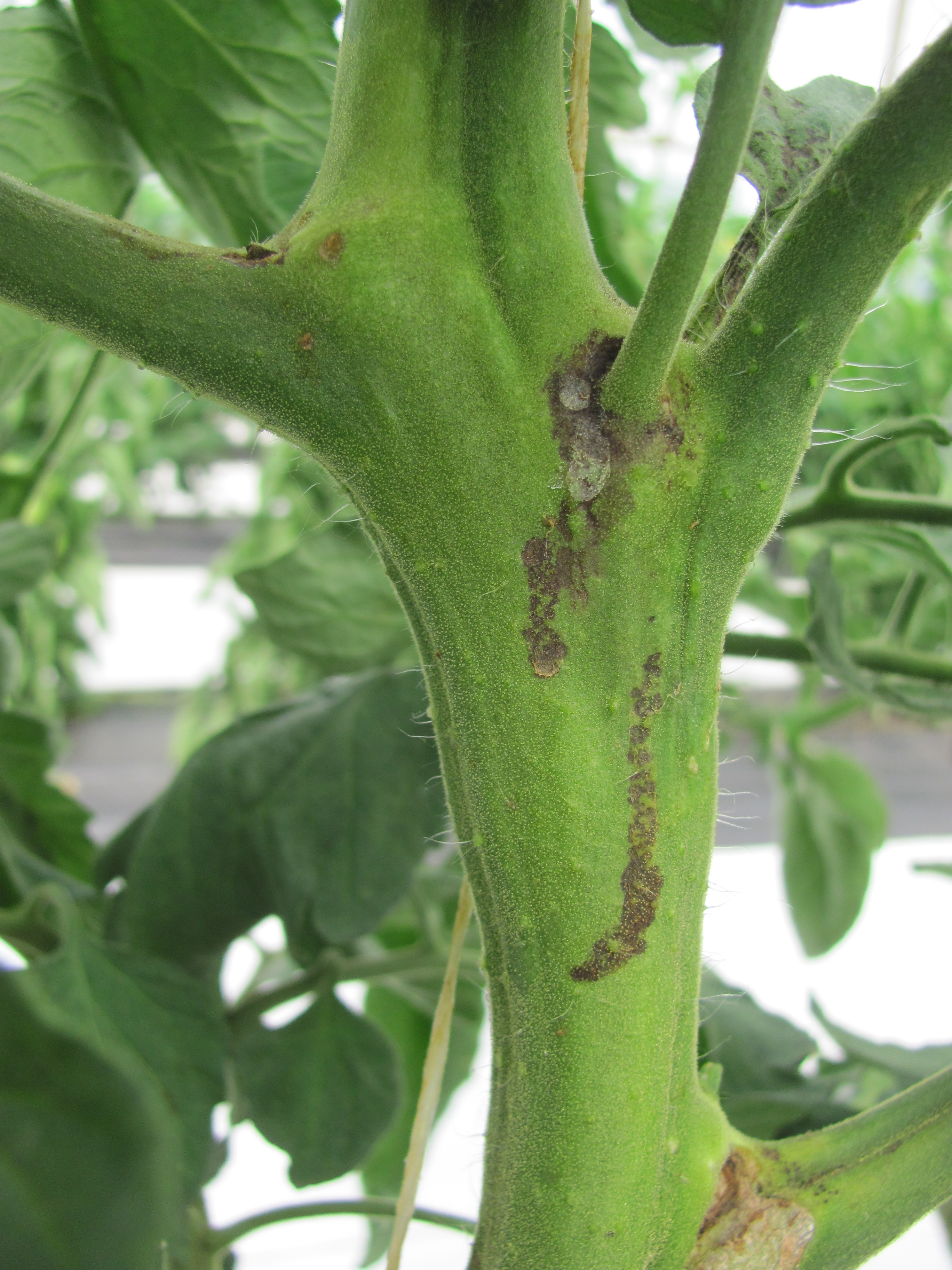 Figure 1. Initial symptoms of pith necrosis of tomato may appear to be minor necrosis on the stem.
 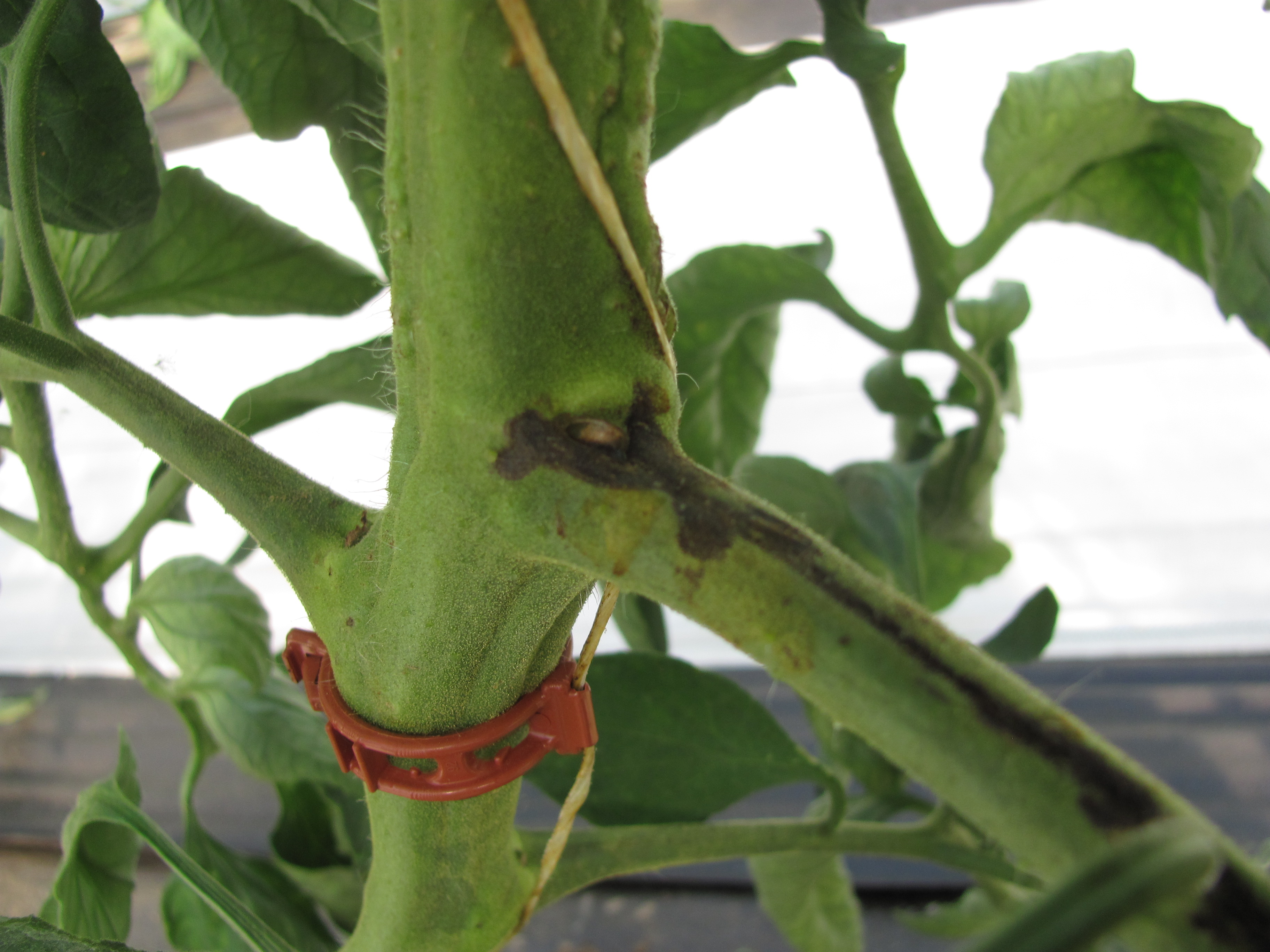 Figure 2. Dark pith necrosis lesion on tomato stem.
 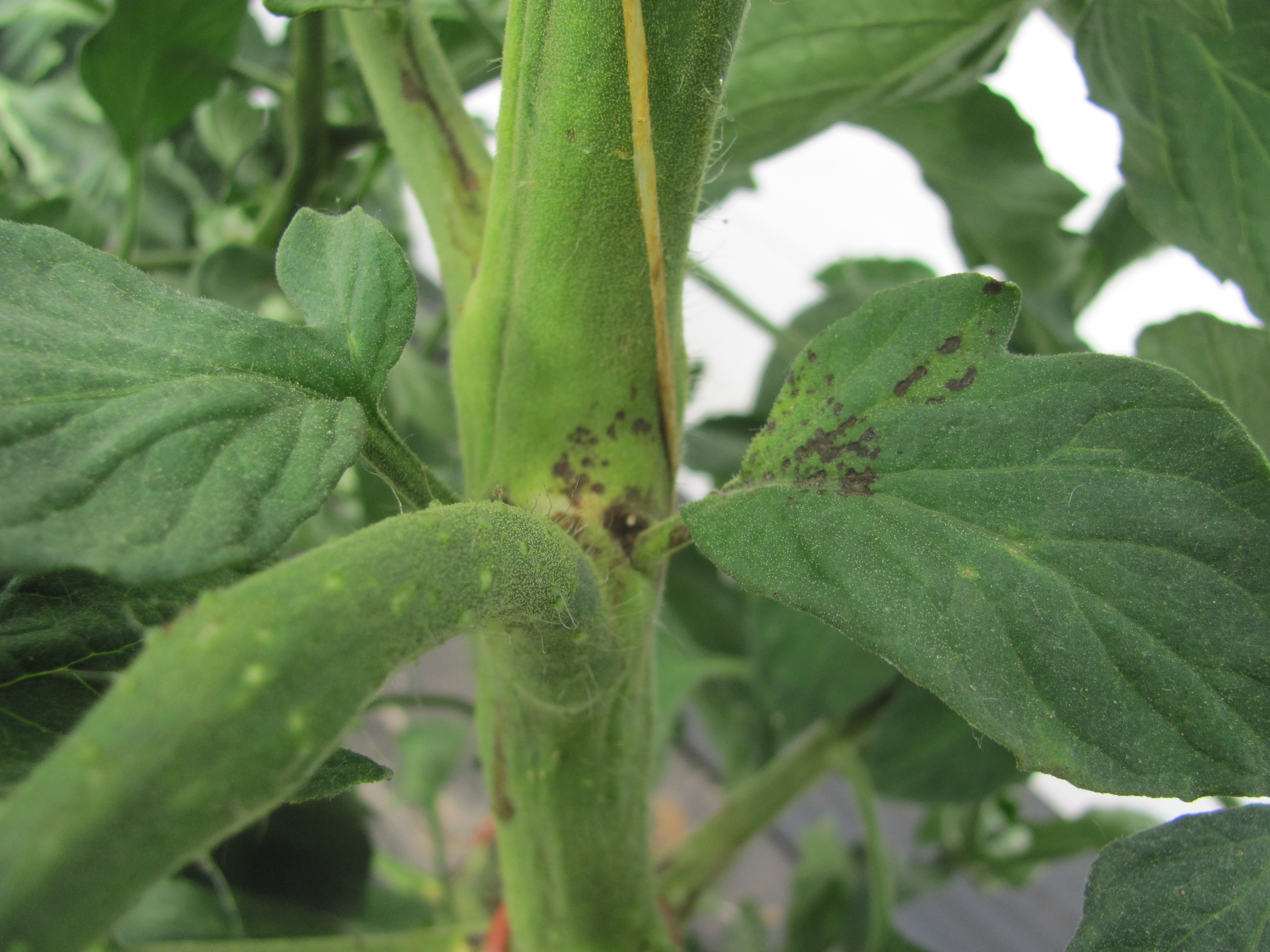 Figure 3. Early symptoms of pith necrosis of tomato.
 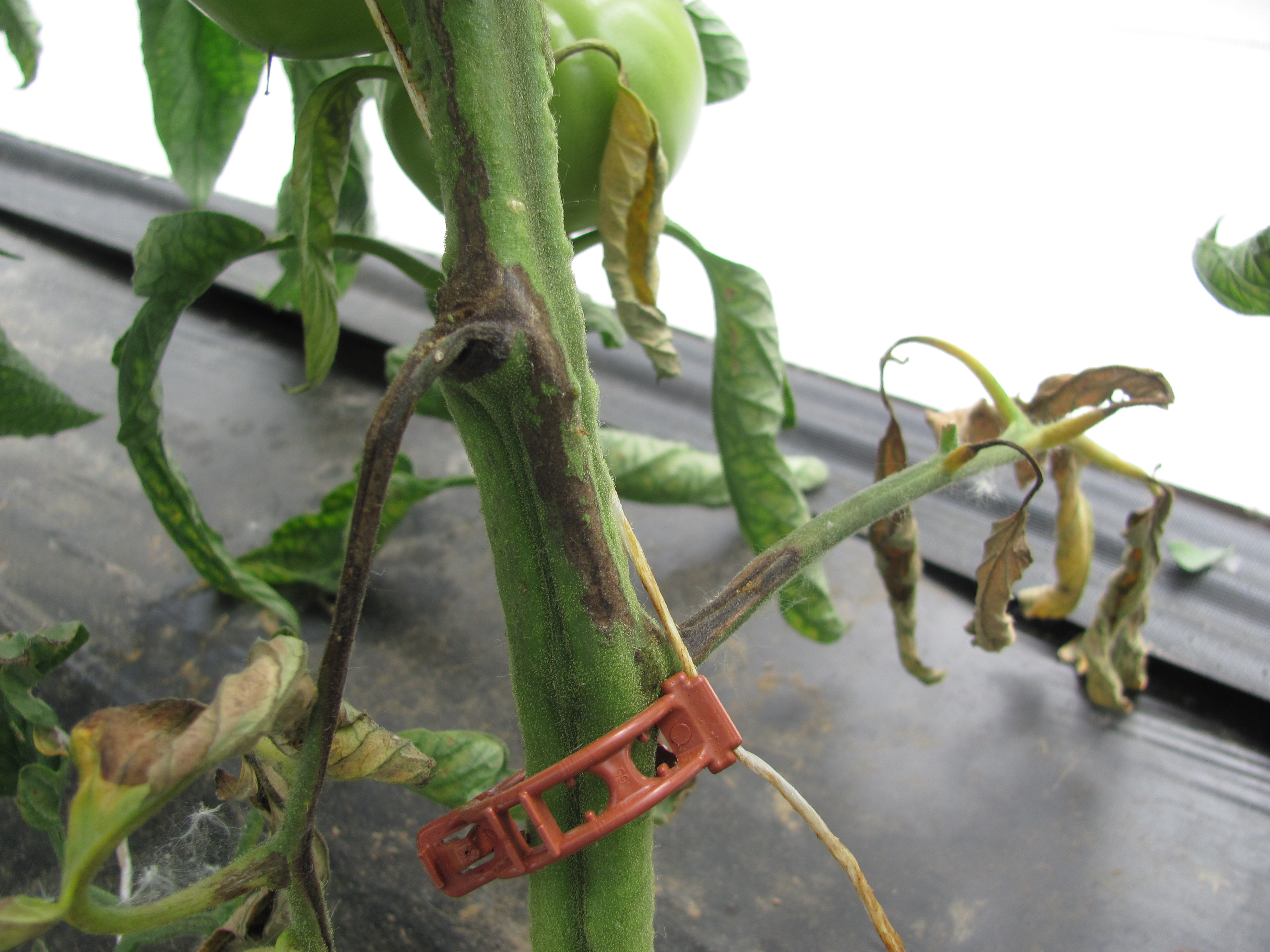 Figure 4. A dark lesion of tomato pith necrosis extends along the stem and leaf petioles.
 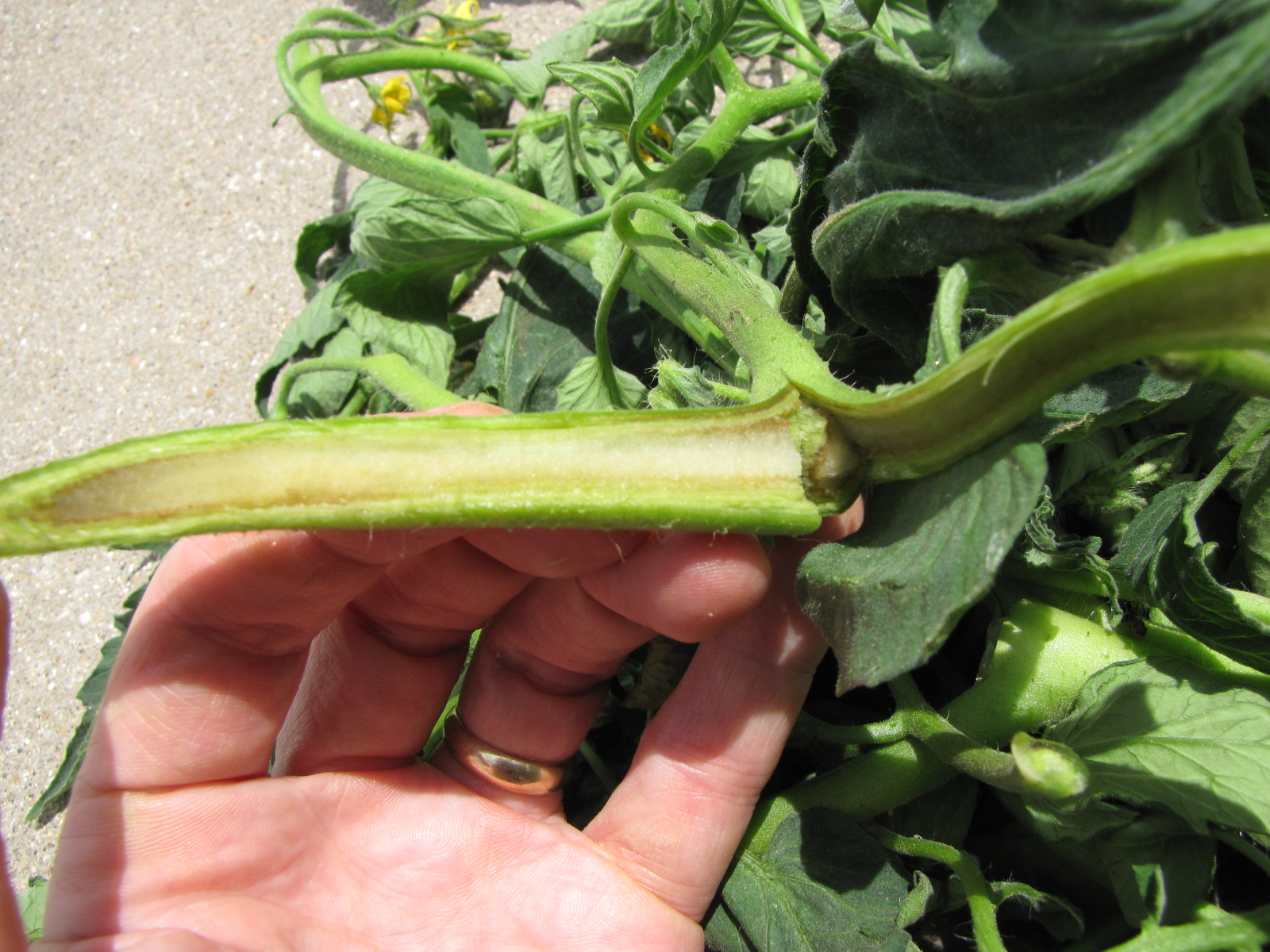 Figure 5. Pith necrosis of tomato can sometimes cause internal stem discoloration as seen here.
 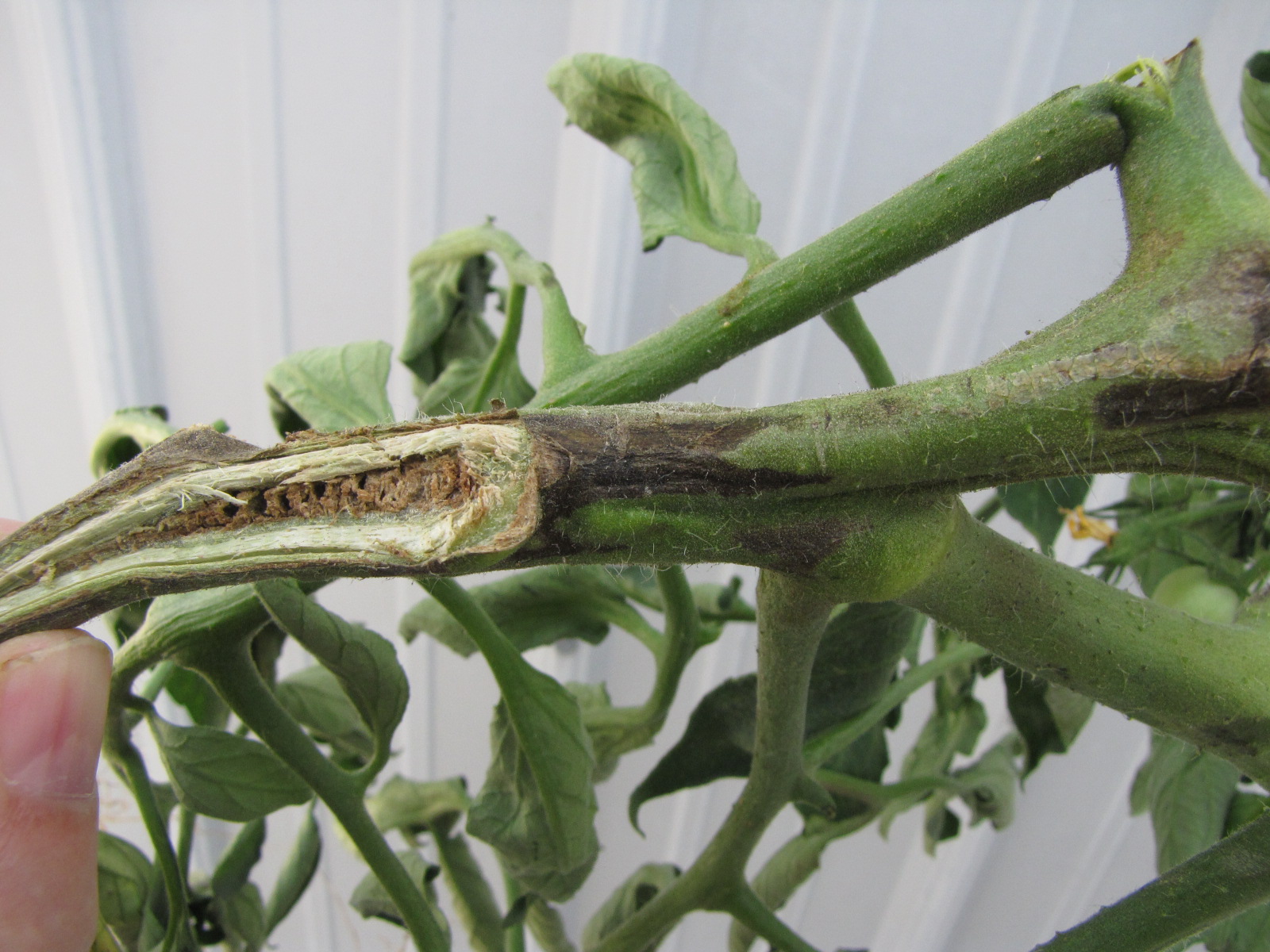 Figure 6. Dark necrosis on stem and chambered pith caused by tomato pith necrosis.
 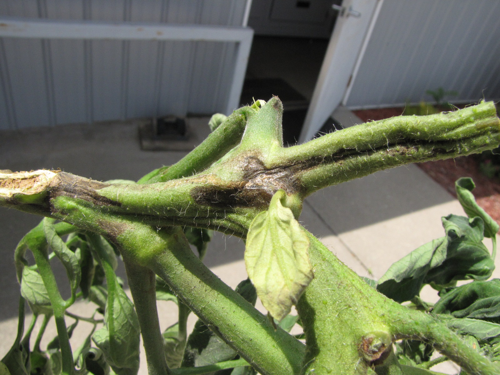 Figure 7. Tomato pith necrosis.
 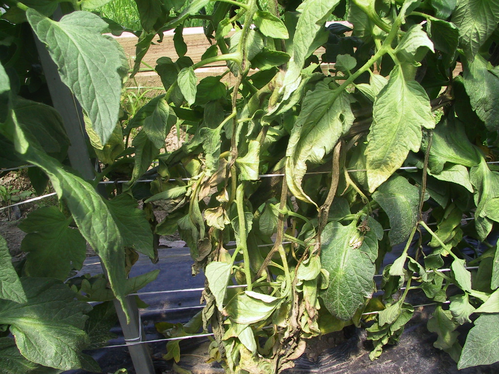 Figure 8. A portion of a tomato plant with symptoms of pith necrosis has begun to wilt due to stem lesions.
 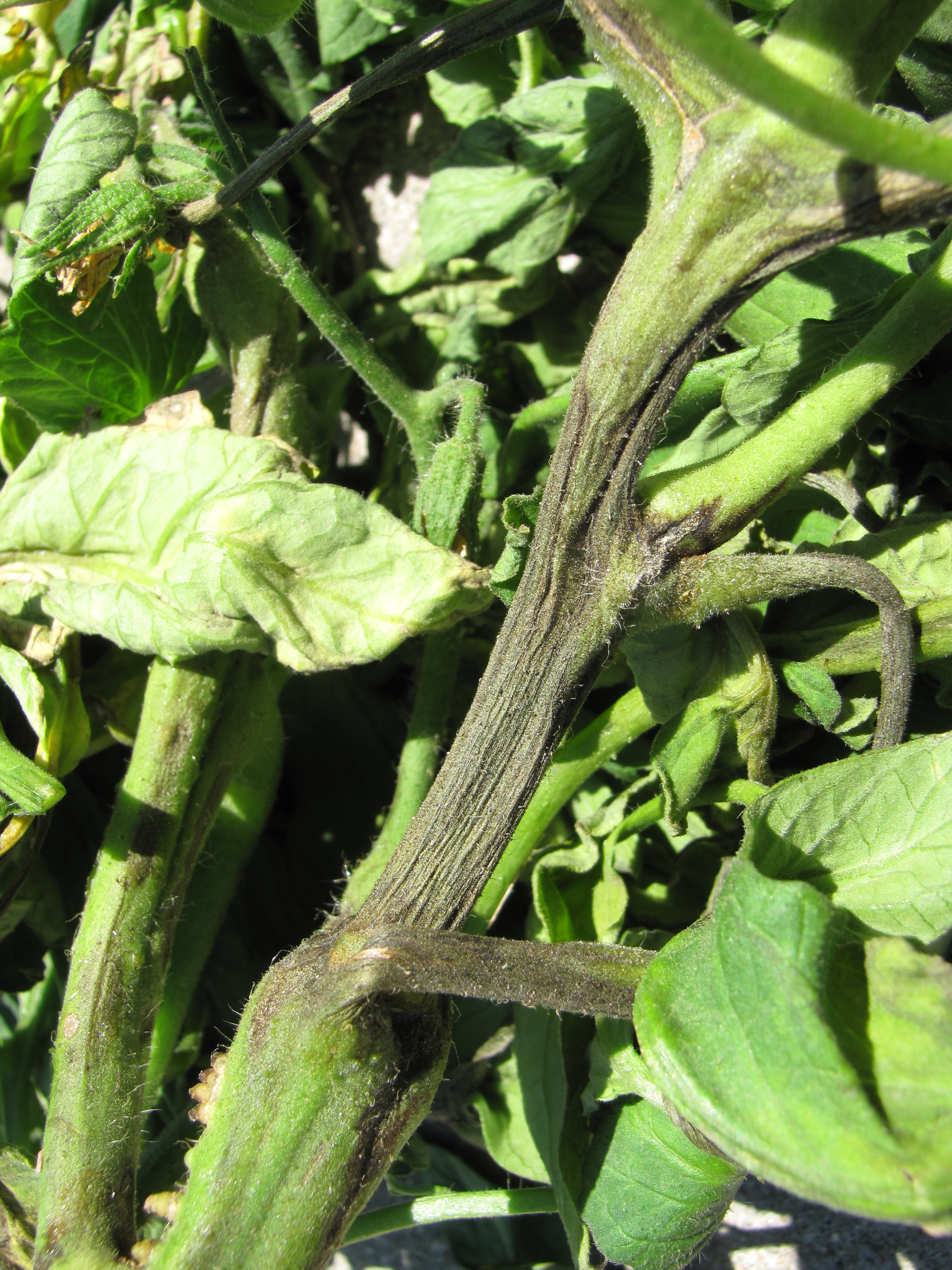 Figure 9. Withered stem due to tomato pith necrosis.
 Powdery mildew
Powdery mildew-easily recognized by the white talc-like lesions on leaves and stems. More often observed in greenhouses or high tunnels than in the field. Not usually a problem.
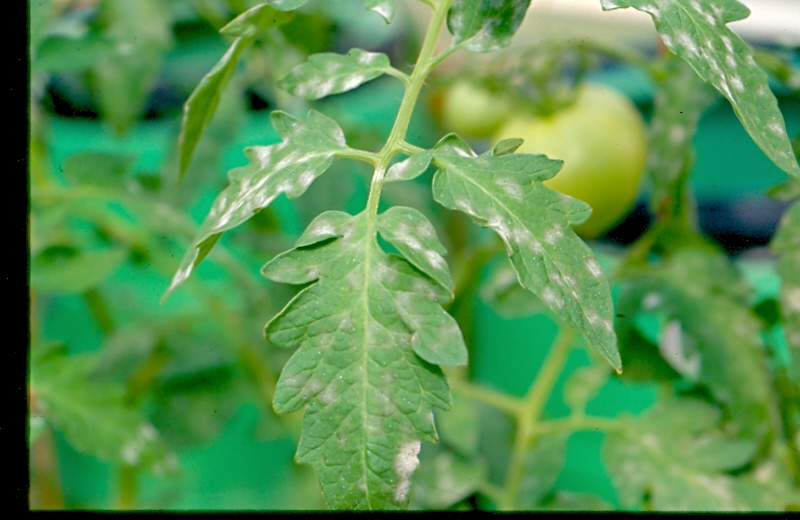 Figure 1. Powdery mildew of tomato. Lesions of powdery mildew have talc-like appearance.
 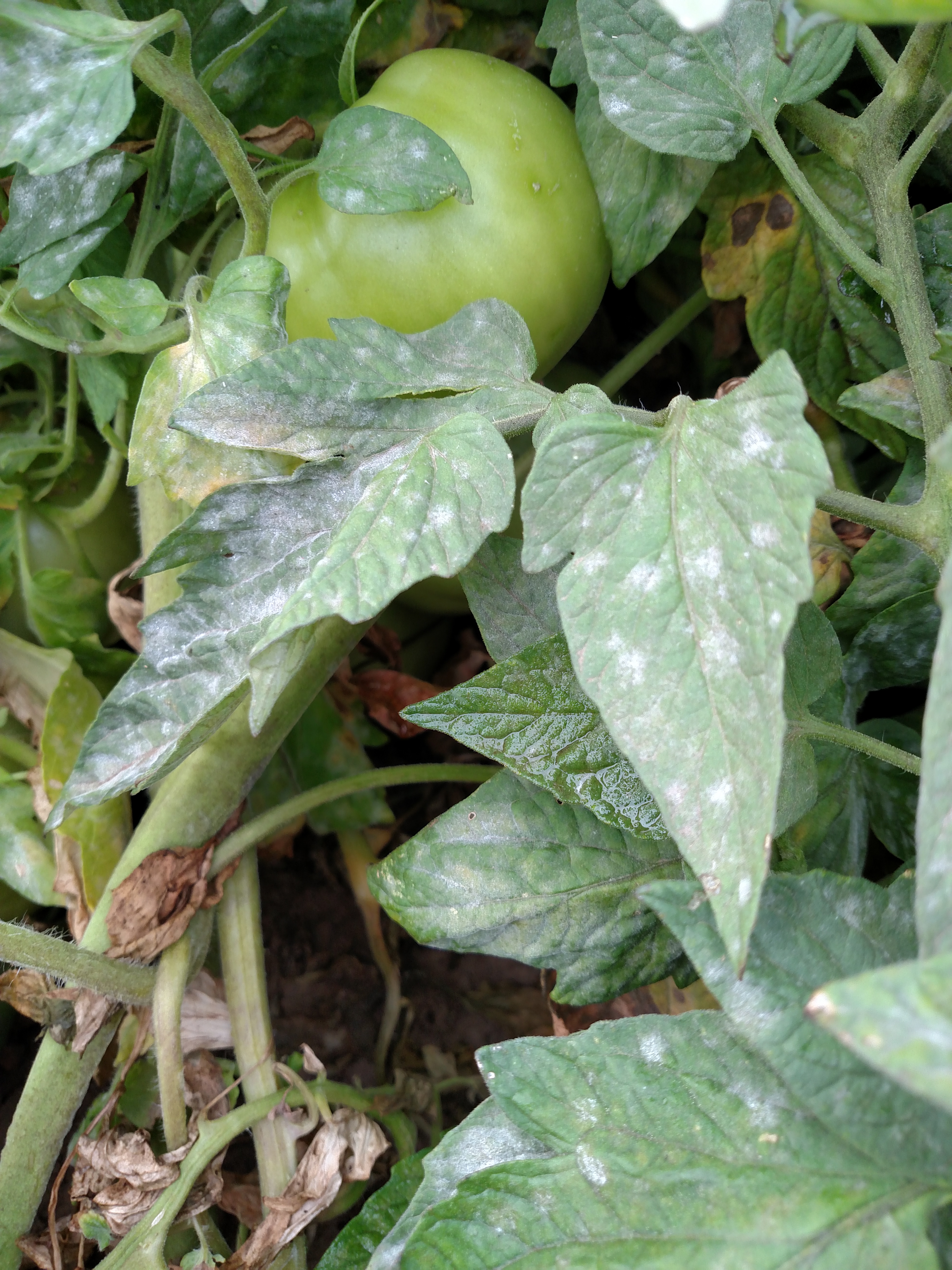 Figure 2. Powdery mildew of tomatoes has covered much of the leaf surfaces shown here.
 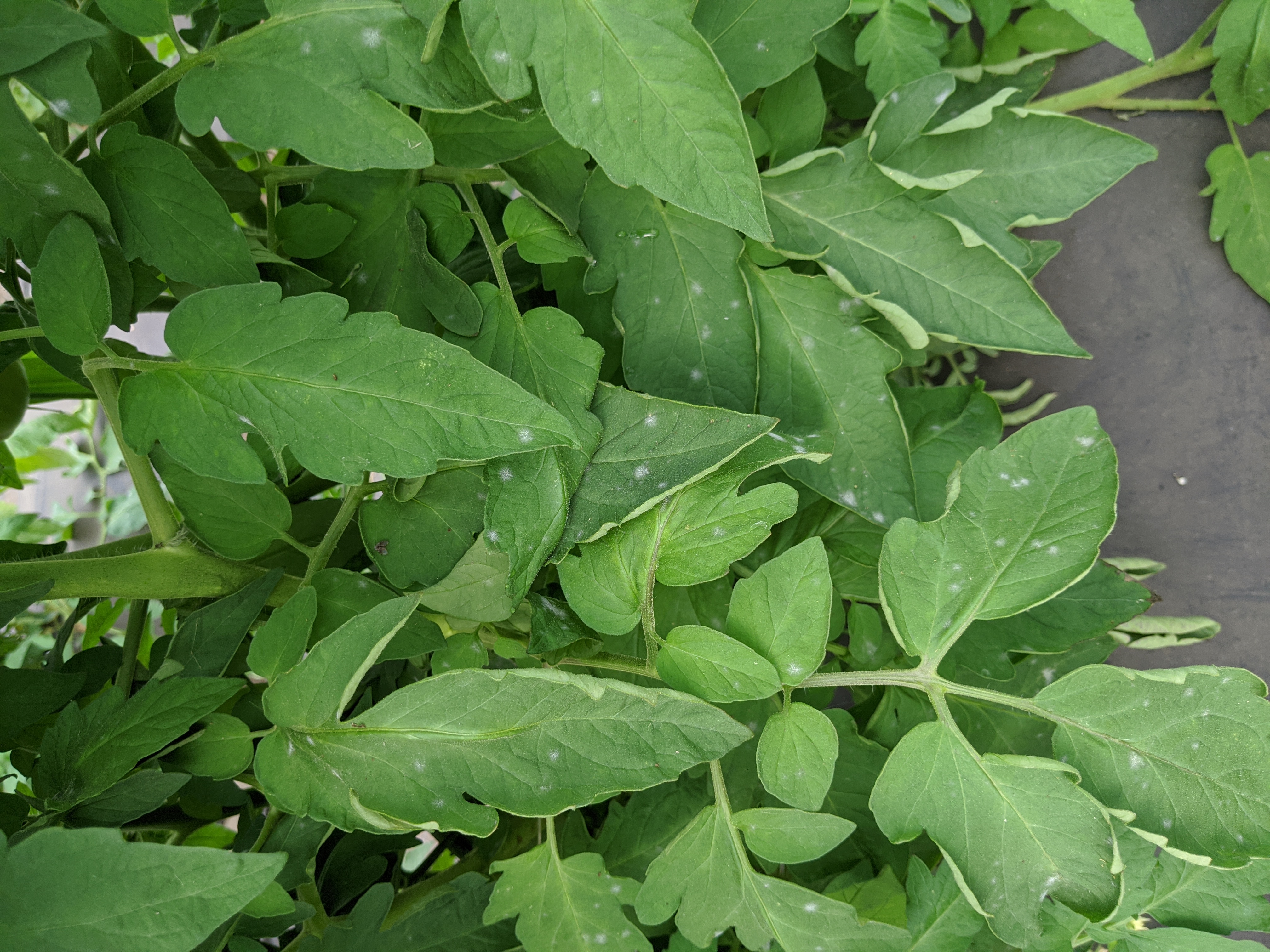 Figure 3. A relatively mild outbreak of powdery mildew of tomato.
 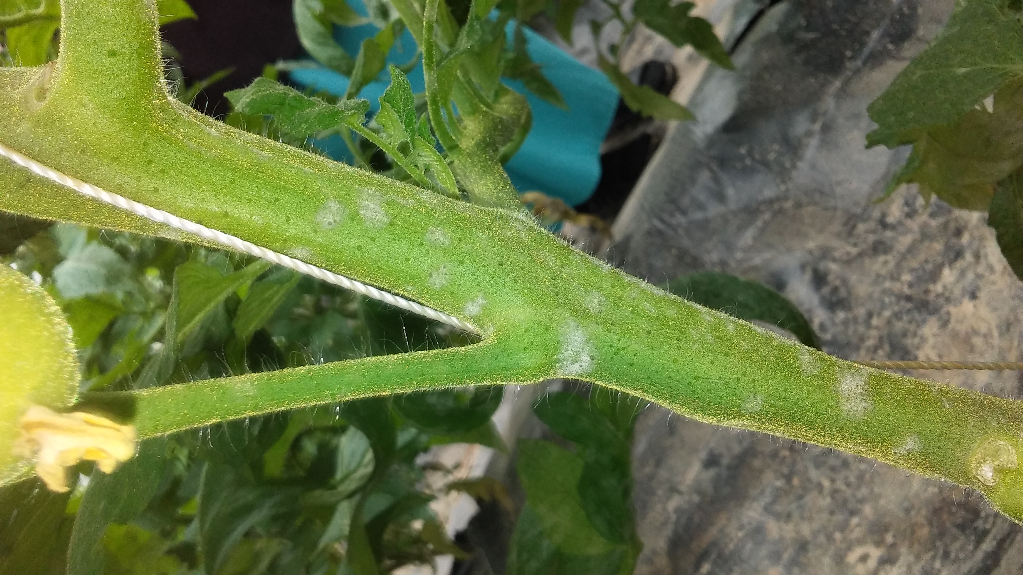 Figure 4. Lesions of powdery mildew of tomato on stem.
 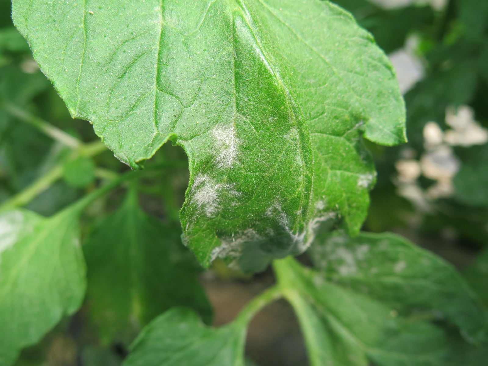 Figure 5. A close up of powdery mildew on tomato leaves.
 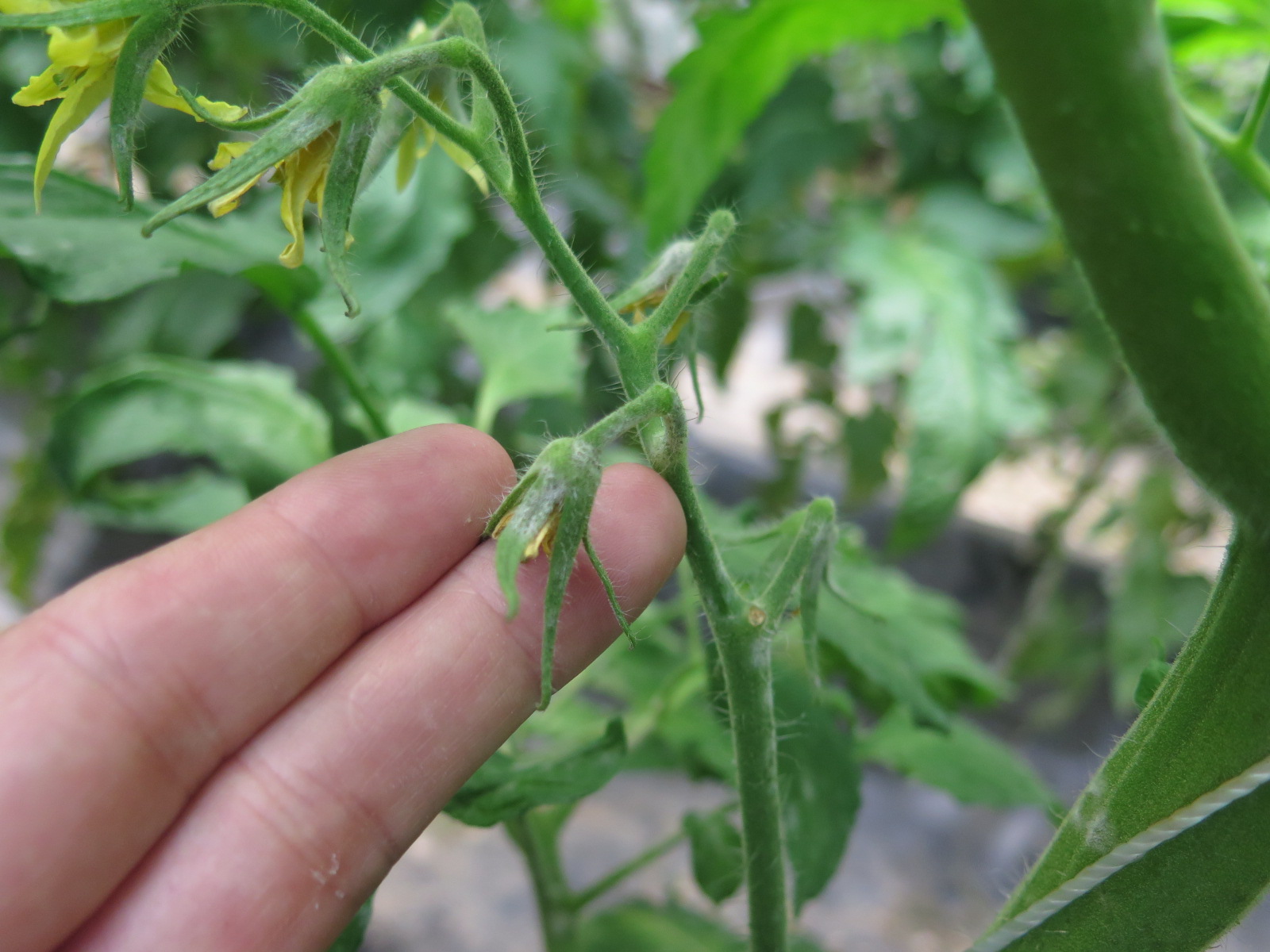 Figure 6. Powdery mildew of tomato on flower sepals.
 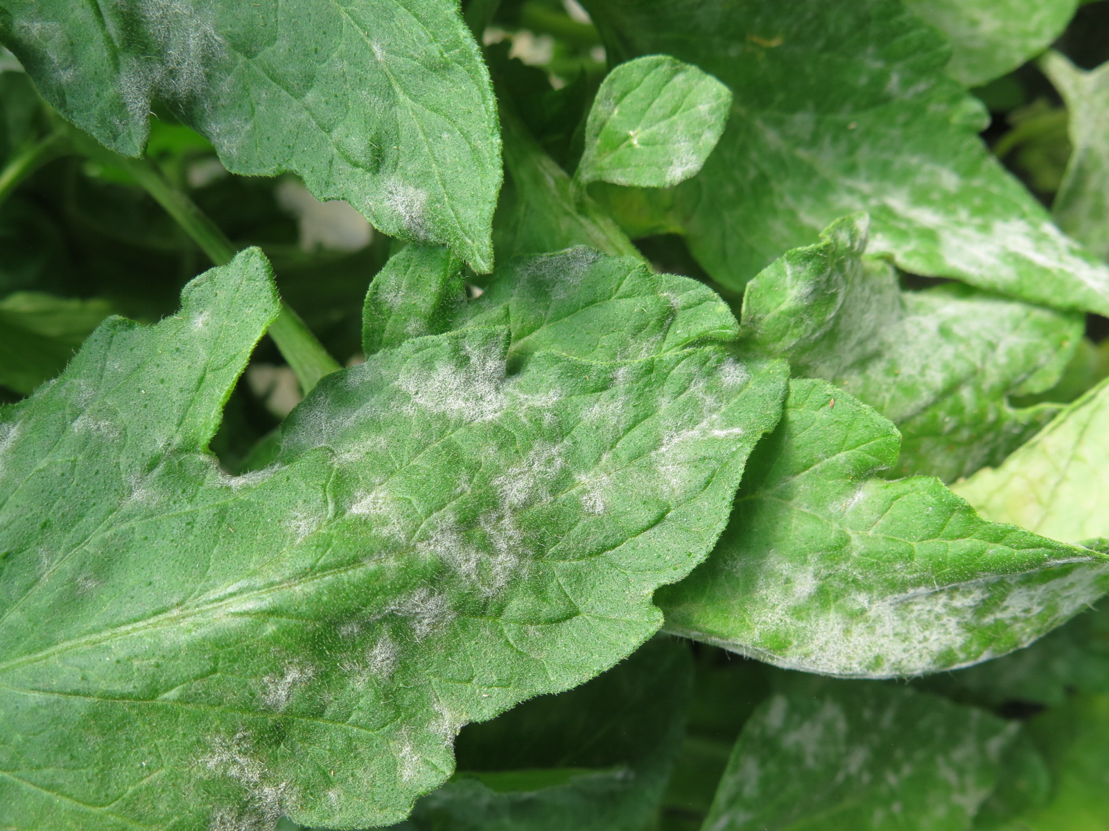 Figure 7. Powdery mildew of tomato.
 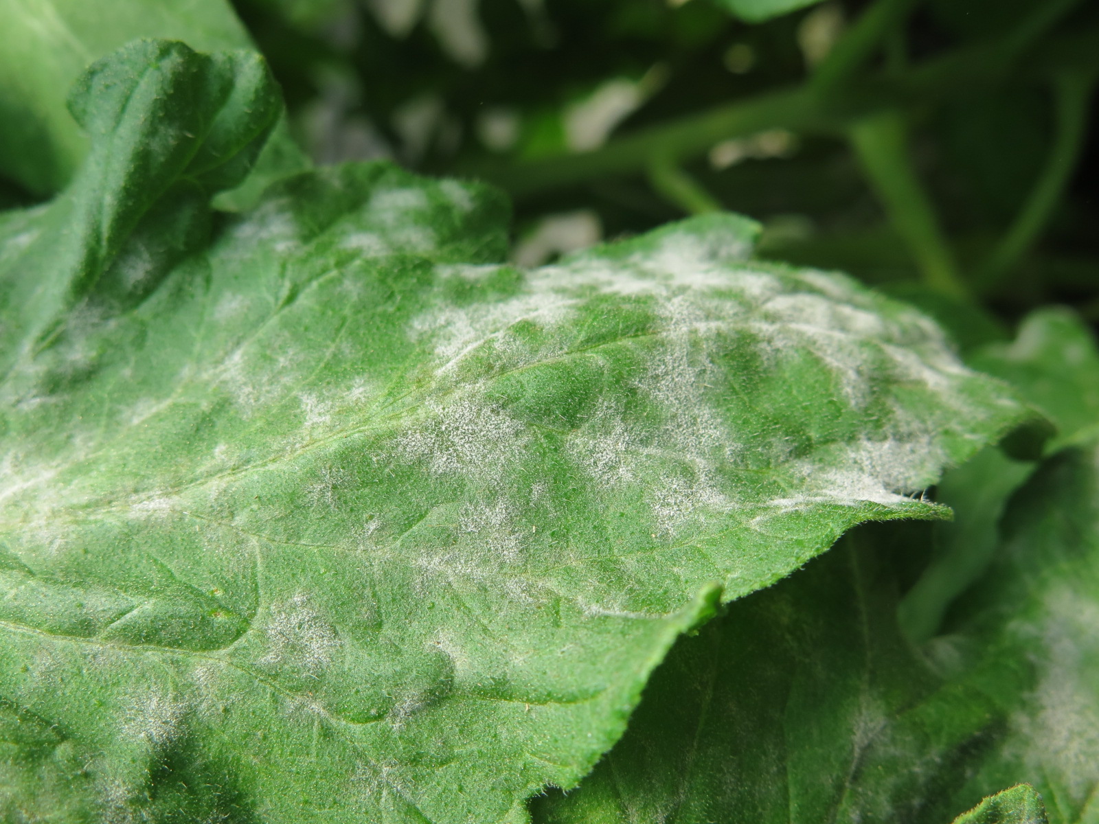 Figure 8. Powdery mildew of tomato.
 Septoria leaf spot
Septoria leaf spot-One of the most common diseases of tomato in the field. Symptoms occur first on older leaves. Lesions are medium brown with gray centers. Dark, fungal structures (pycnidia) can often be observed in center of lesions. Lesions may have chlorotic margins.
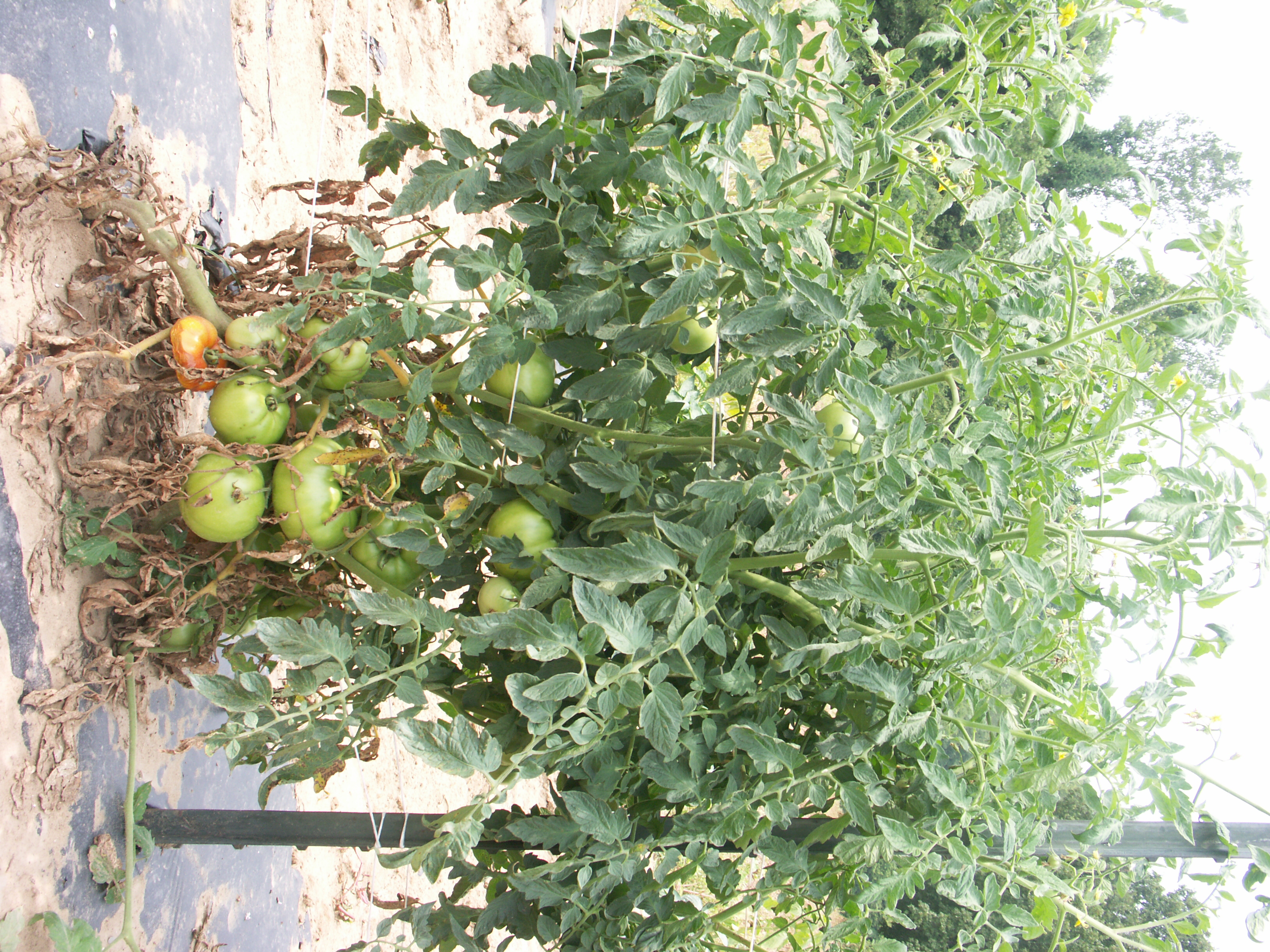 Figure 1. Septoria leaf spot of tomato is more severe on older leaves. Thus, older leaves are often the first to show symptoms.
 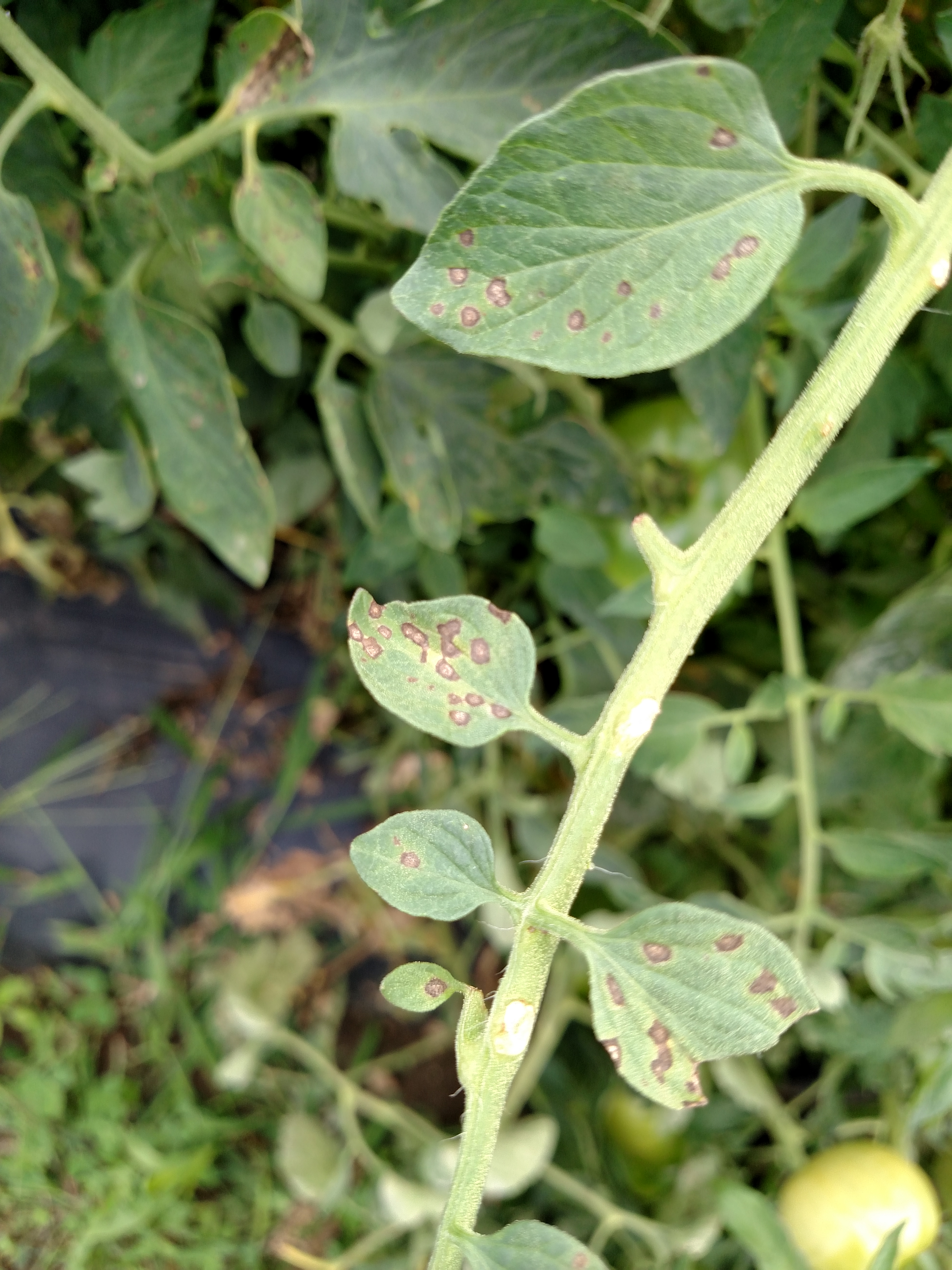 Figure 2. Lesions of Septoria leaf spot are often dark brown on the inside margin.
 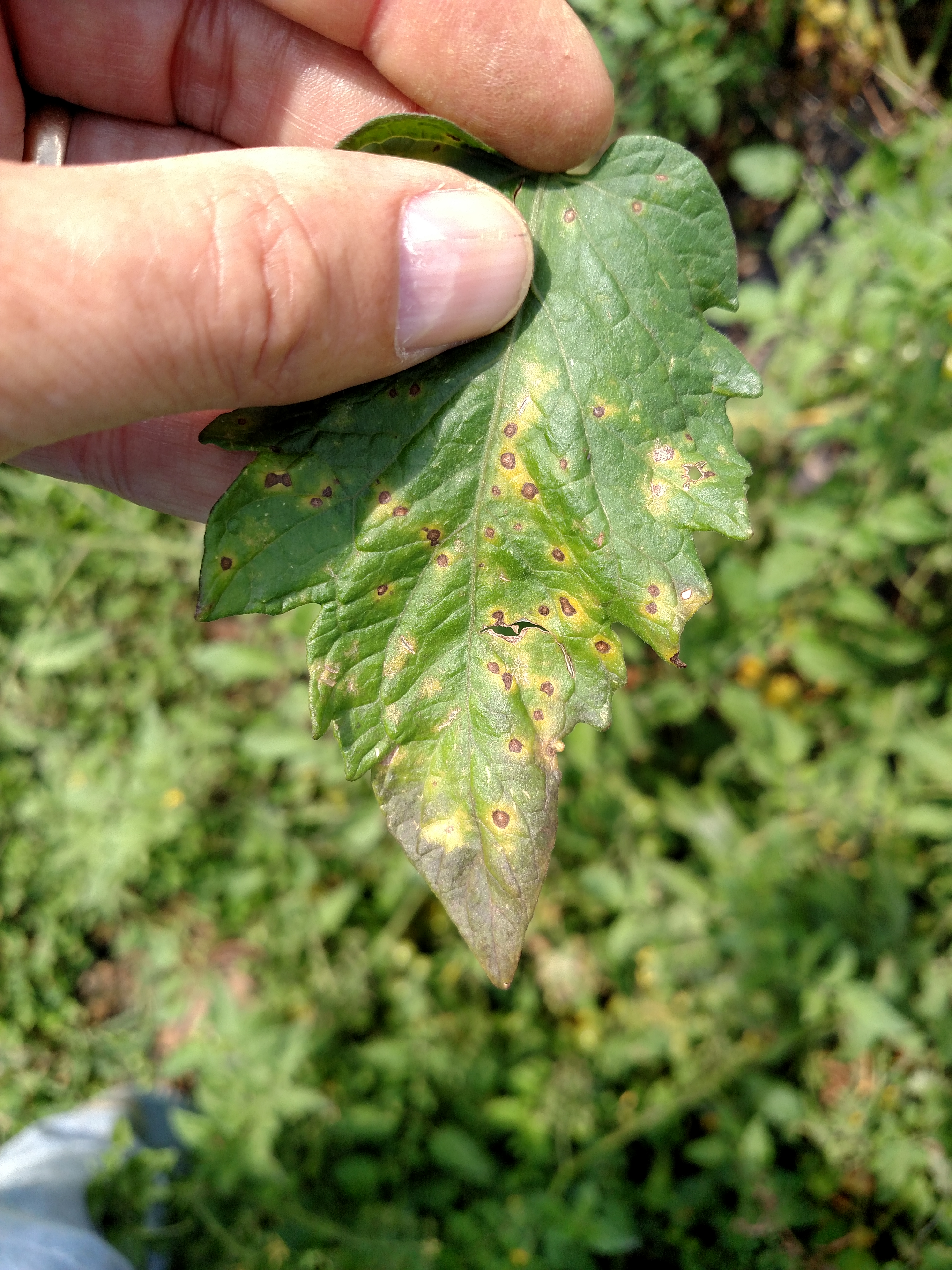 Figure 3. Septoria leaf spot of tomato. Note brown margin and gray center of lesions.
 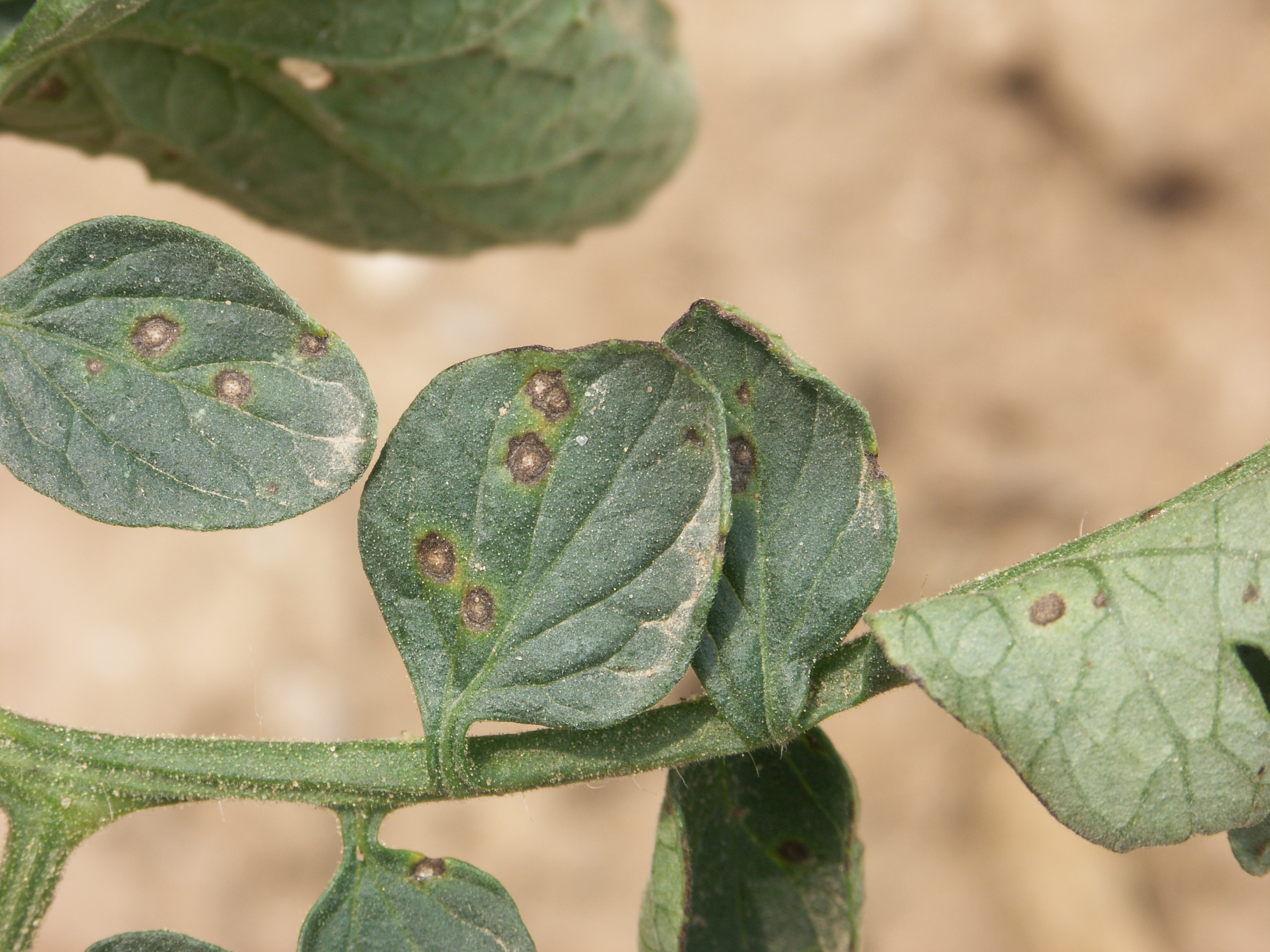 Figure 4. Septoria leaf spot of tomato. Note gray center of lesion and gray margin. Dark, fungal bodies may visible in the lesion center with 10X hand lens.
 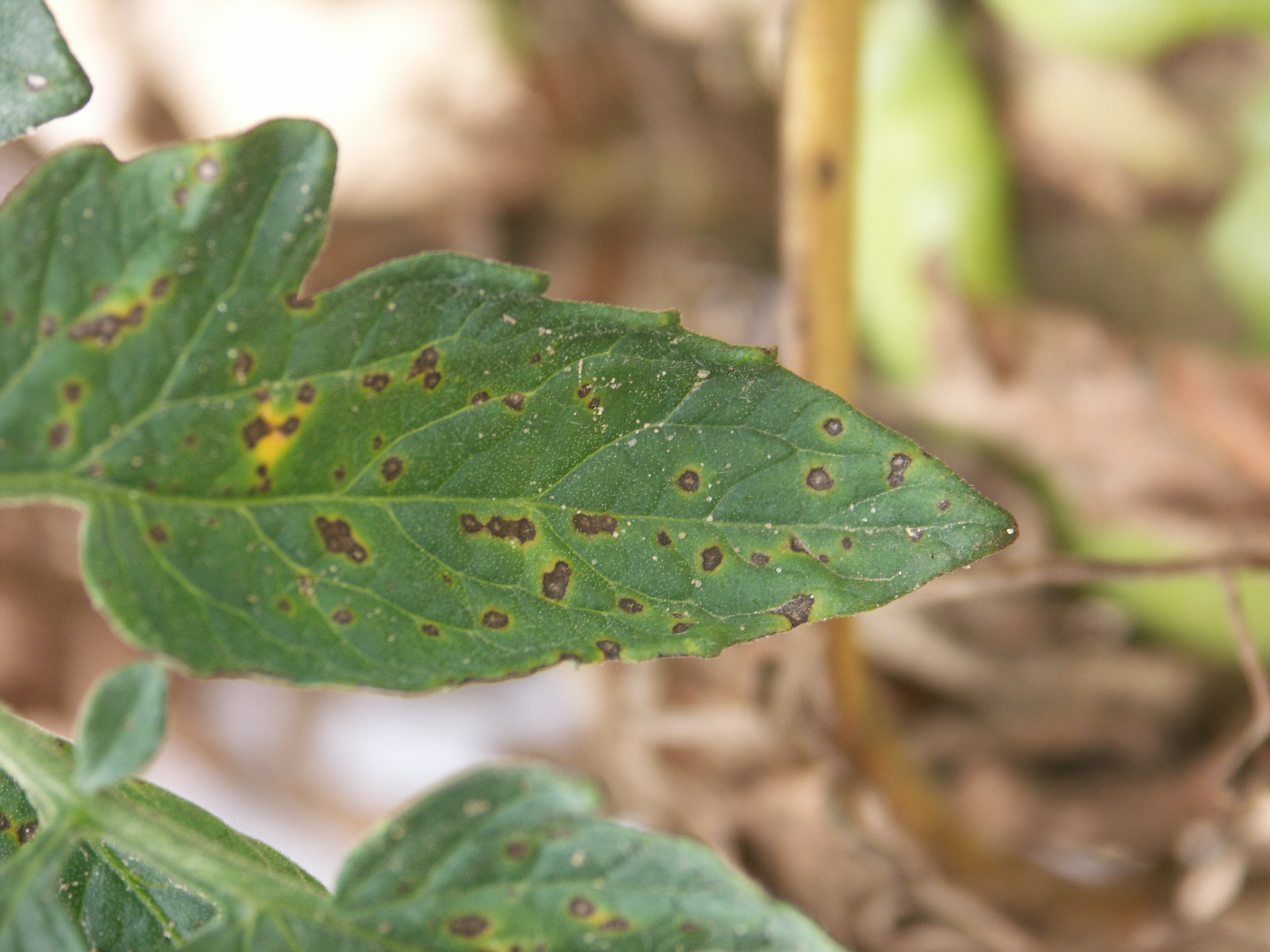 Figure 5. Septoria leaf spot of tomato.
 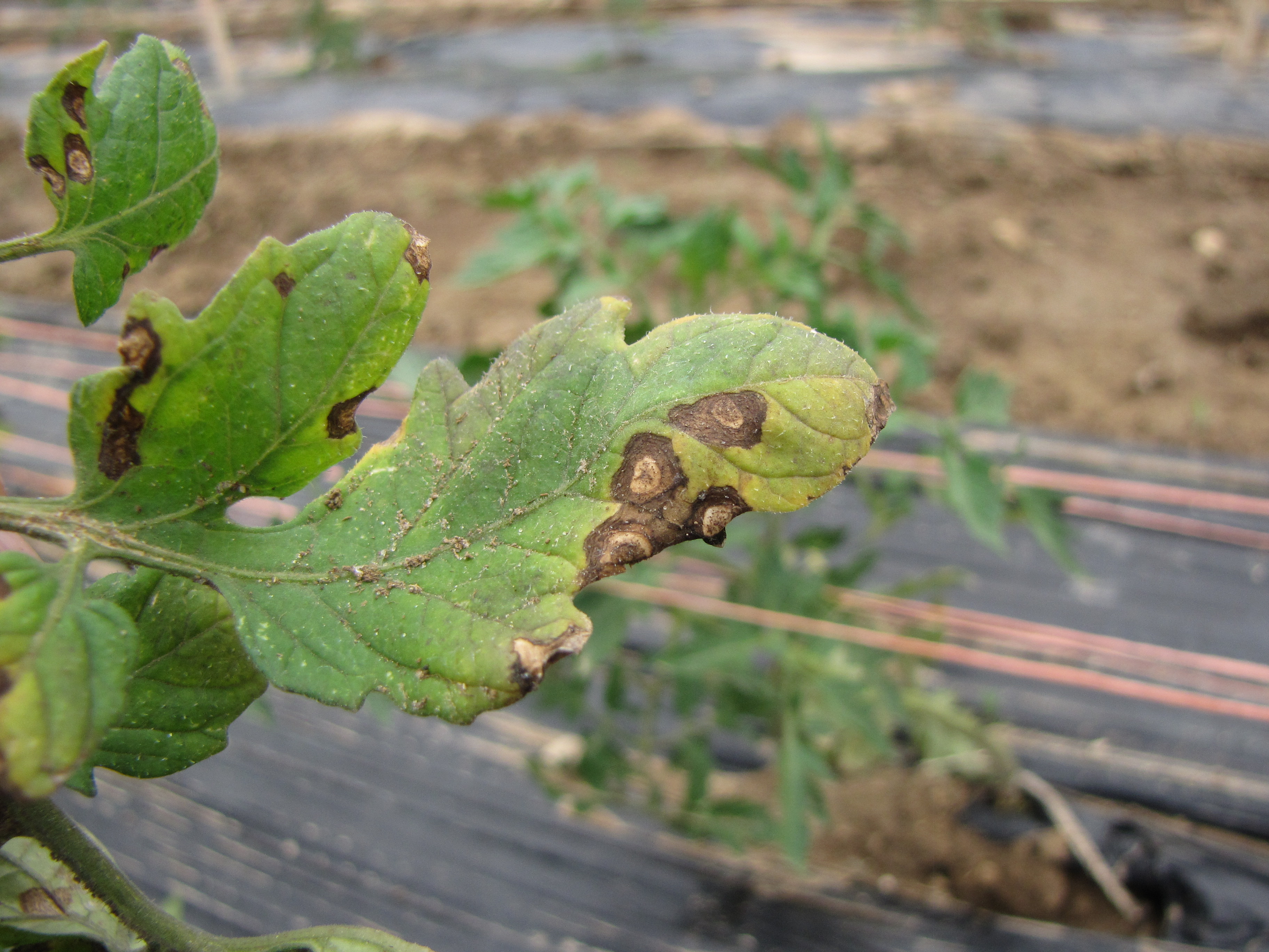 Figure 6. Lesions of Septoria leaf spot on a very susceptible variety. Look for dark fungal bodies in center of lesion.
 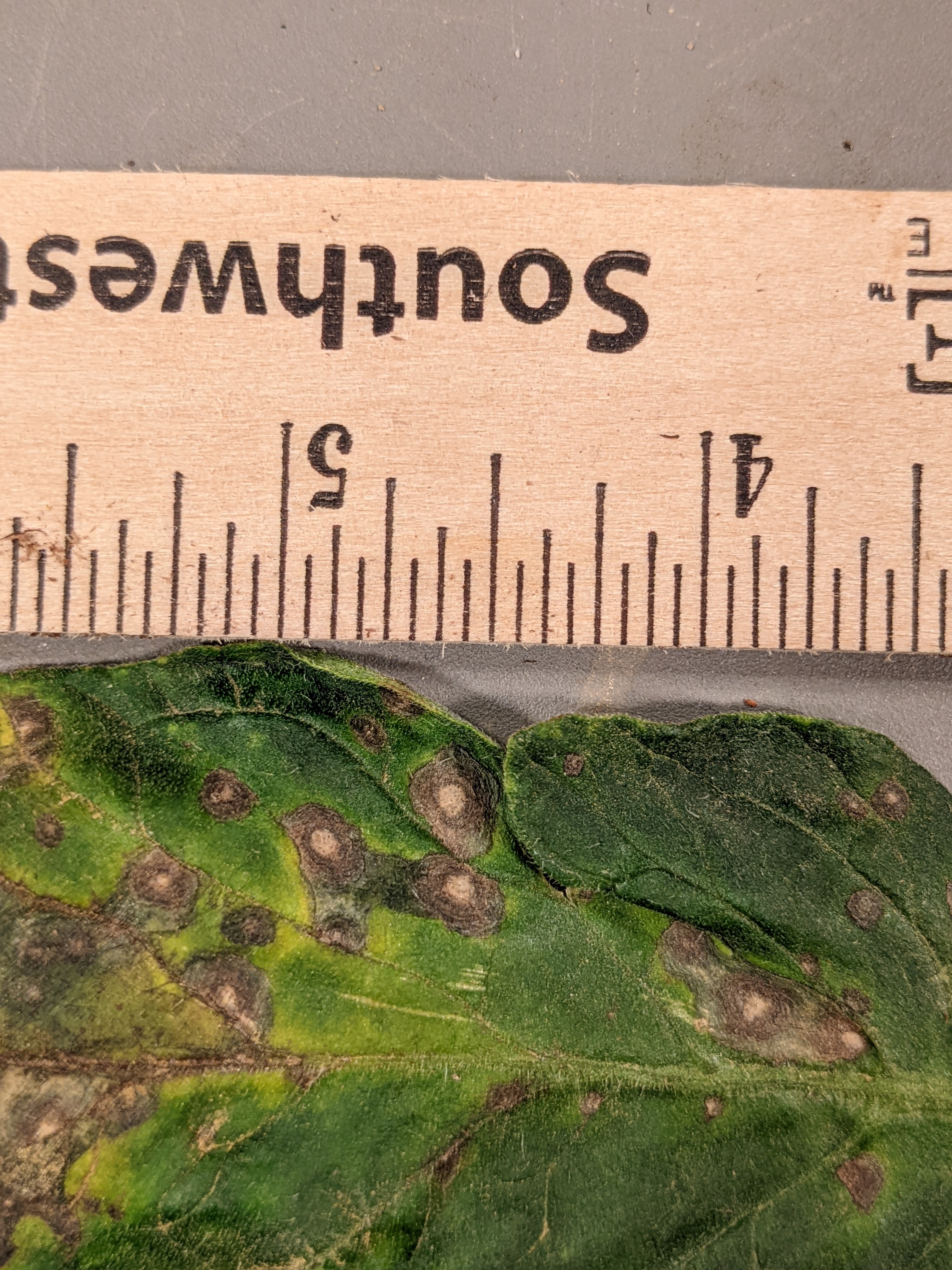 Figure 7. Septoria leaf spot of tomato.
 Southern blight
Southern blight-Often the initial symptom of this disease is the sudden wilt and decline of the plant. Closer observation will show a lesion at soil level. Lesions will have small light brown fungal structures known as sclerotia. Causal fungus will affect many other crops. Not common in Indiana.
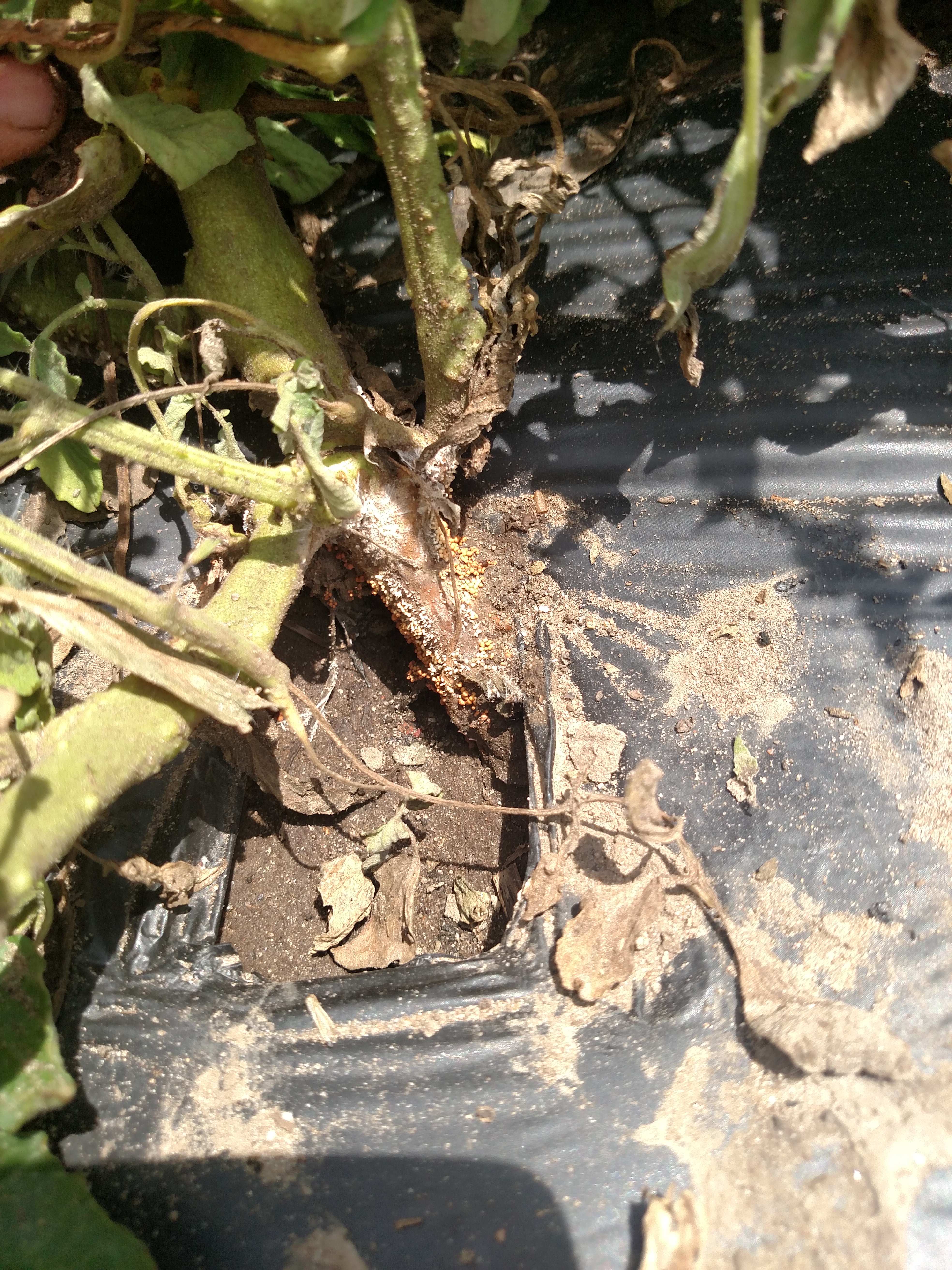 Figure 1. Southern blight can be recognized by the small sclerotia at the base of the stem. These lesions will eventually result in the wilting of the plant.
 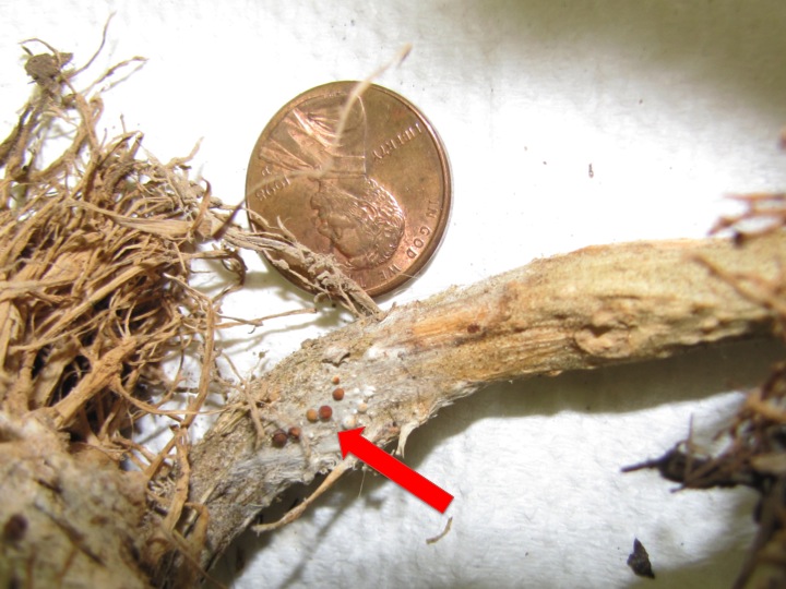 Figure 2. Close up of southern blight of tomato lesions. Sclerotia become darker as they age. Red arrow indicates sclerotia.
 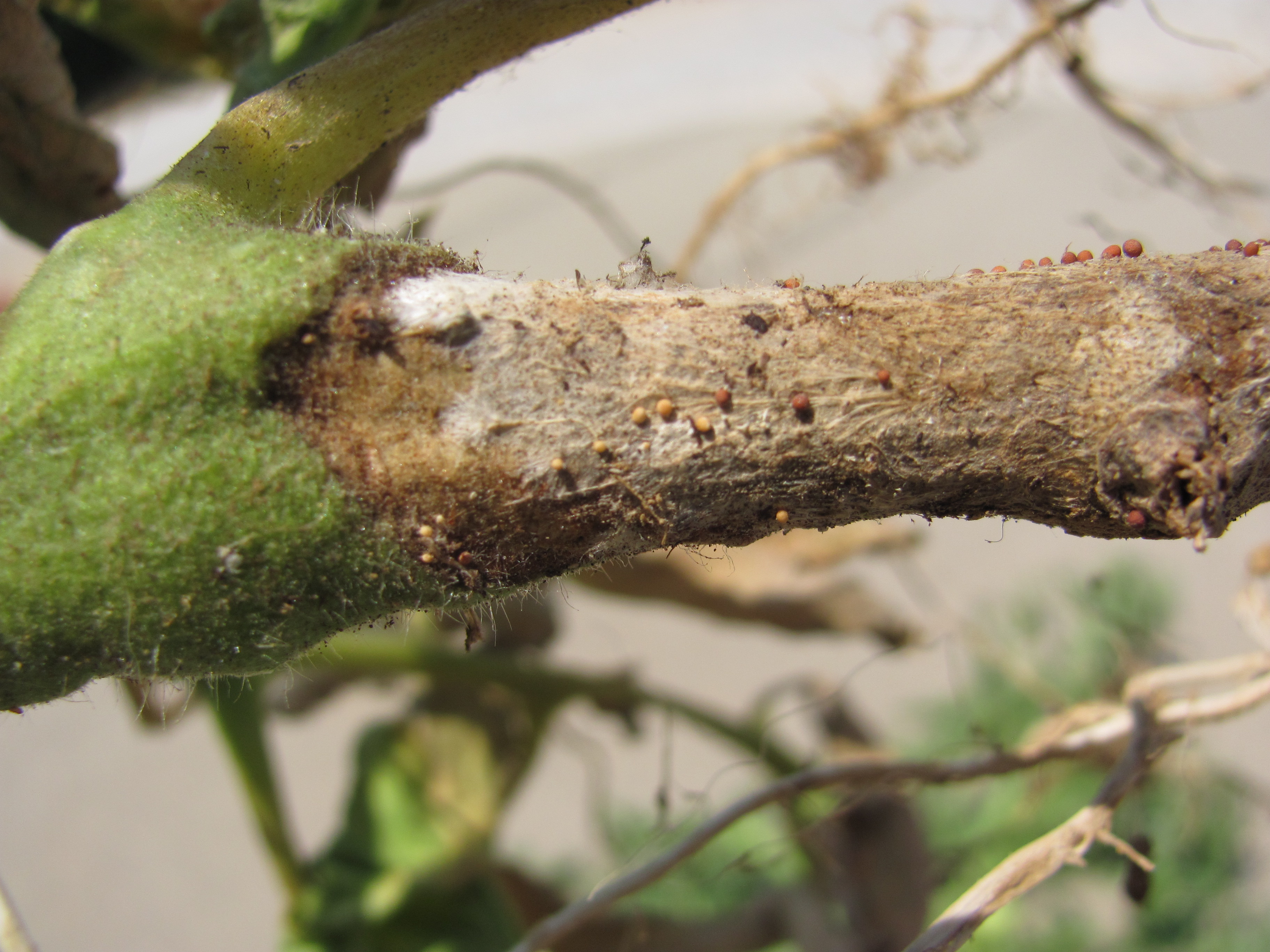 Figure 3. Southern blight of tomato.
 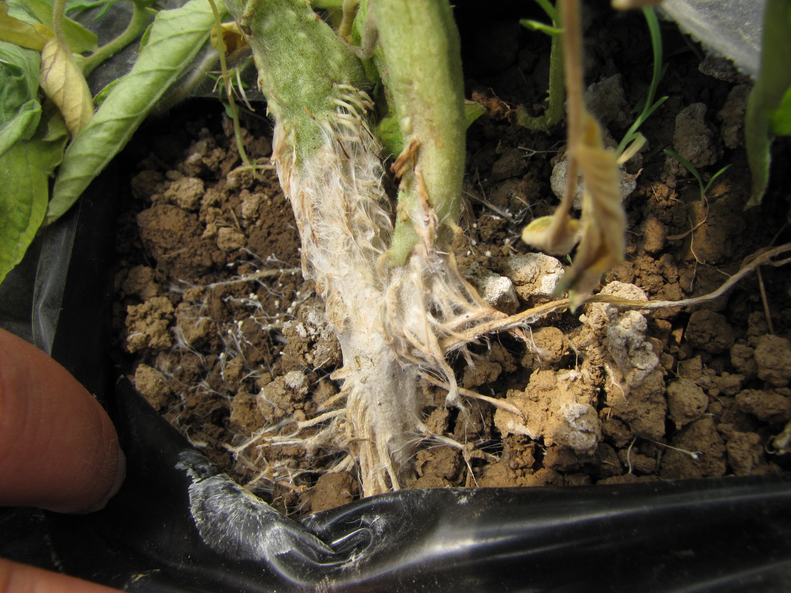 Figure 4. Base of plant with southern blight of tomato. Note sclerotia.
 Target spot
Target spot-Lesions often have a ring structure. Not common in Indiana. Casual fungus may affect other crops as well.
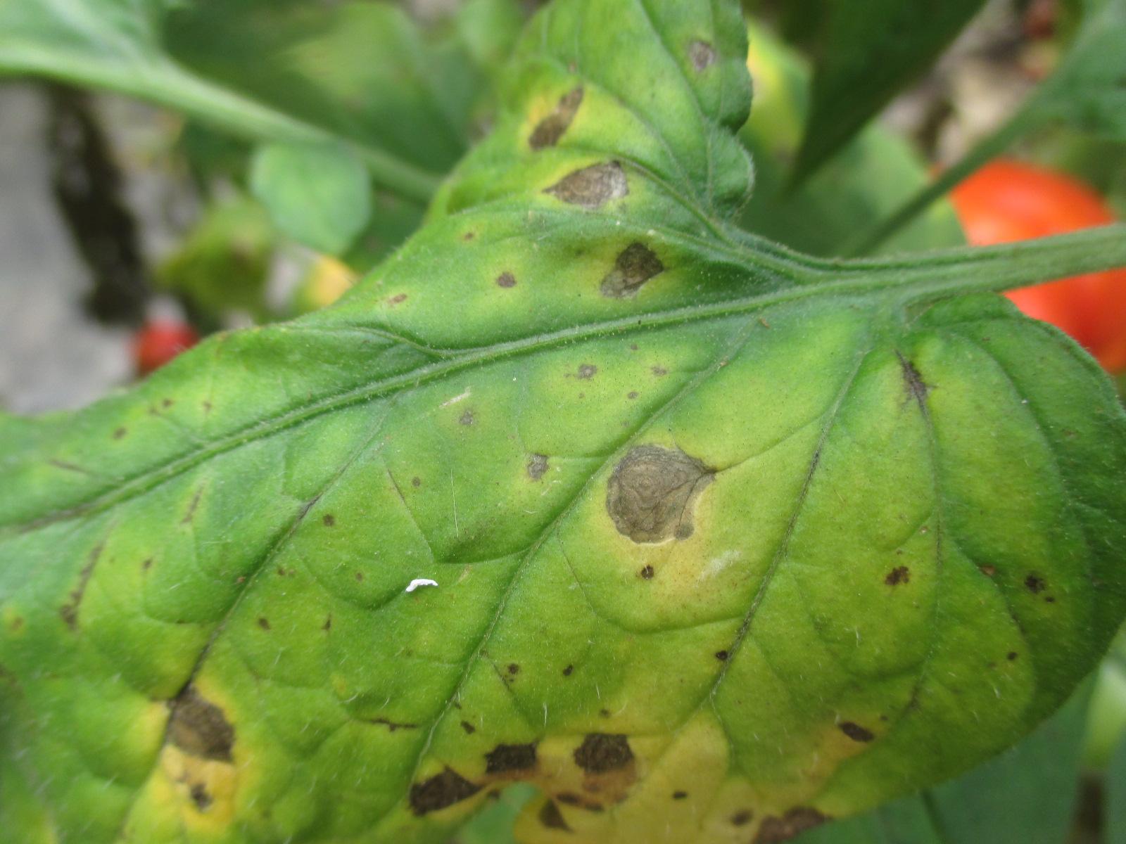 Figure 1. Target spot of tomato (Photo by Wenjing Guan).
 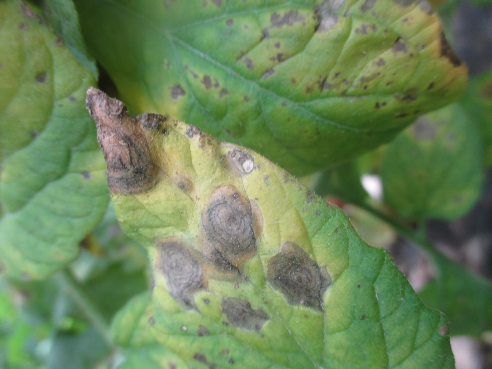 Figure 2. Target spot of tomato.
 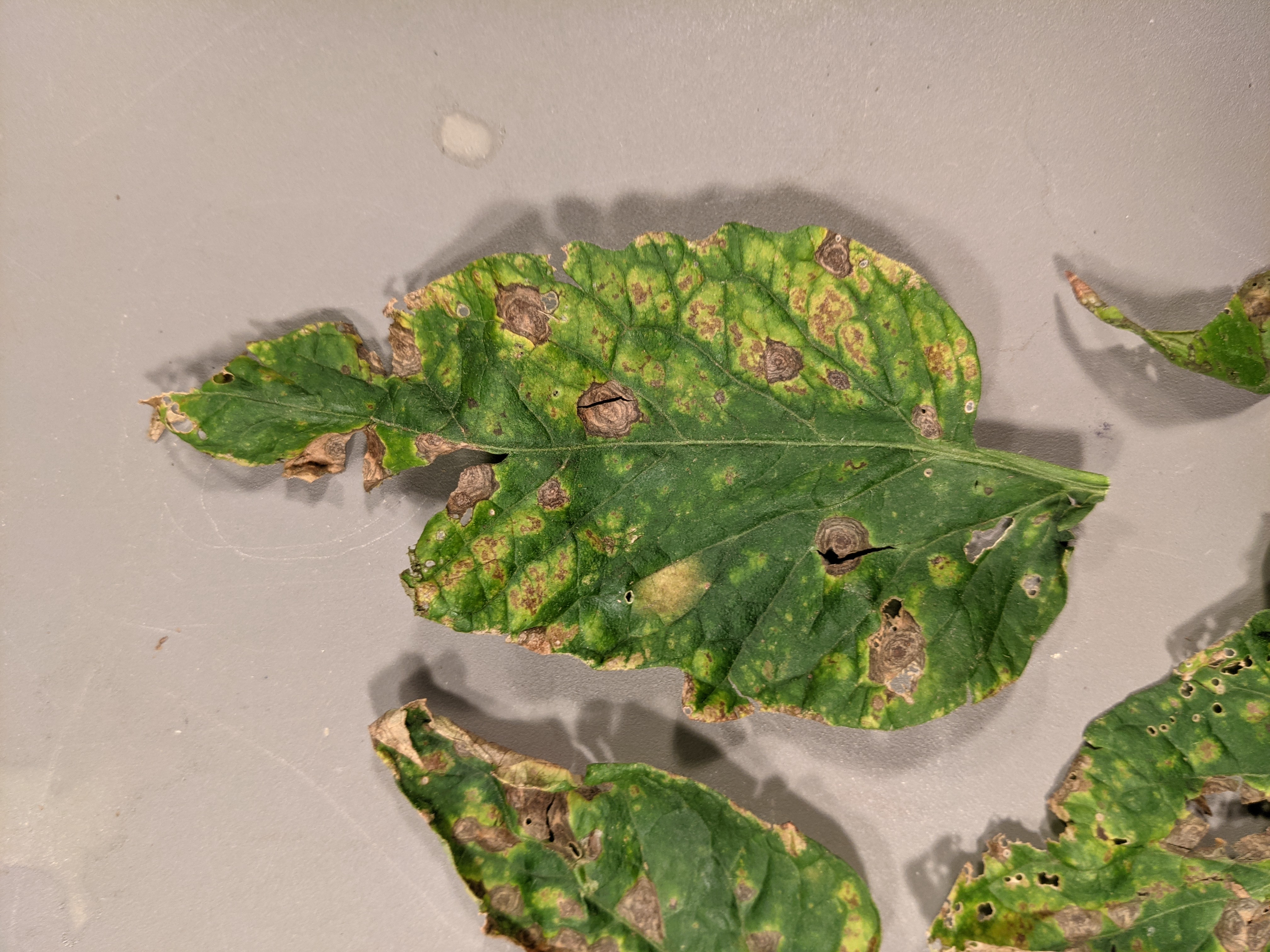 Figure 3. Target spot of tomato.
 Tomato spotted wilt virus
Tomato spotted wilt virus-may be associated with diverse symptoms. Symptoms may include stunting, chlorosis, round lesions, and discolored fruit. Since the disease is transmitted by thrips, observations of these insects or feeding damage may help with diagnosis. Often the result of tomato production alongside flower plugs in a greenhouse or high tunnel.
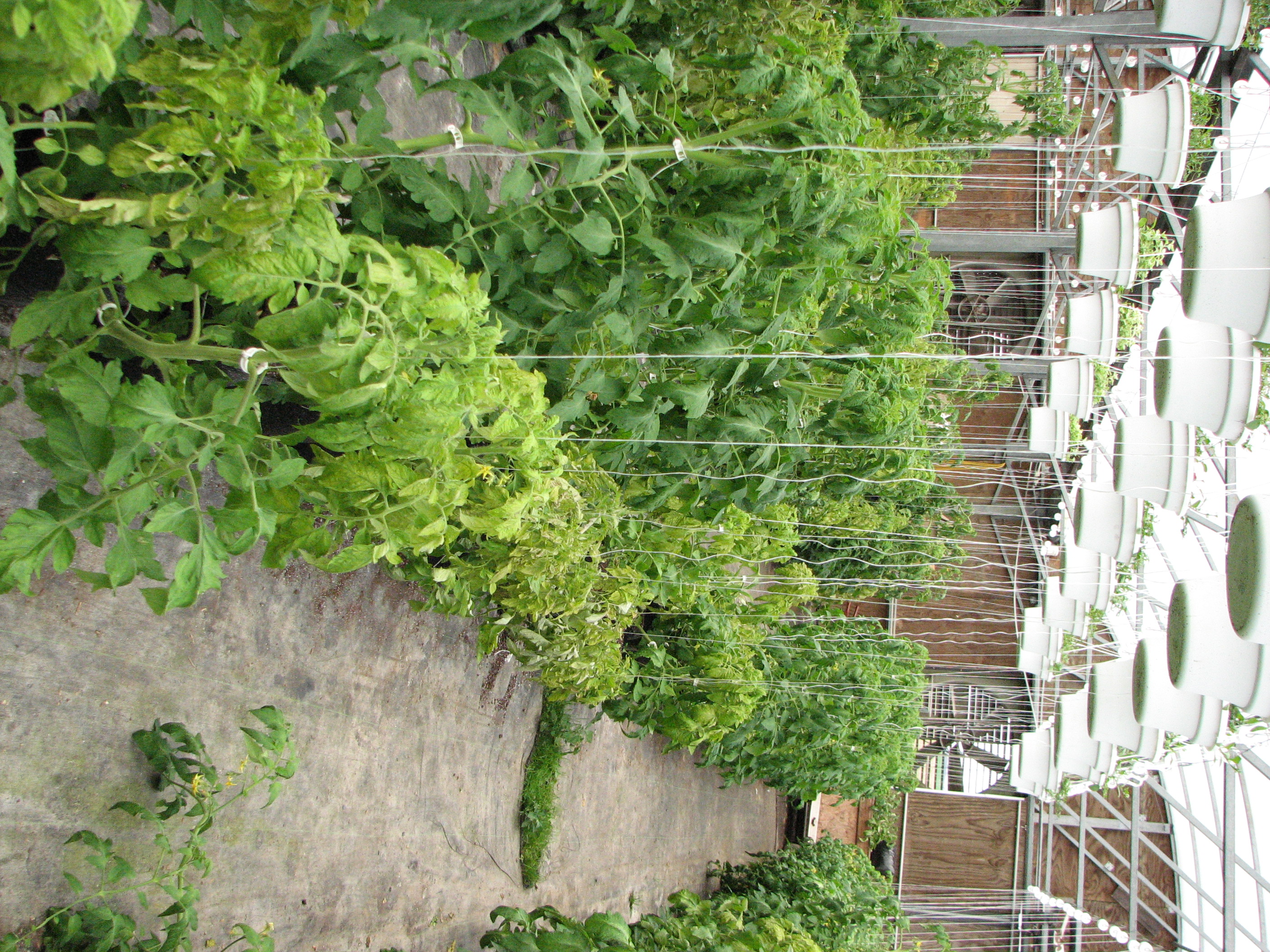 Figure 1. Symptoms of tomato spotted wilt virus include stunting such can be seen in the tomatoes on the right. Note the baskets of hanging flowers in the greenhouse.
 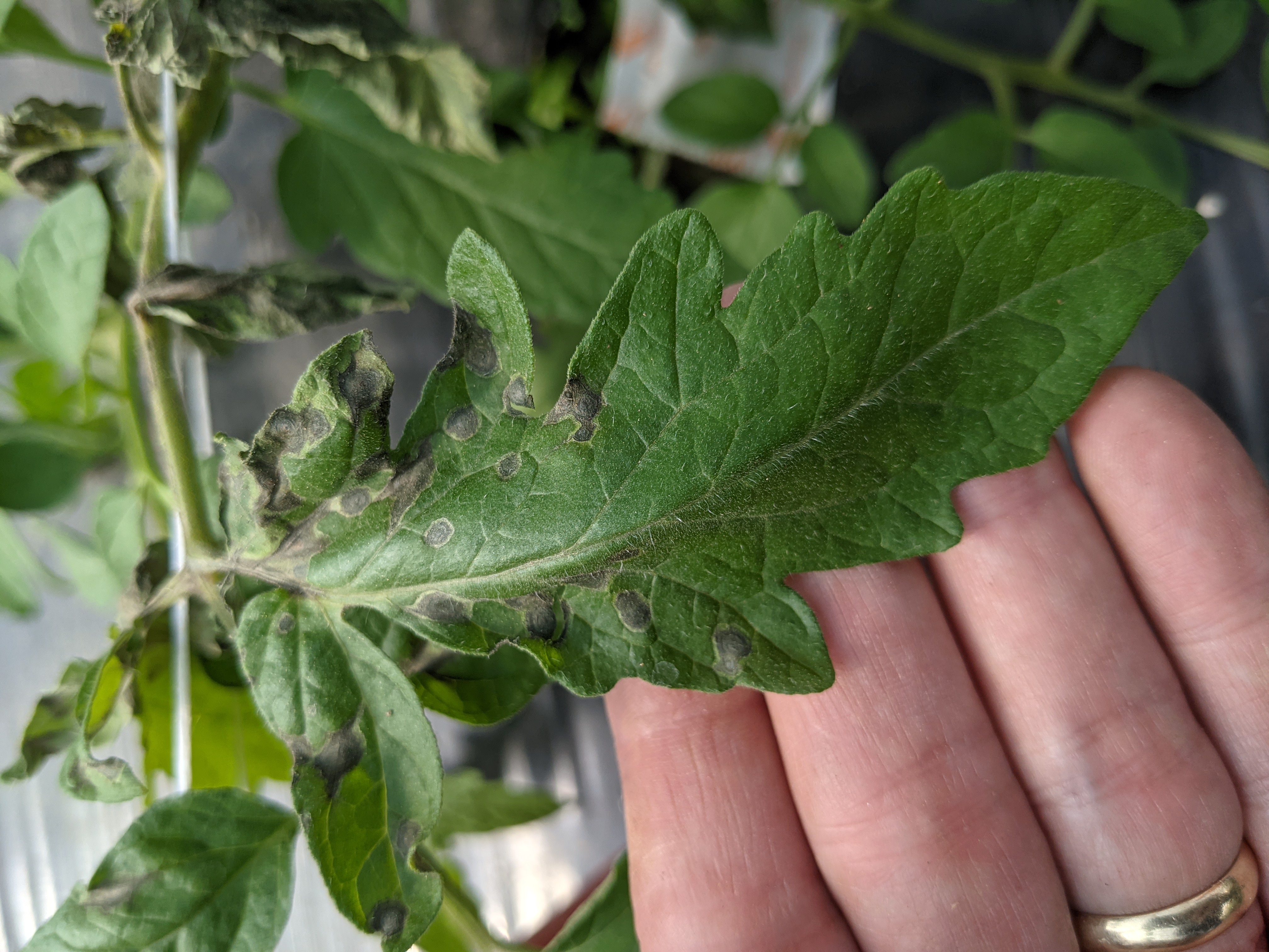 Figure 2. Tomato spotted wilt virus includes a necrotic ring spot lesion.
 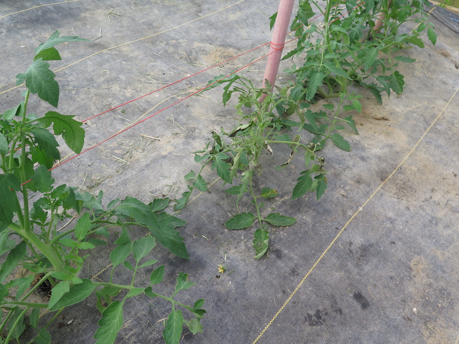 Figure 3. Tomato in center of photo is wilting due to tomato spotted wilt virus.
 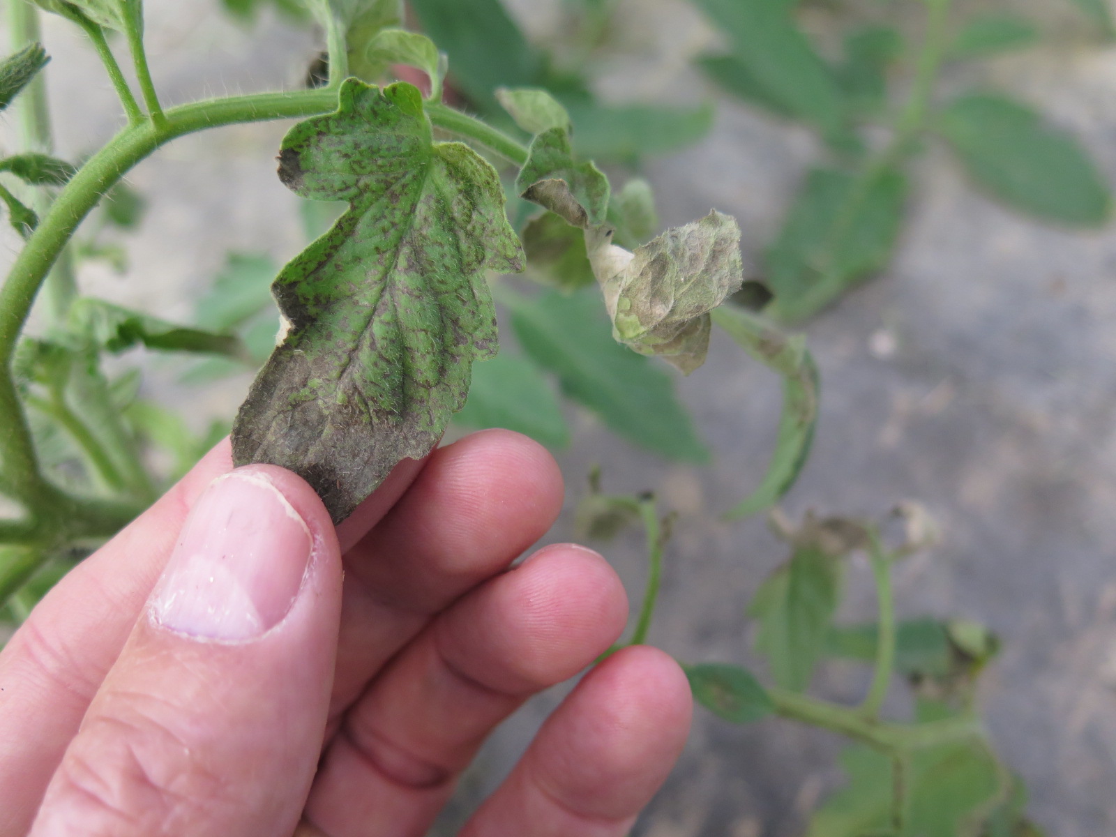 Figure 4. Tomato spotted wilt virus on tomato.
 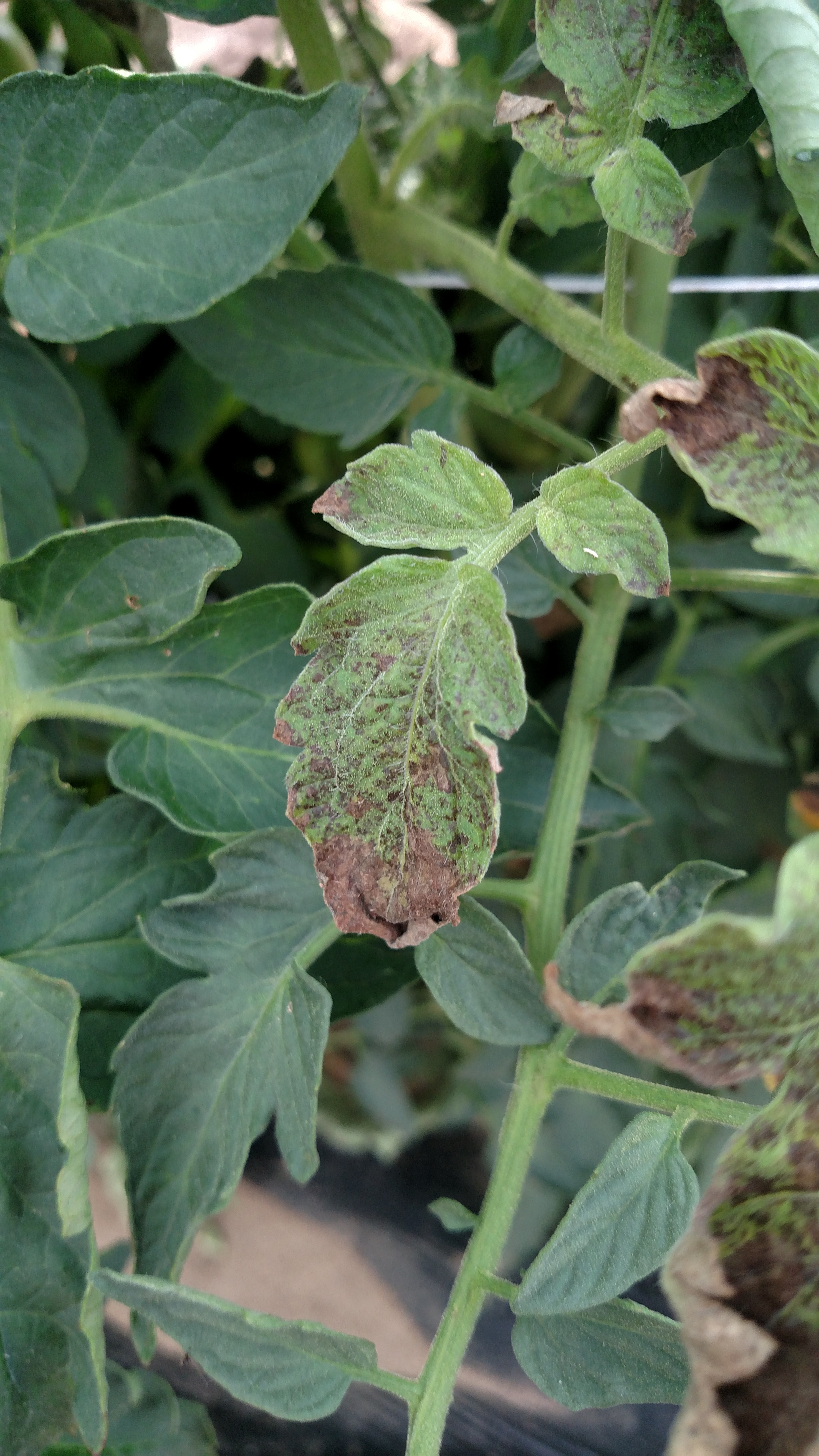 Figure 5. Tomato spotted wilt virus on tomato.
 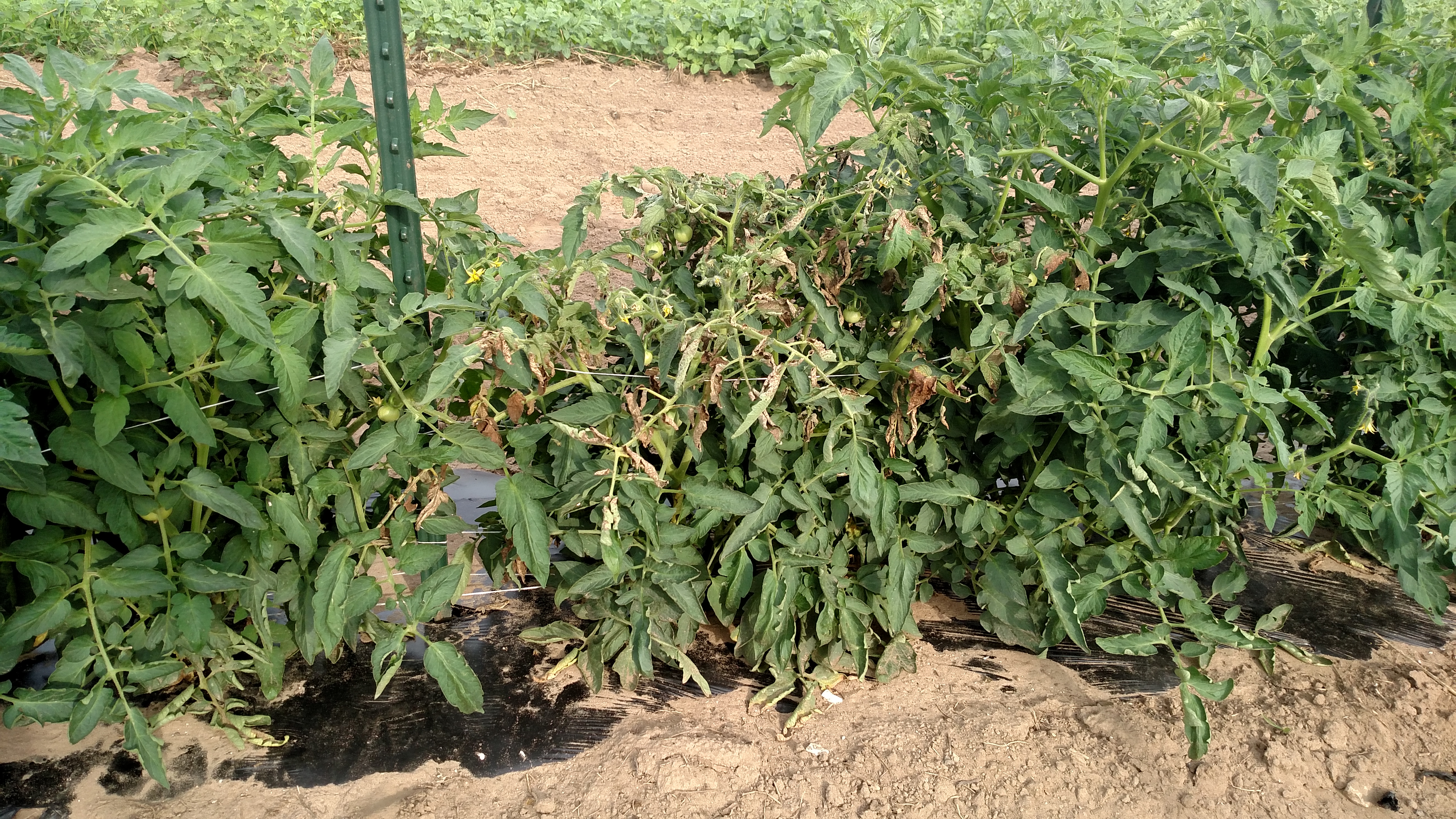 Figure 6. Center plant is stunted and wilted due to tomato spotted wilt virus.
 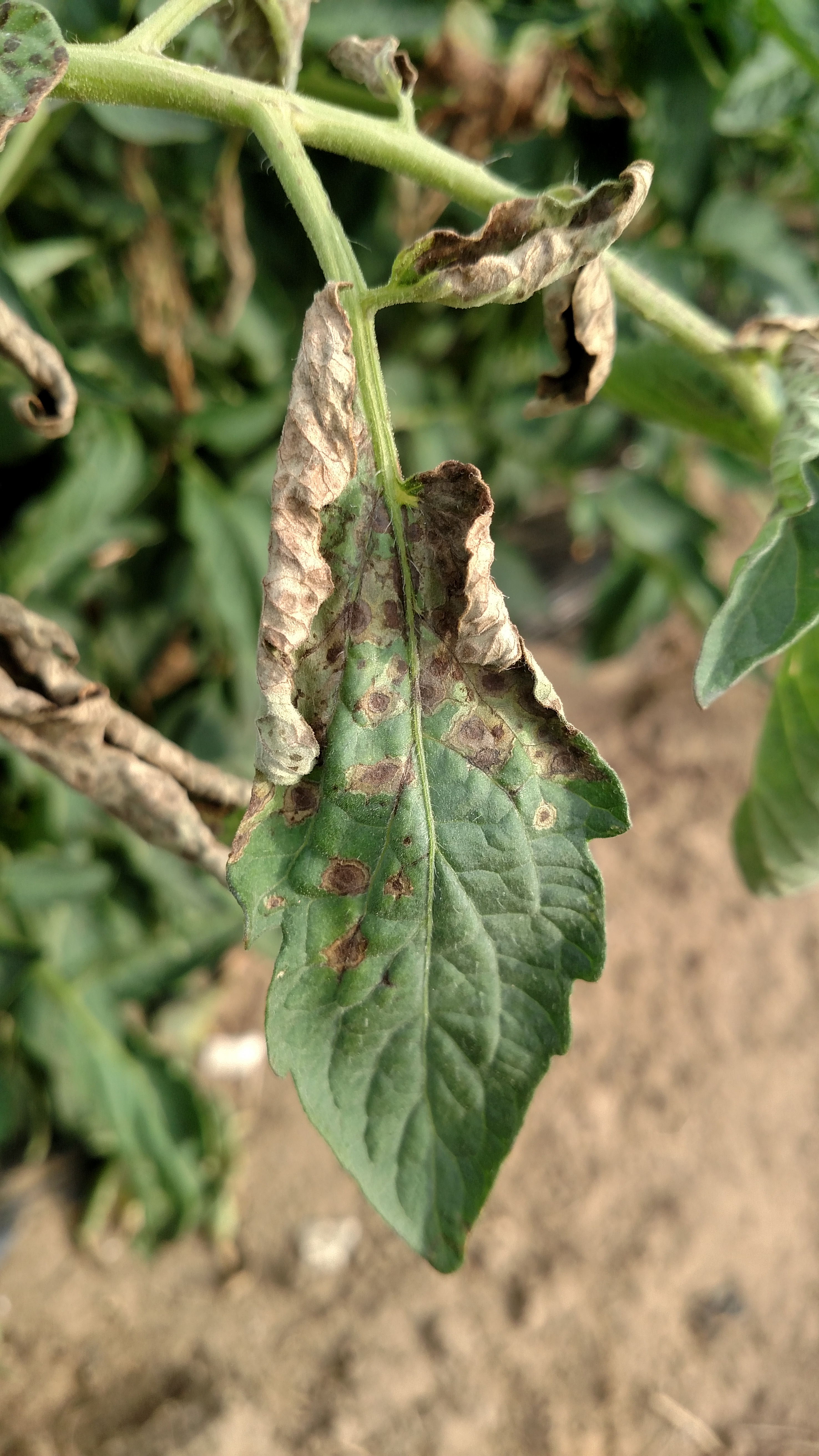 Figure 7. Tomato spotted wilt virus on tomato.
 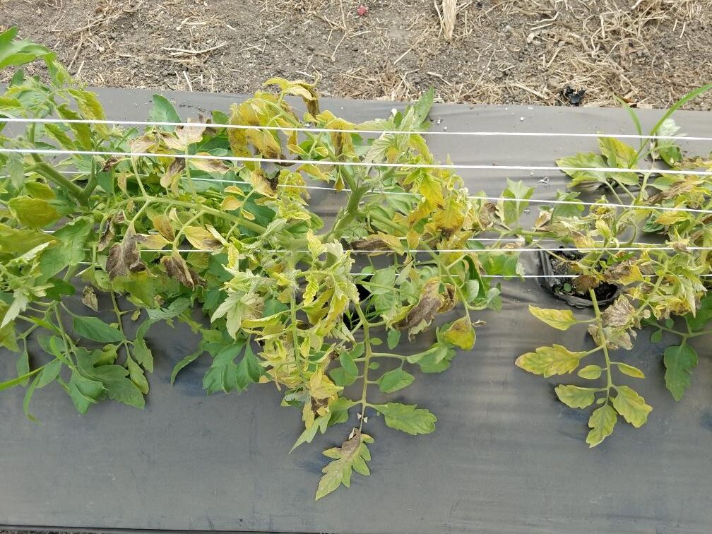 Figure 8. Chlorosis due to tomato spotted wilt virus.
 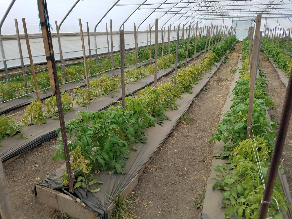 Figure 9. Wide spread symptoms of tomato spotted wilt symptoms in greenhouse.
 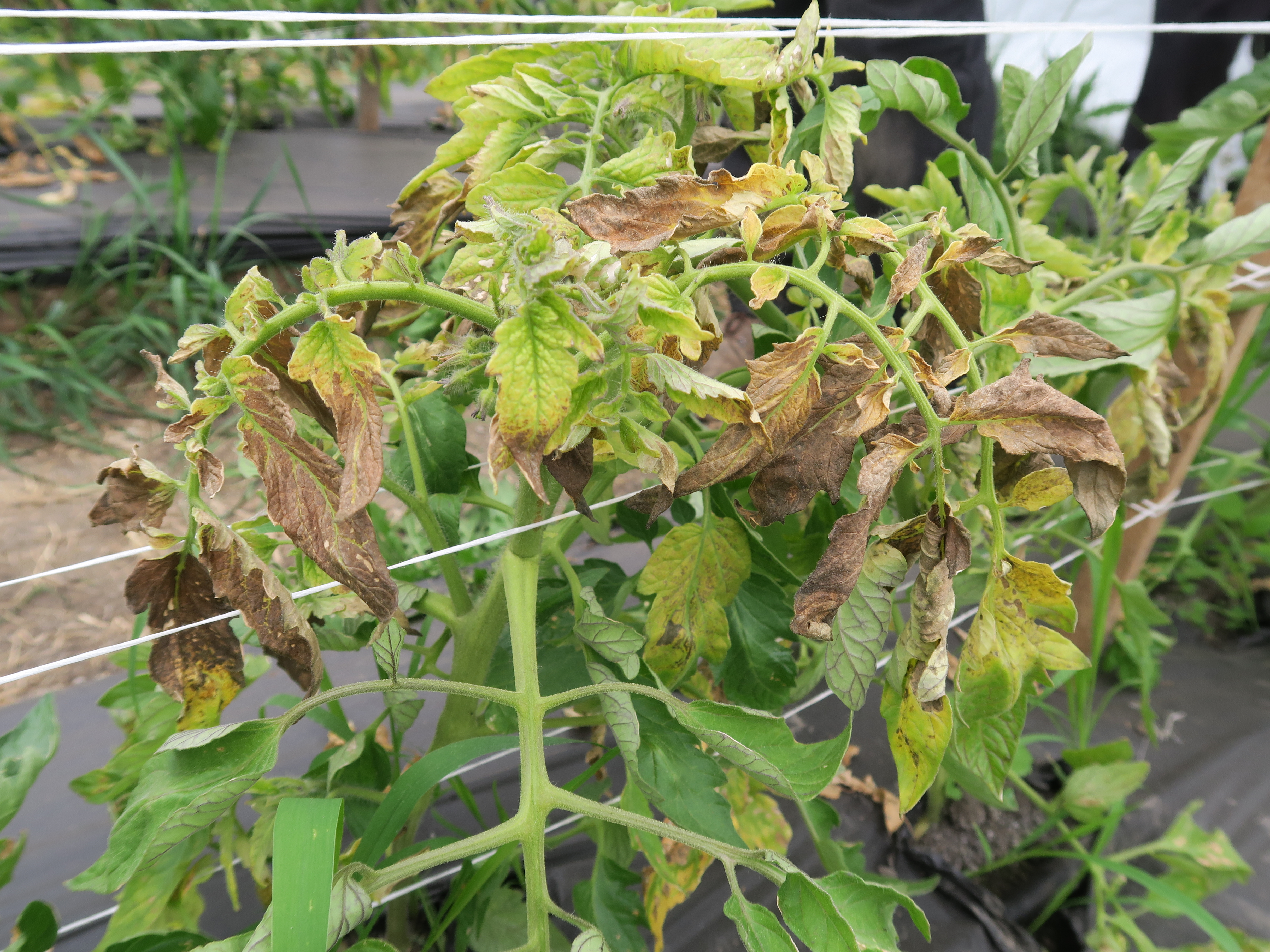 Figure 10. Tomato spotted wilt virus on tomato.
 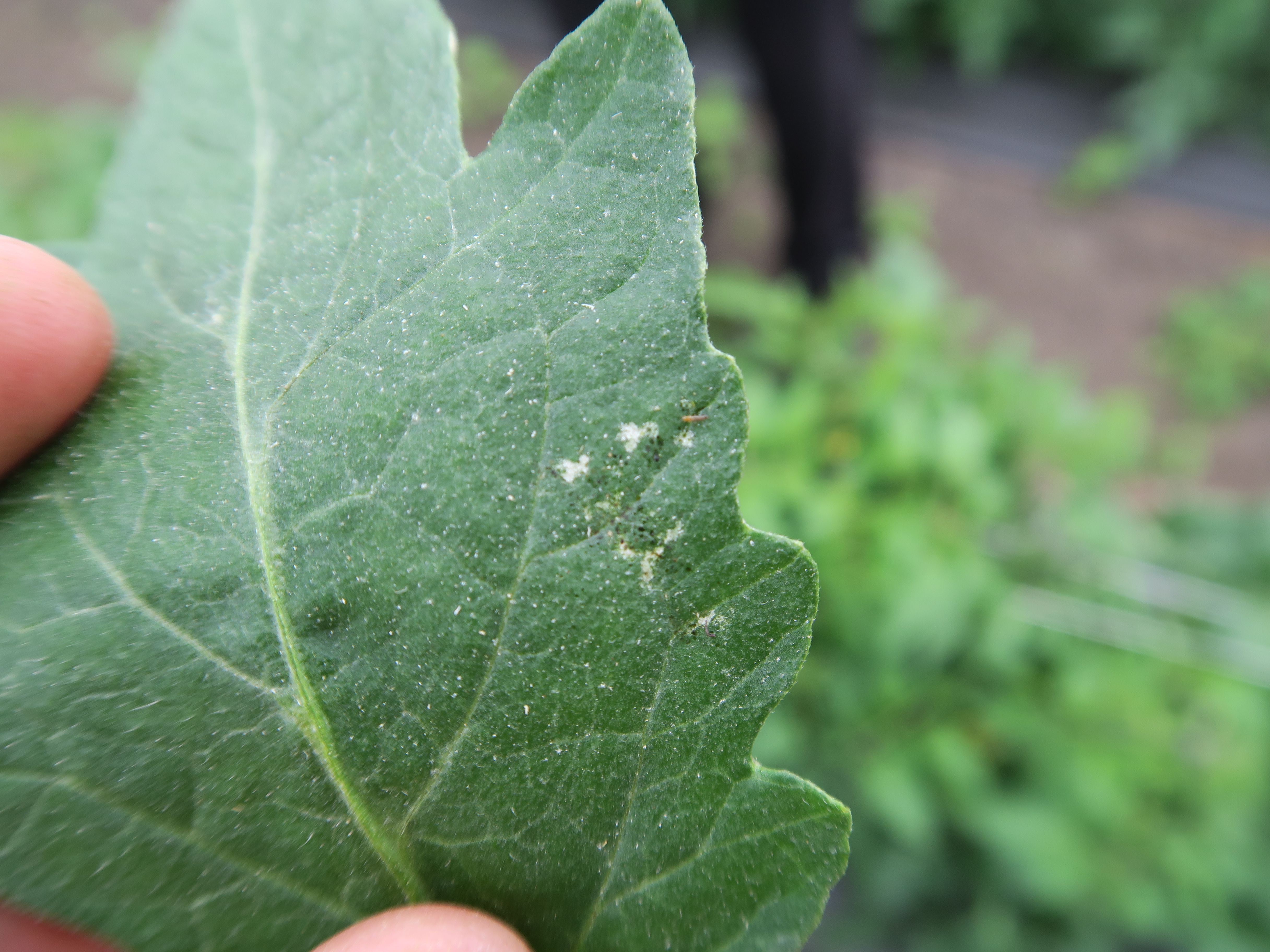 Figure 11. Thrips feeding on tomato leaf.
 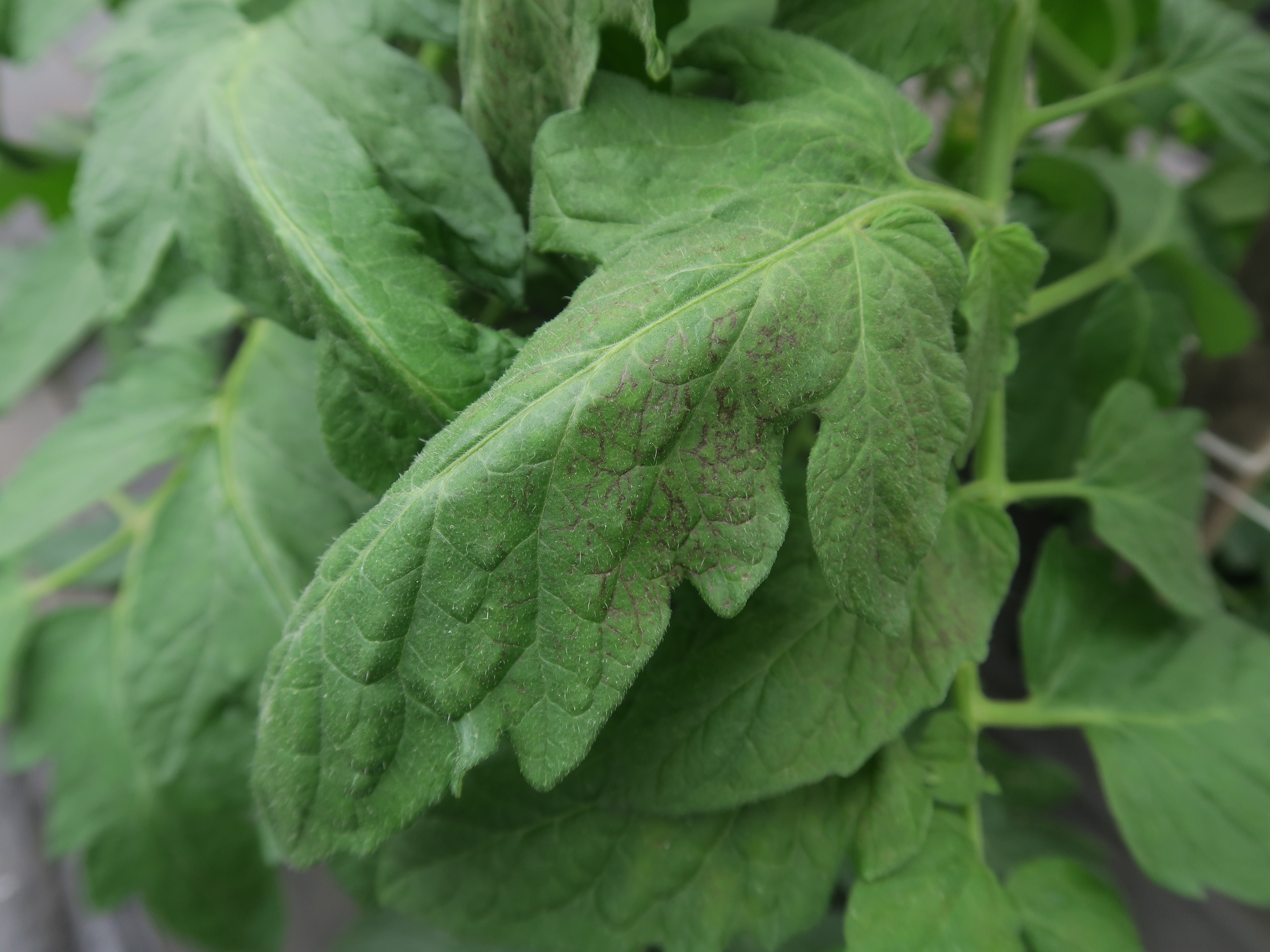 Figure 12. Necrosis due to tomato spotted wilt virus on tomato leaf.
 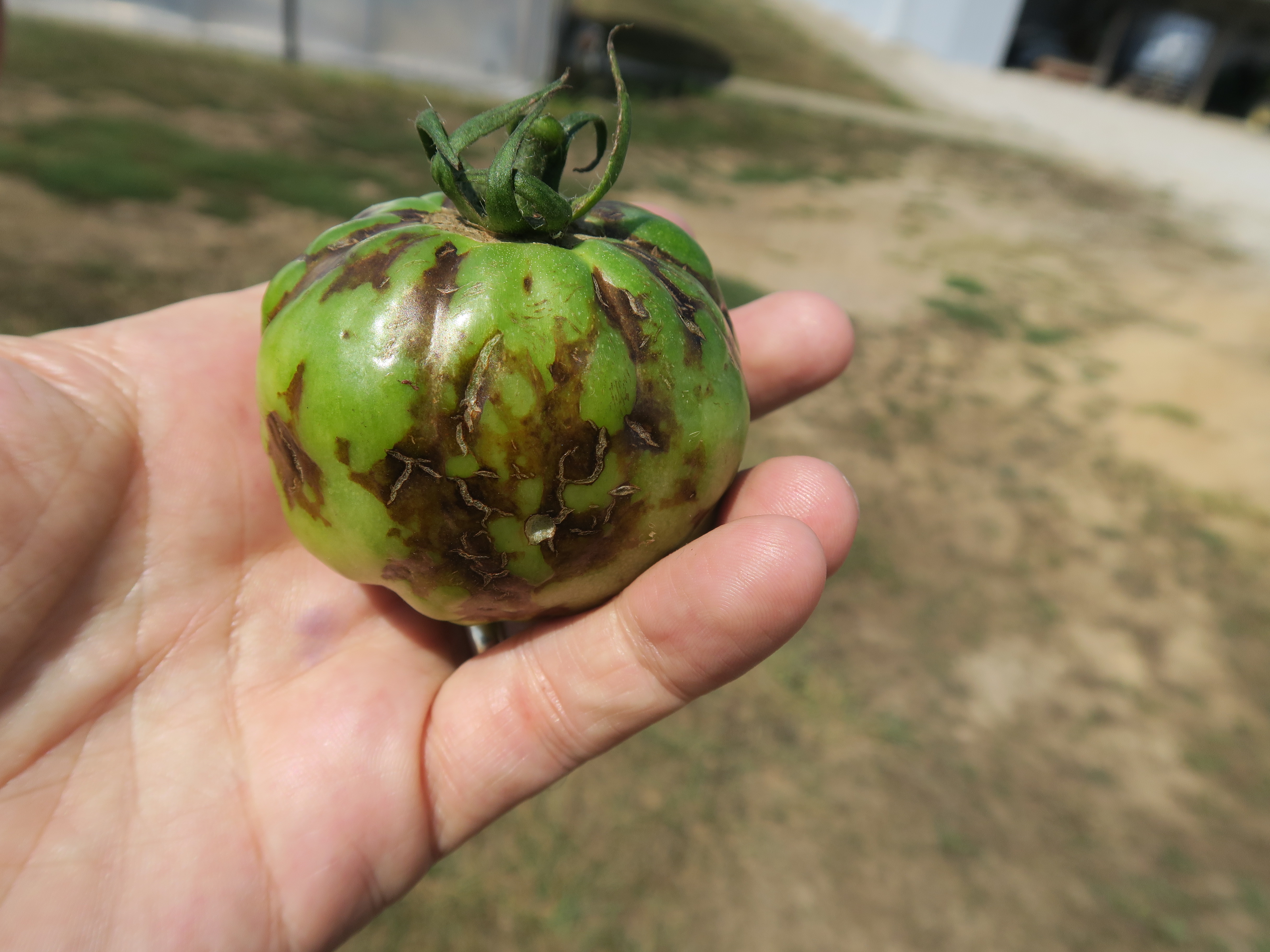 Figure 13. Symptoms of tomato spotted wilt virus on tomato fruit.
 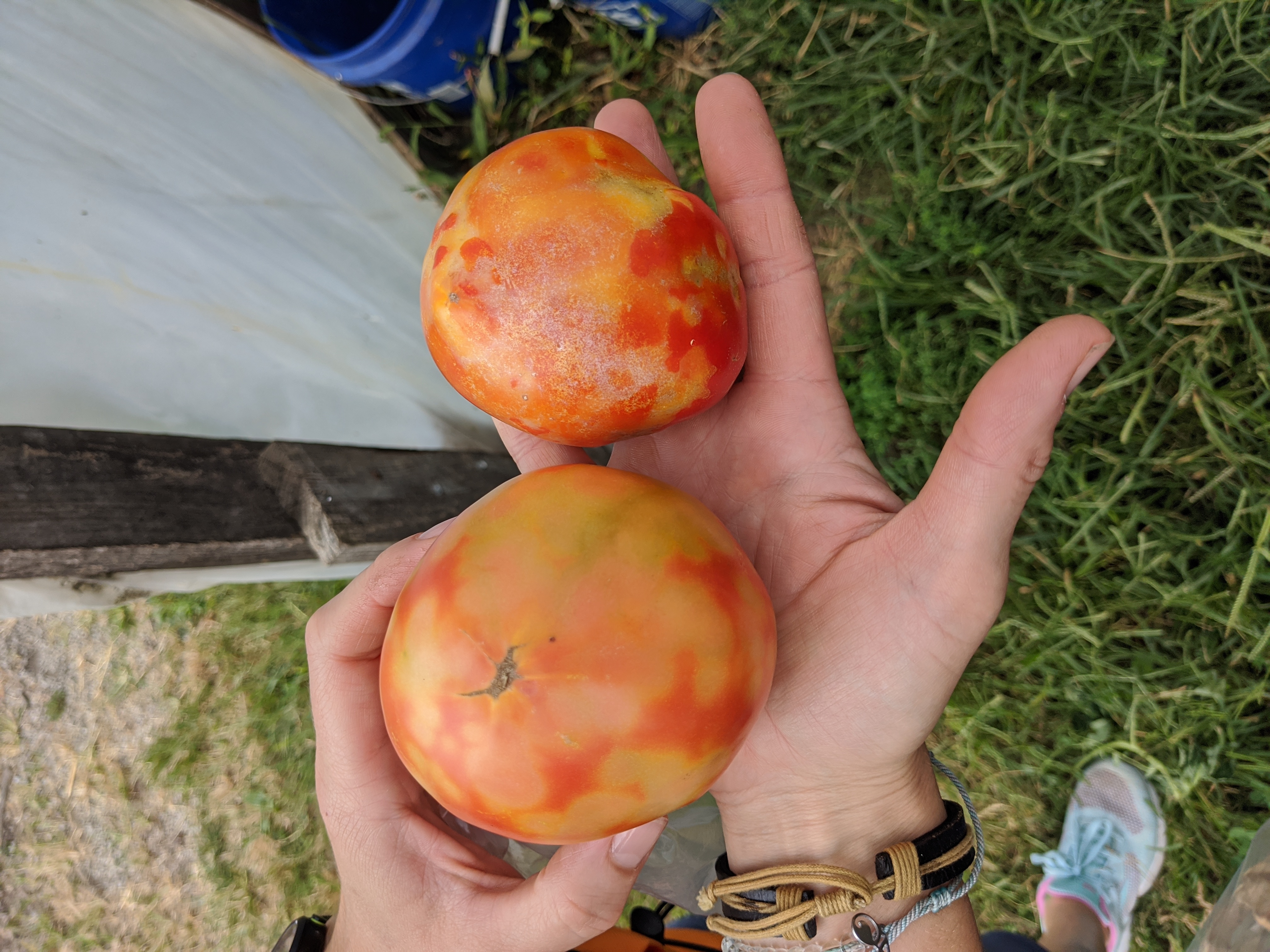 Figure 14. Symptoms of tomato spotted wilt virus on tomato fruit.
 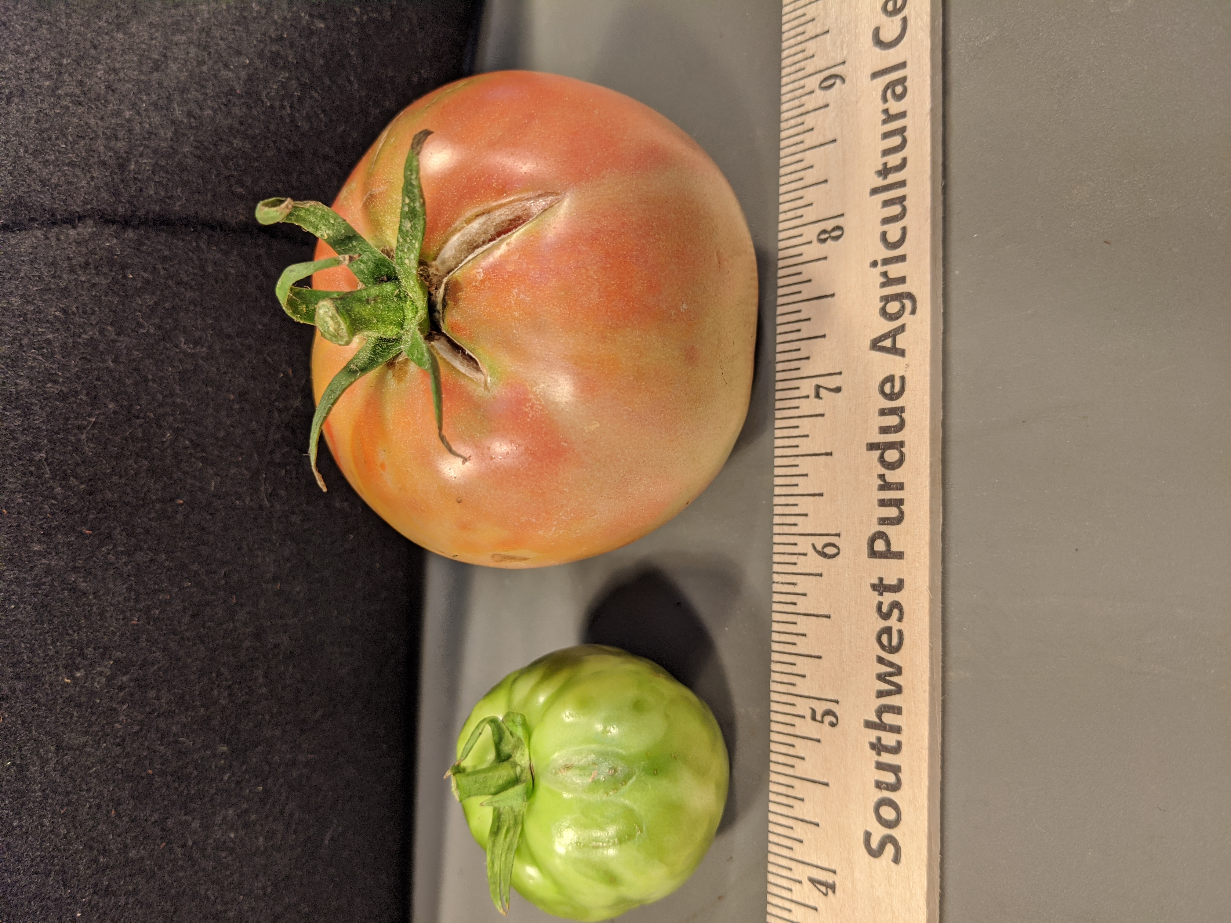 Figure 15. Tomato spotted wilt virus on tomato fruit.
 Walnut allelopathy
Walnut allelopathy-Roots of black walnut trees are associated with a chemical that can interfere with the growth of many plants including tomatoes. Not an infectious disease.
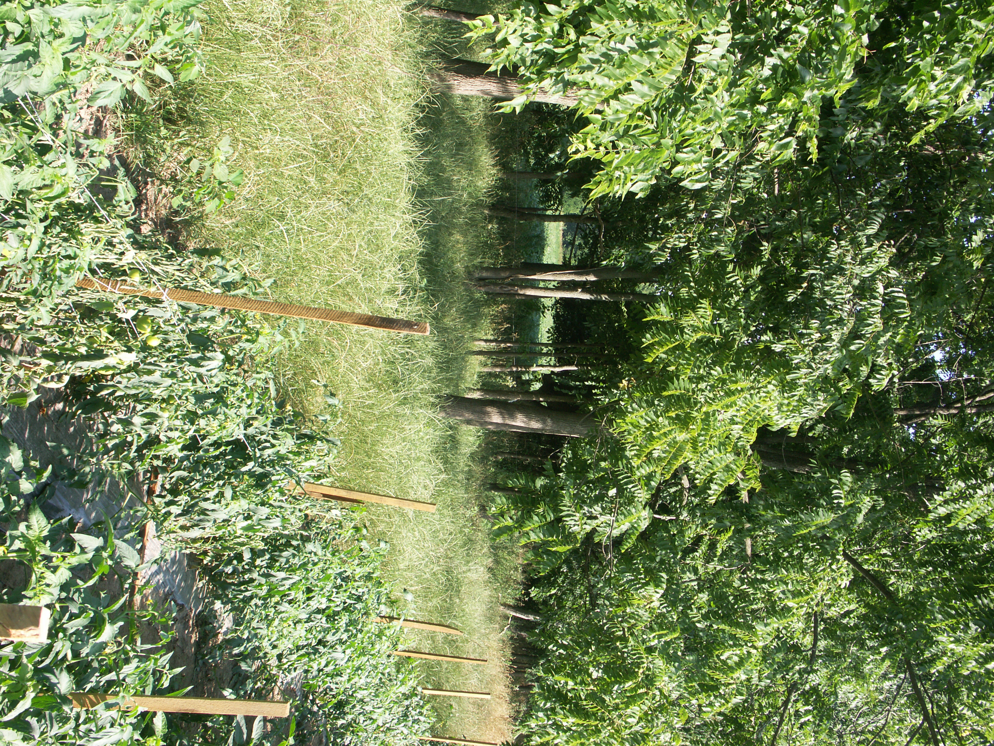 Figure 1. Walnut allelopathy. Note wilting tomato plants in the foreground adjacent to walnut trees
 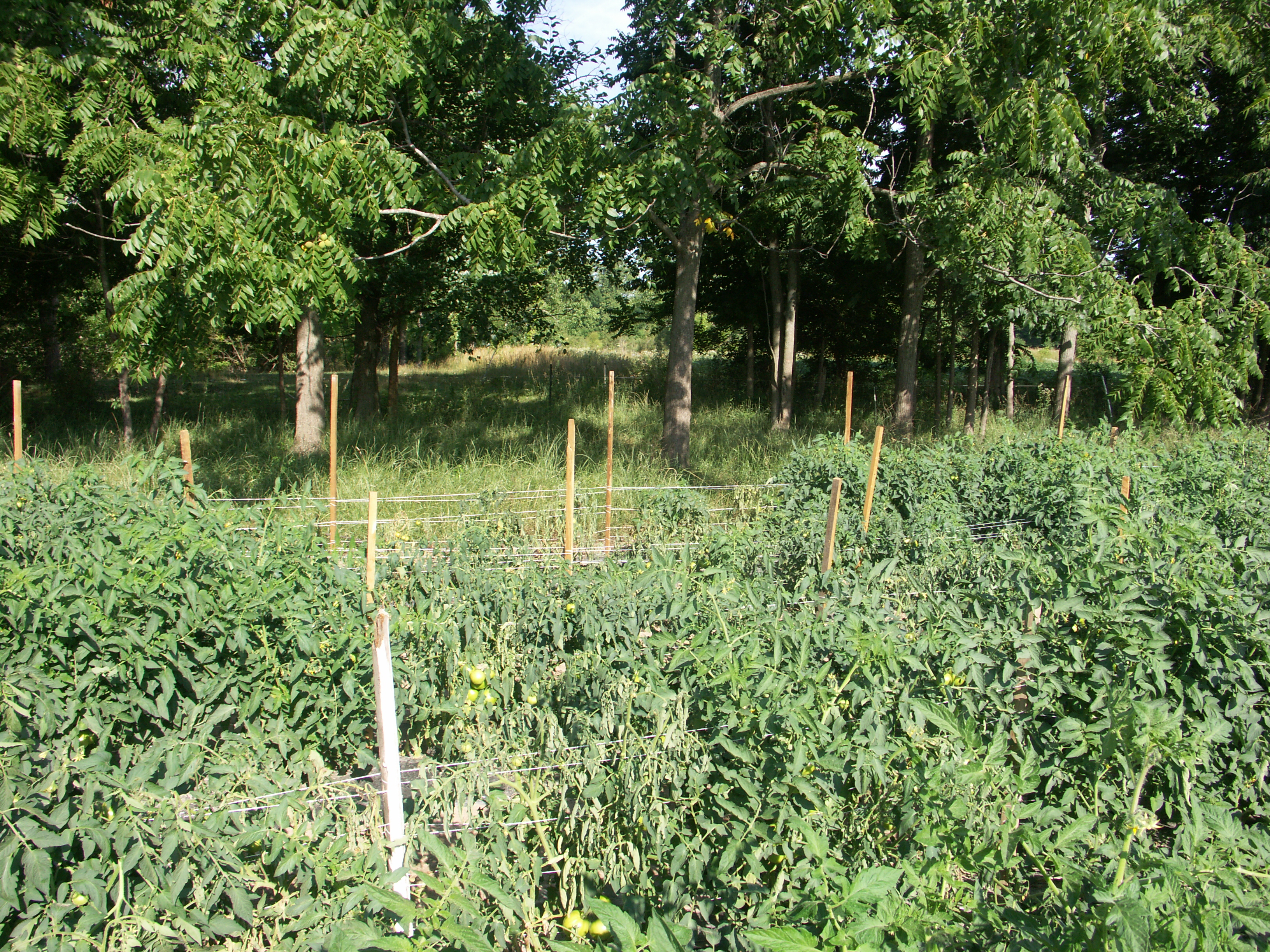 Figure 2. Walnut allelopathy affecting tomato plants.
 White mold
White mold-Also known as timber rot when affecting tomato. Portions of the plant may begin to wilt due to lesions on stems. Dark, irregularly shaped sclerotia may be found inside or outside stems and are often associated with a white mold. Fruit may also be affected. Huge host range. More common in greenhouses than in fields.
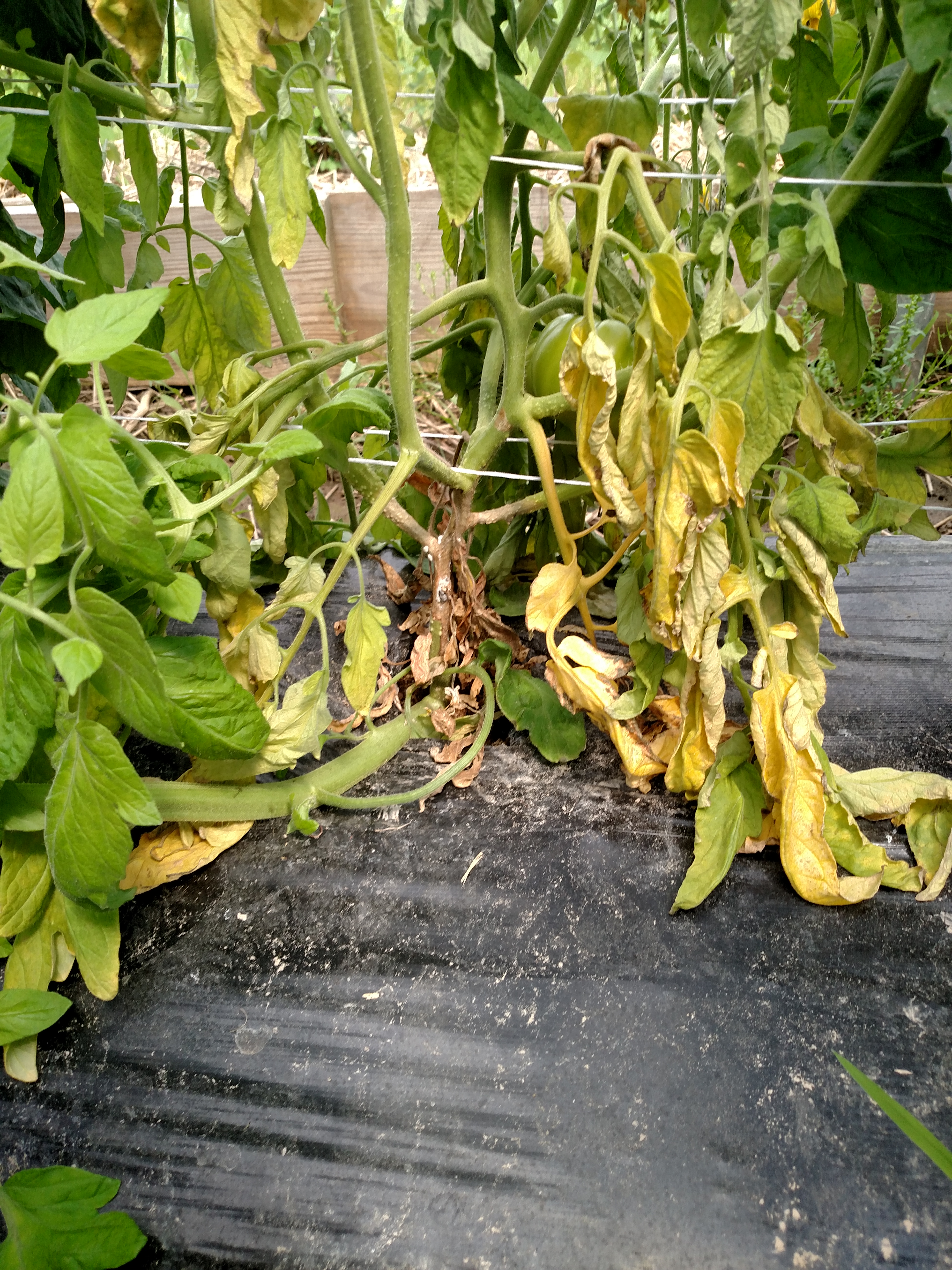 Figure 1. White mold (timber rot) of tomato. Lesion as base of plant leads to wilt.
 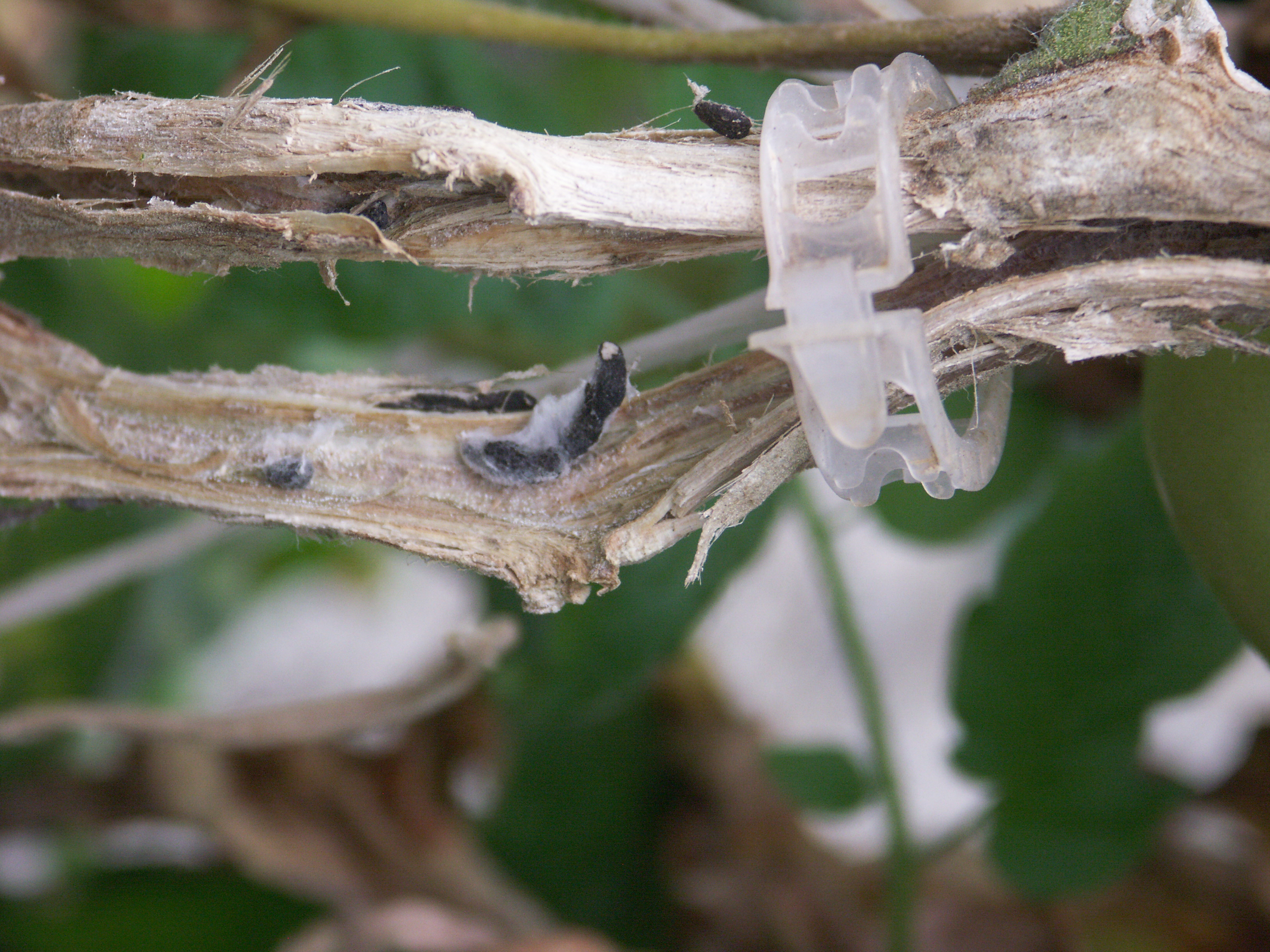 Figure 2. White mold of tomato. Note dry, brittle appearance of stem and dark, irregular sclerotia in and on stem.
 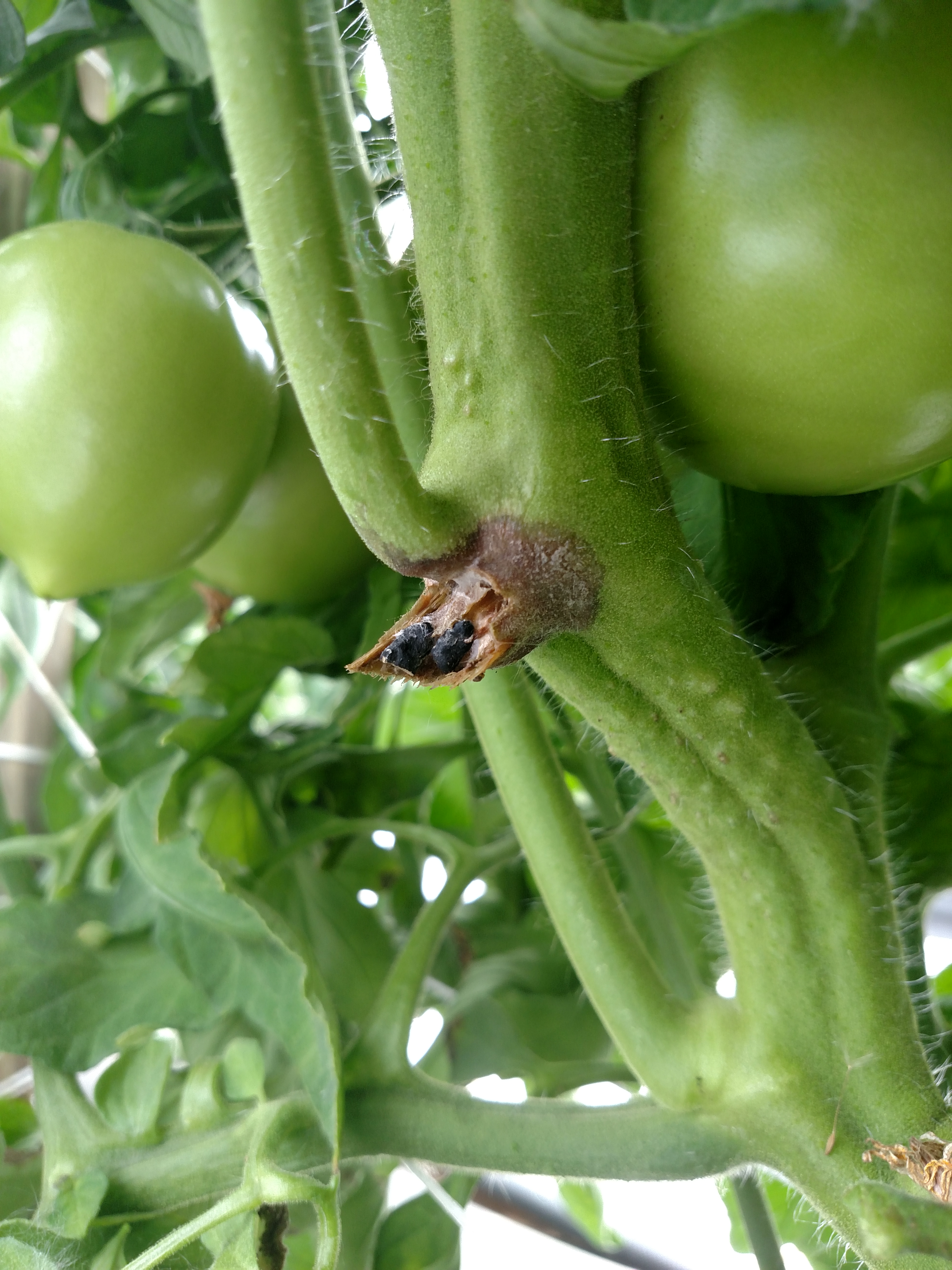 Figure 3. White mold of tomato. Stem has been torn away to reveal sclerotia.
 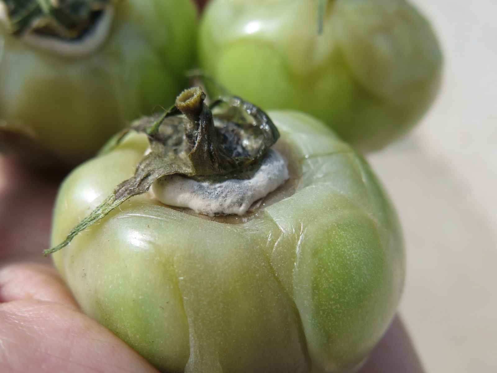 Figure 4. White mold of tomato fruit.
 Zippering
Zippering-This is not an infectious disorder. The scar that appears along the fruit is caused when a flower drags along the embryo (fruit) surface as it enlarges.
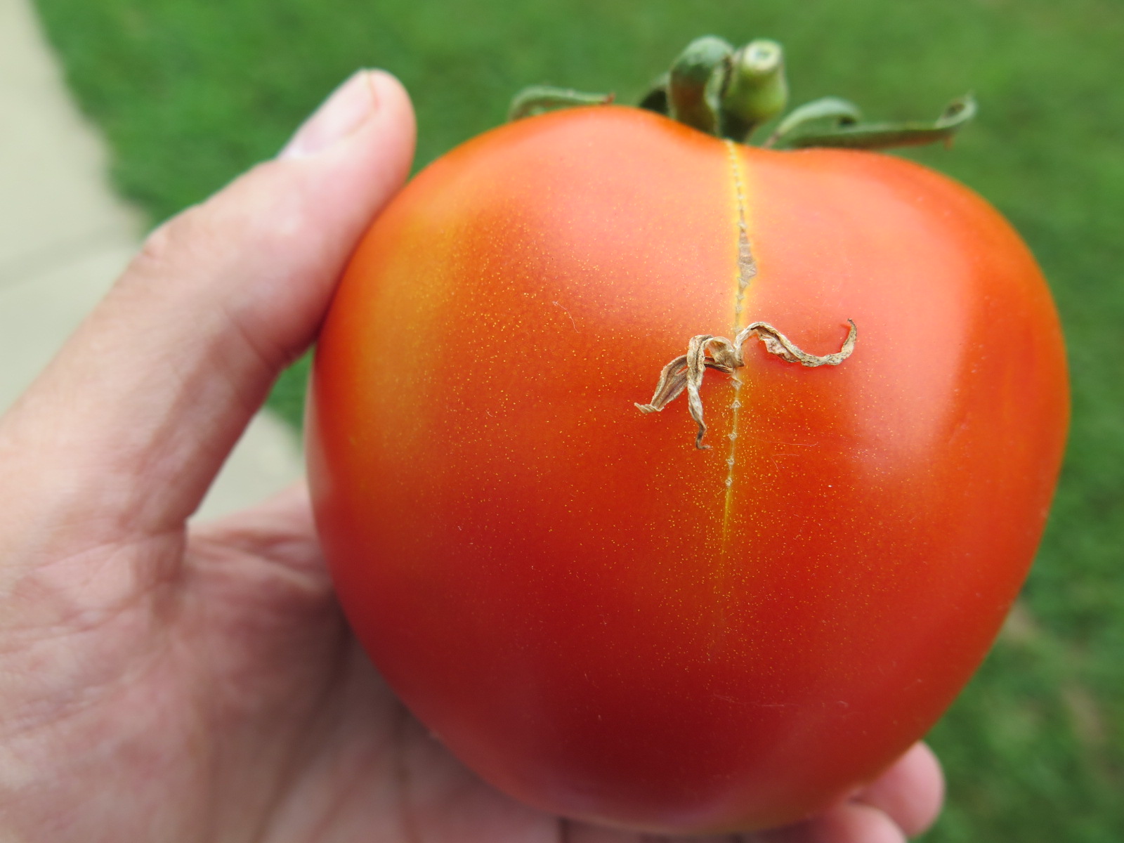 Figure 1. Zipper scar on tomato. Note flower adhering to fruit.
 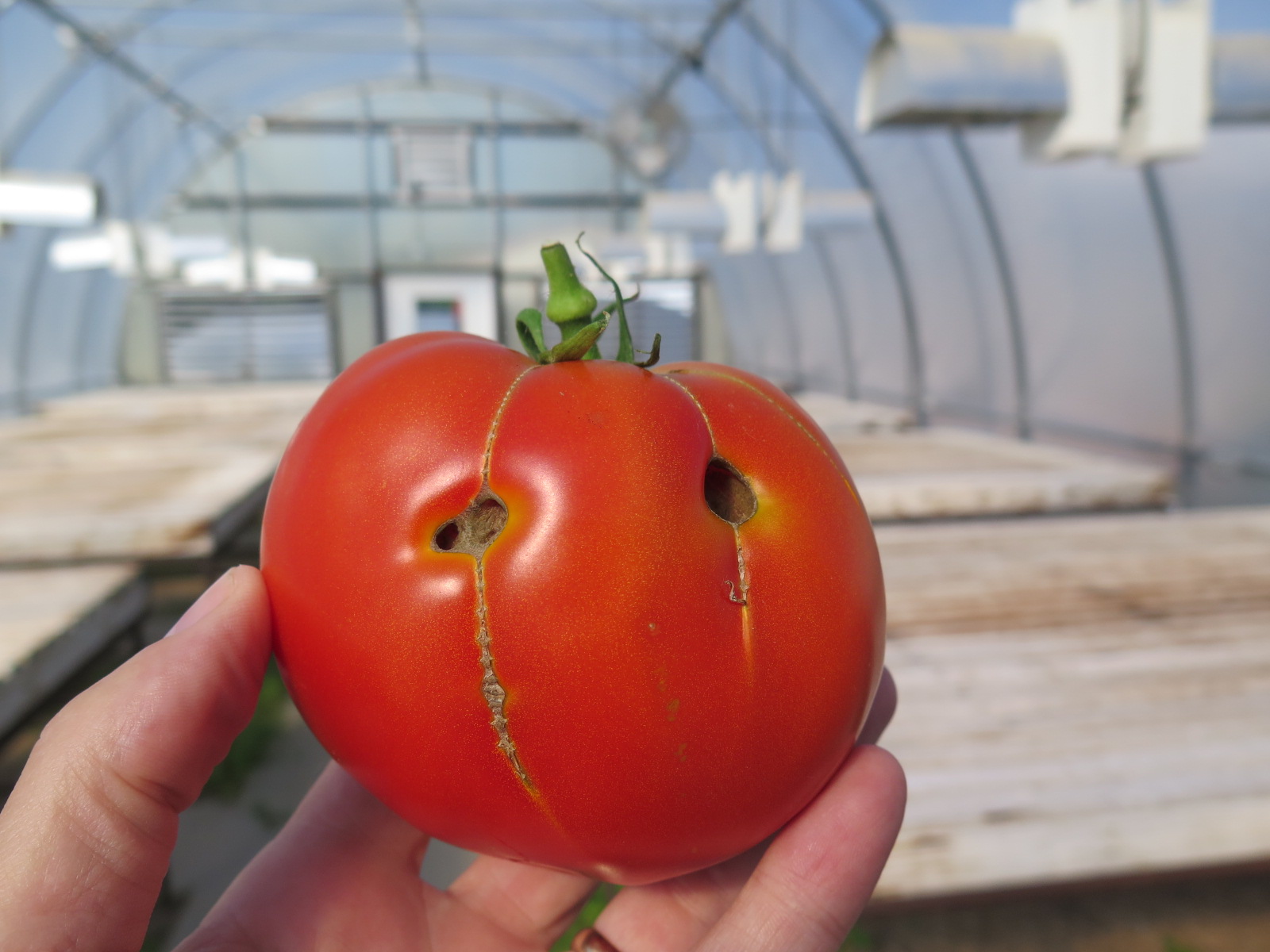 Figure 2. More severe zipper scar has opened up tomato fruit.
 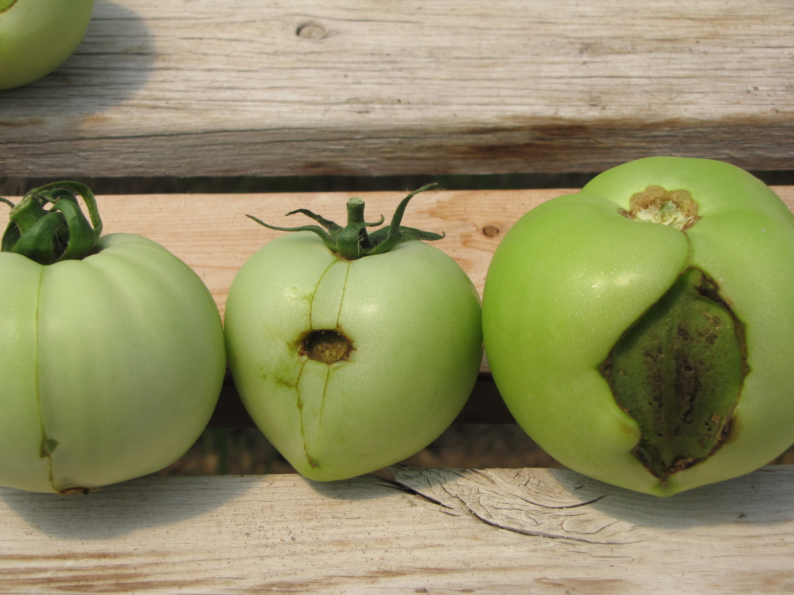 Figure 3. A range of zipper scar symptoms on tomato.