

watermelon diseases
In most years, Indiana ranks among the top handful of states in the US in watermelon production. While some farms will produce watermelon for retail or auction, much watermelon production is for wholesale for midsized and large operations. The most common foliar diseases are anthracnose and gummy stem blight. Fusarium wilt and Phytophthora blight may be the most important soil borne diseases.
Alternaria leaf blight
Alternaria leaf blight-This disease is more common in cantaloupe than watermelon. Note dark, lesions with ring structure. Not an important disease in Indiana.
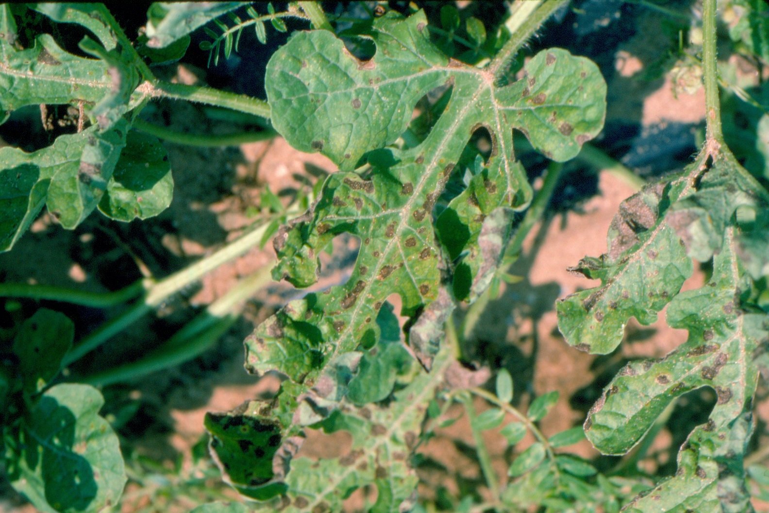 Figure 1. Alternaria leaf blight on watermelon.
 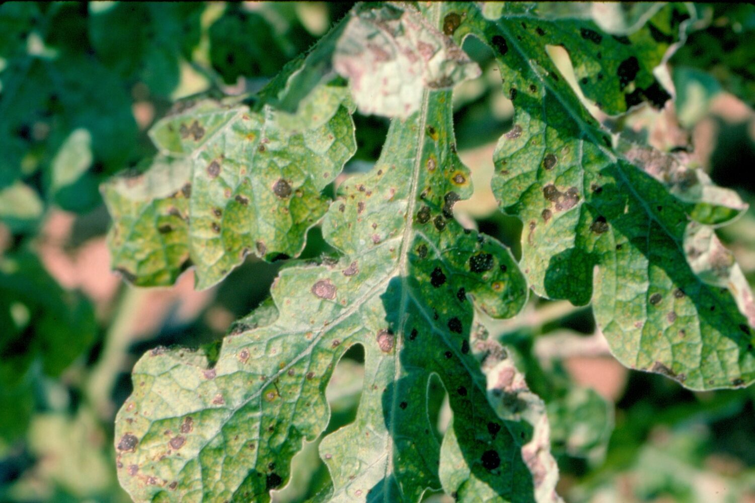 Figure 2. Alternaria leaf blight of watermelon photo showing lesion structures.
 Angular leaf spot
Angular leaf spot-Almost always observed in transplant greenhouses, this disease is seldom observed in the field. Note water-soaked lesions with irregular margins and at least some chlorosis. Disease more common in cool conditions. Not usually an important disease, however, it may be confused with bacterial fruit blotch.
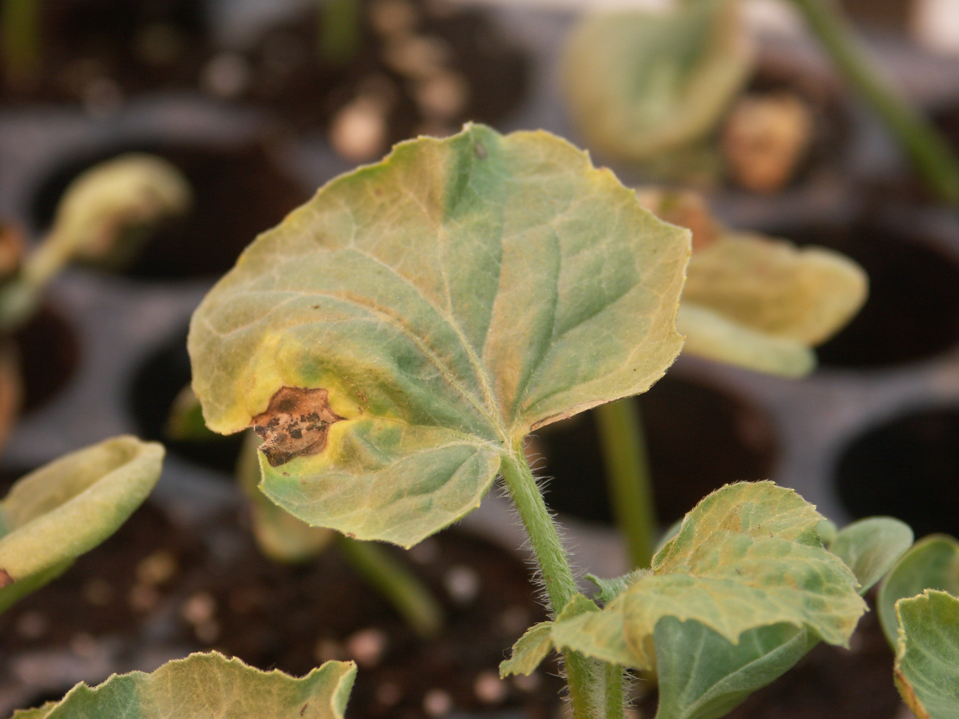 Figure 1. Angular leaf spot of watermelon includes round, light brown necrotic lesion with chlorotic halo.
 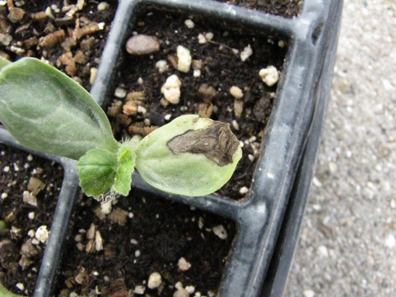 Figure 2. Angular leaf spot on watermelon leaf has caused dark, necrotic lesion on cotyledon.
 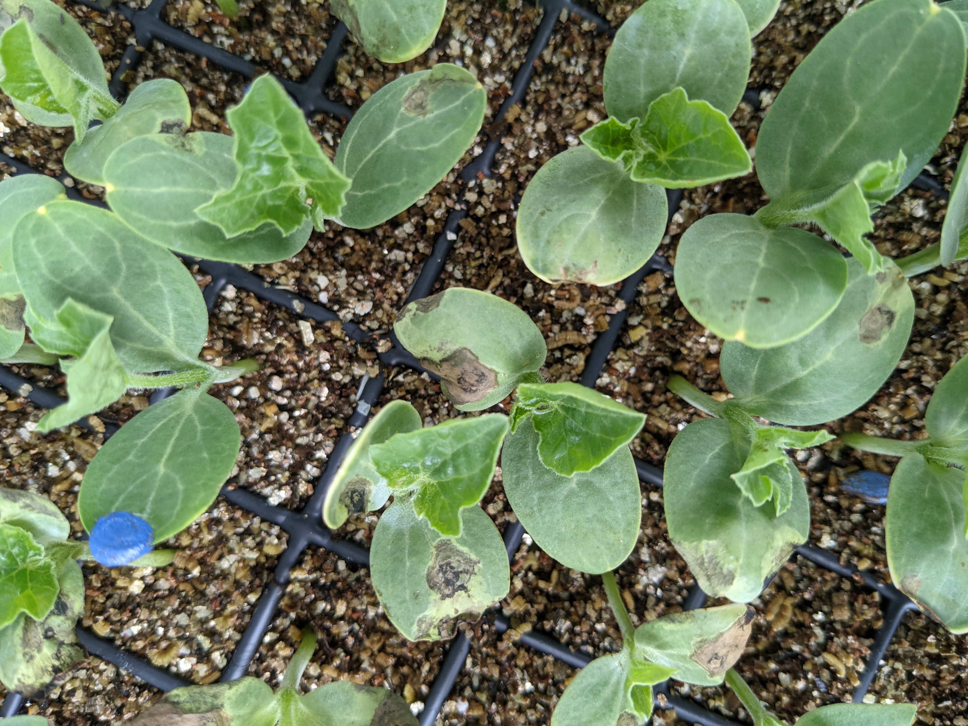 Figure 3. Several watermelon transplants have lesions of angular leaf spot.
 Anthracnose
Anthracnose-One of our most important foliar diseases of watermelon. Lesions on leaves are often irregular and jagged. Lesions on fruit may be sunken; under moist conditions, fruit lesions may be associated with a salmon or orange coloration.
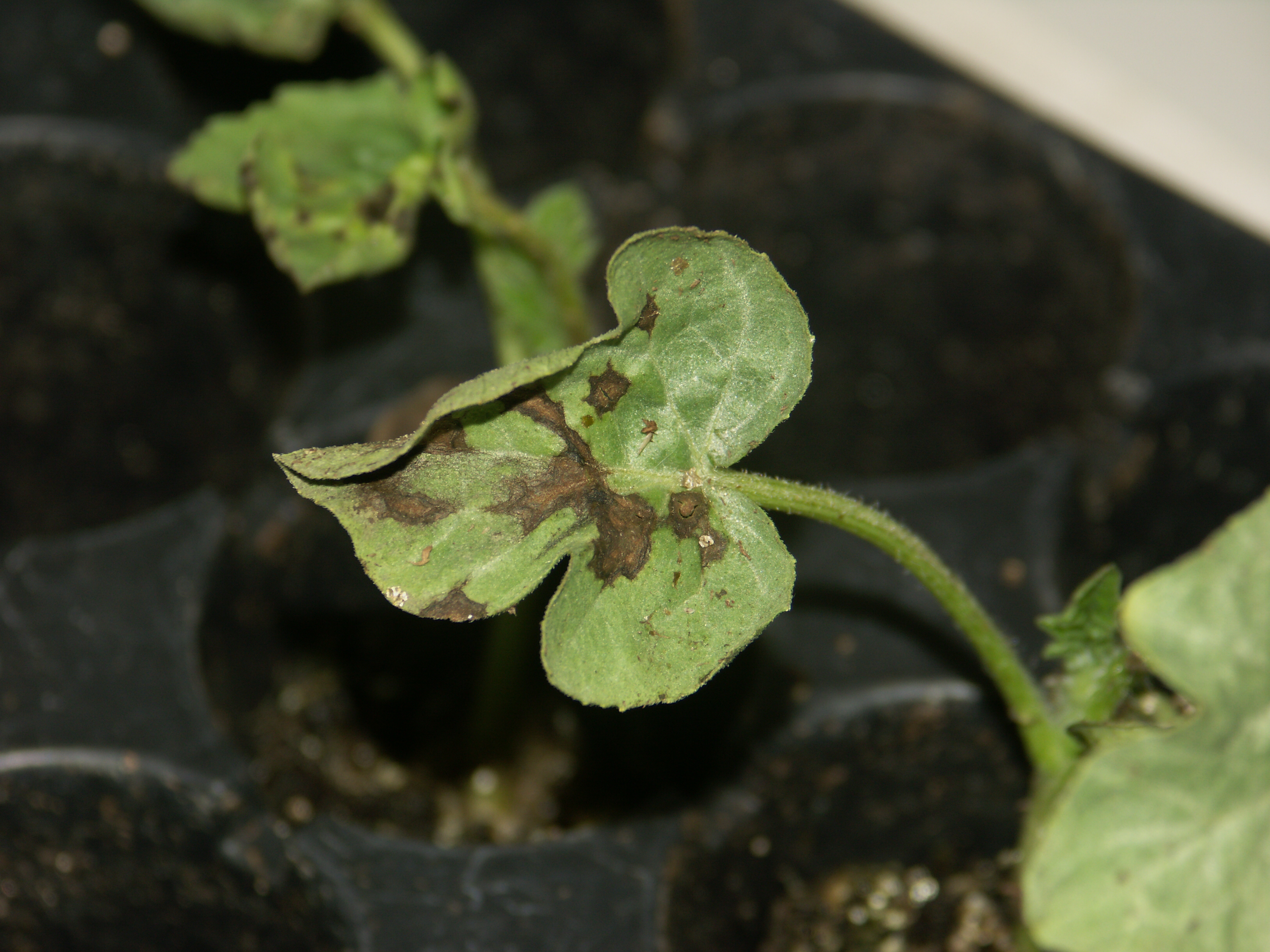 Figure 1. Anthracnose lesion on watermelon transplant. Note angular, jagged lesion.
 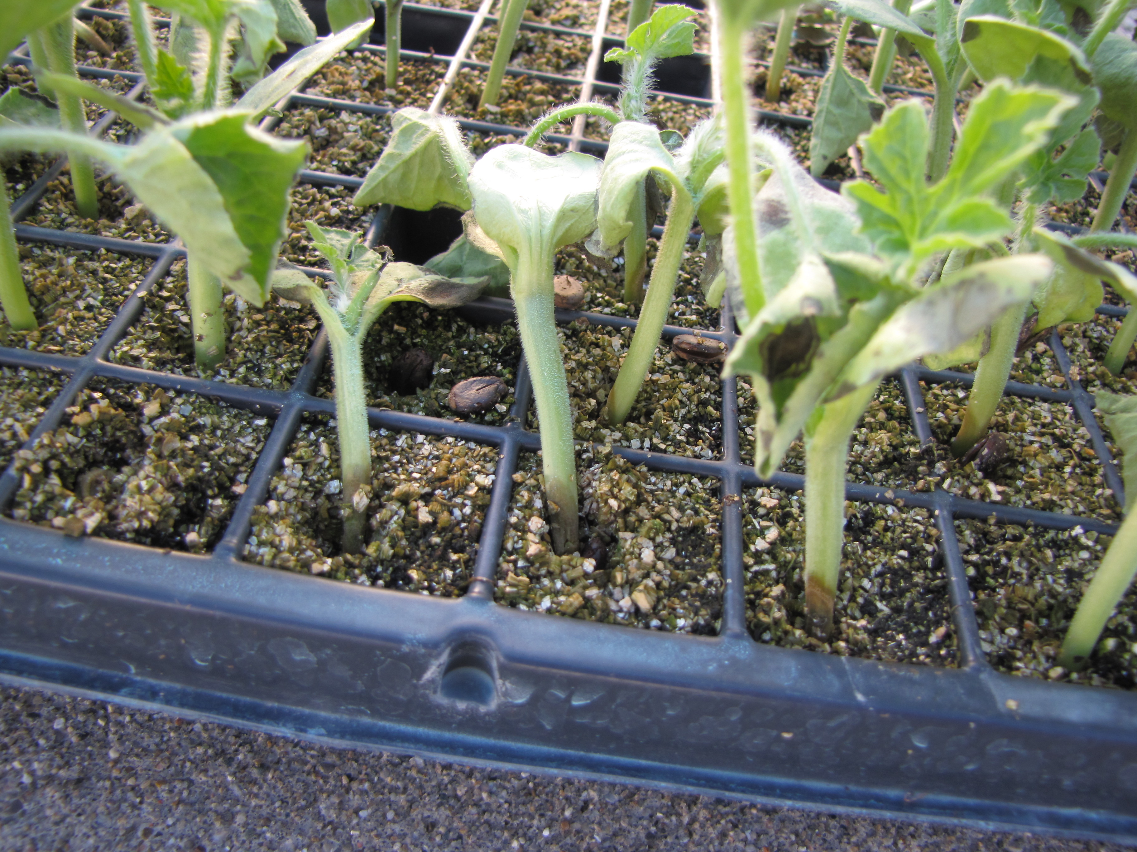 Figure 2. Anthracnose lesions on the hypocotyl of these watermelon transplants has caused the plants to wilt.
 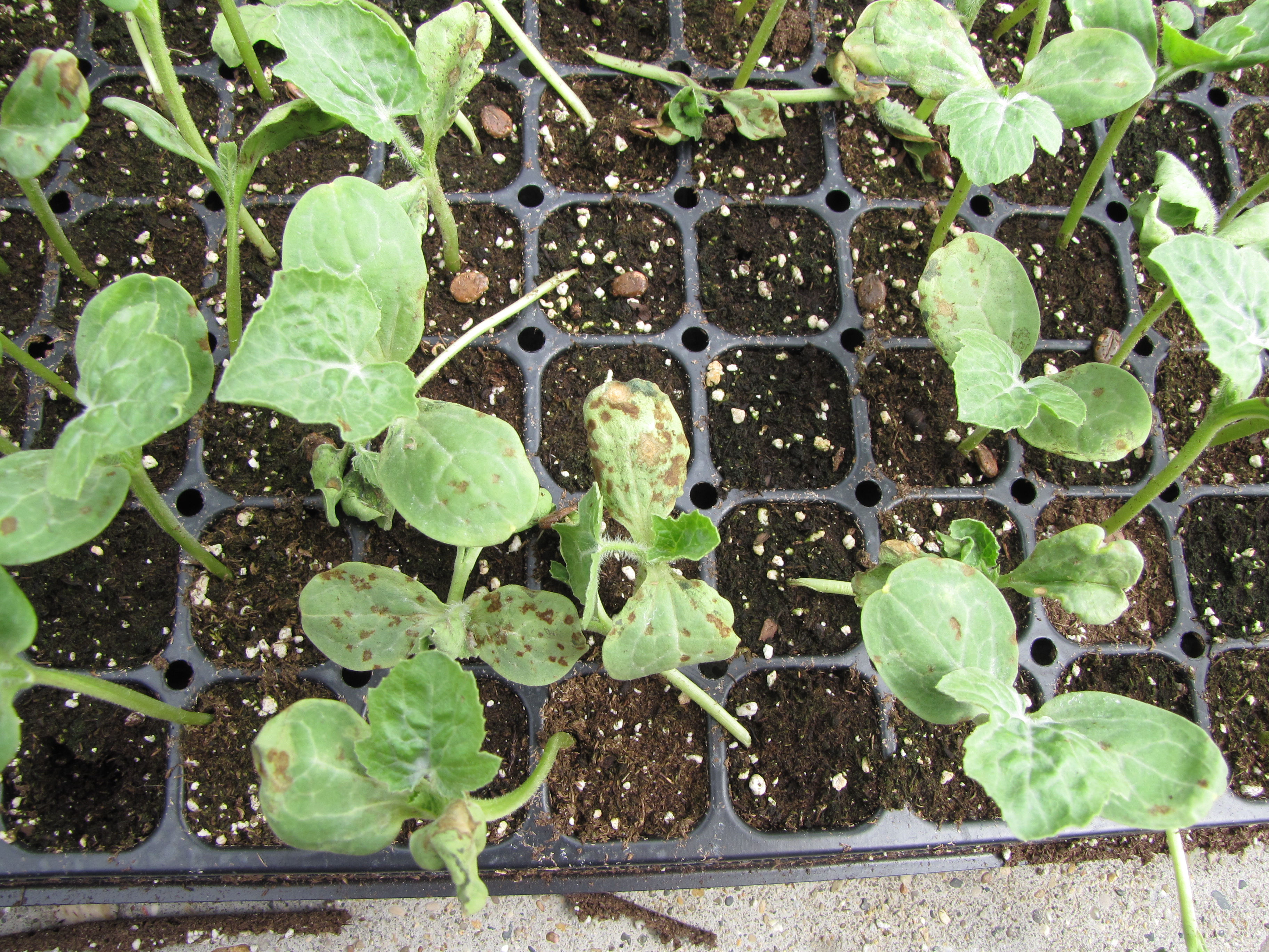 Figure 3. Anthracnose lesion on several watermelon transplants. Lesions are primarily on cotyledons (seed leaves).
 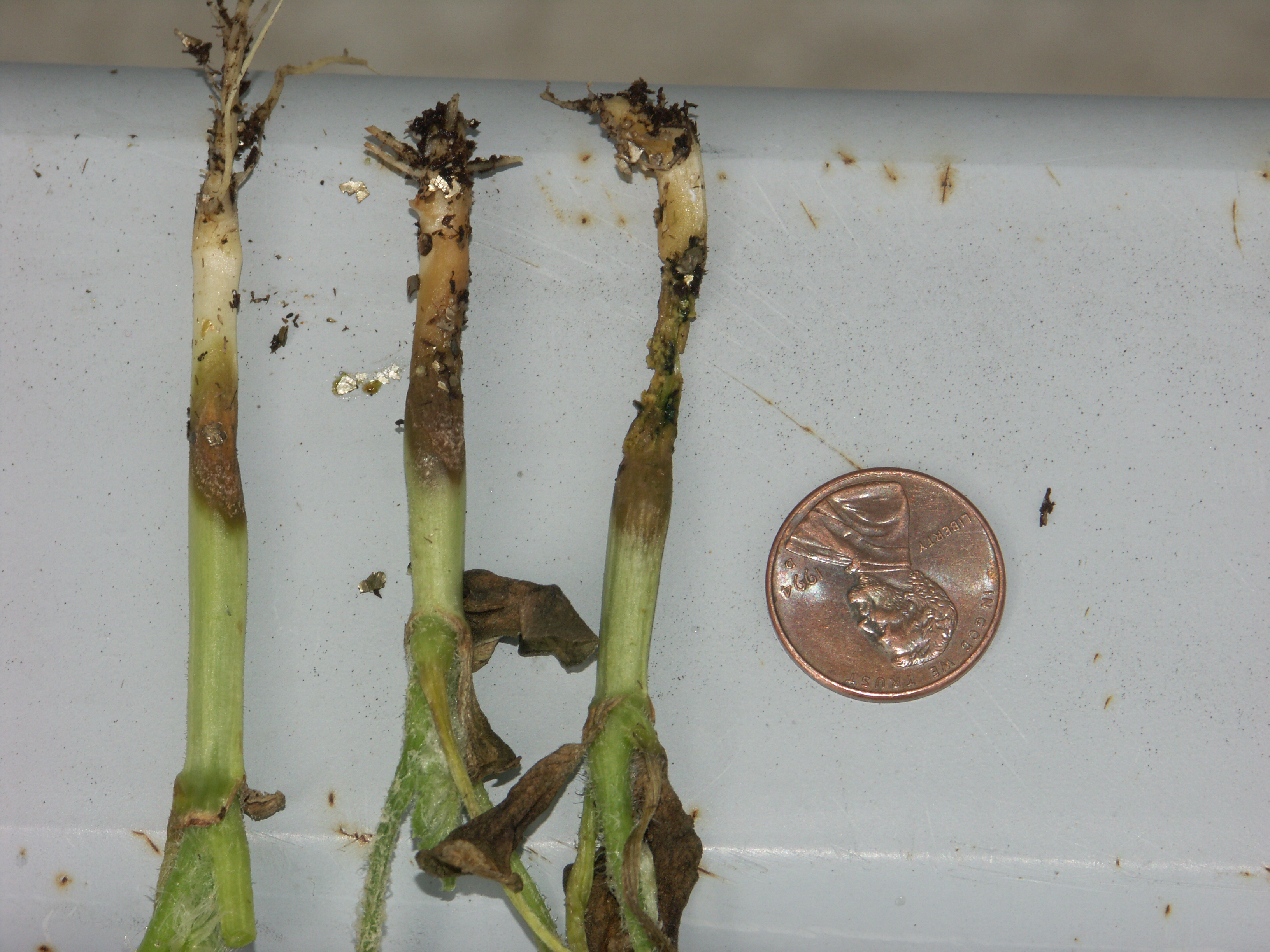 Figure 4. Anthracnose of watermelon occasionally causes lesions on the hypocotyl, between the cotyledon and the soil.
 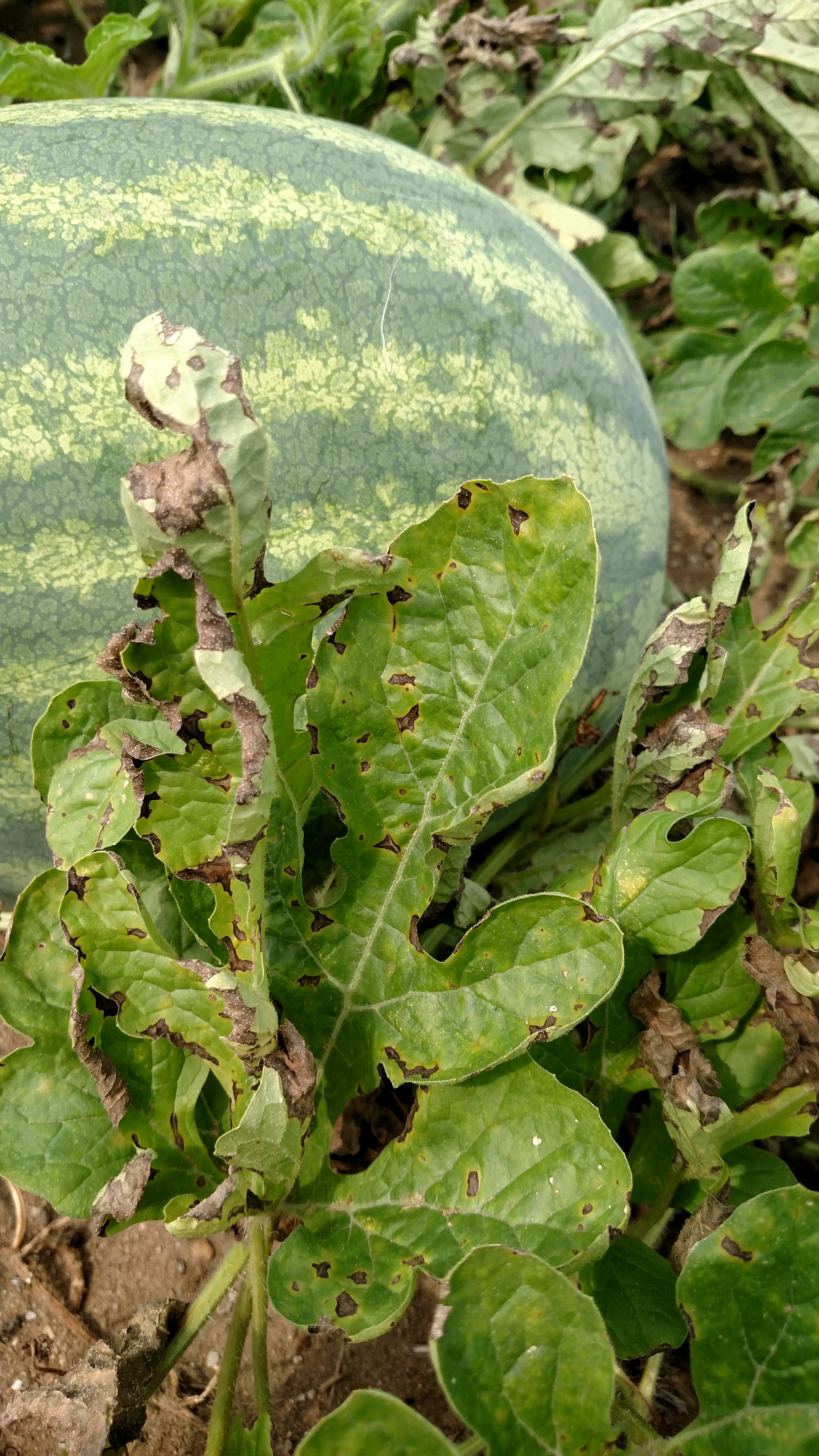 Figure 5. Anthracnose lesions on mature watermelon leaves tend to be angular and jagged.
 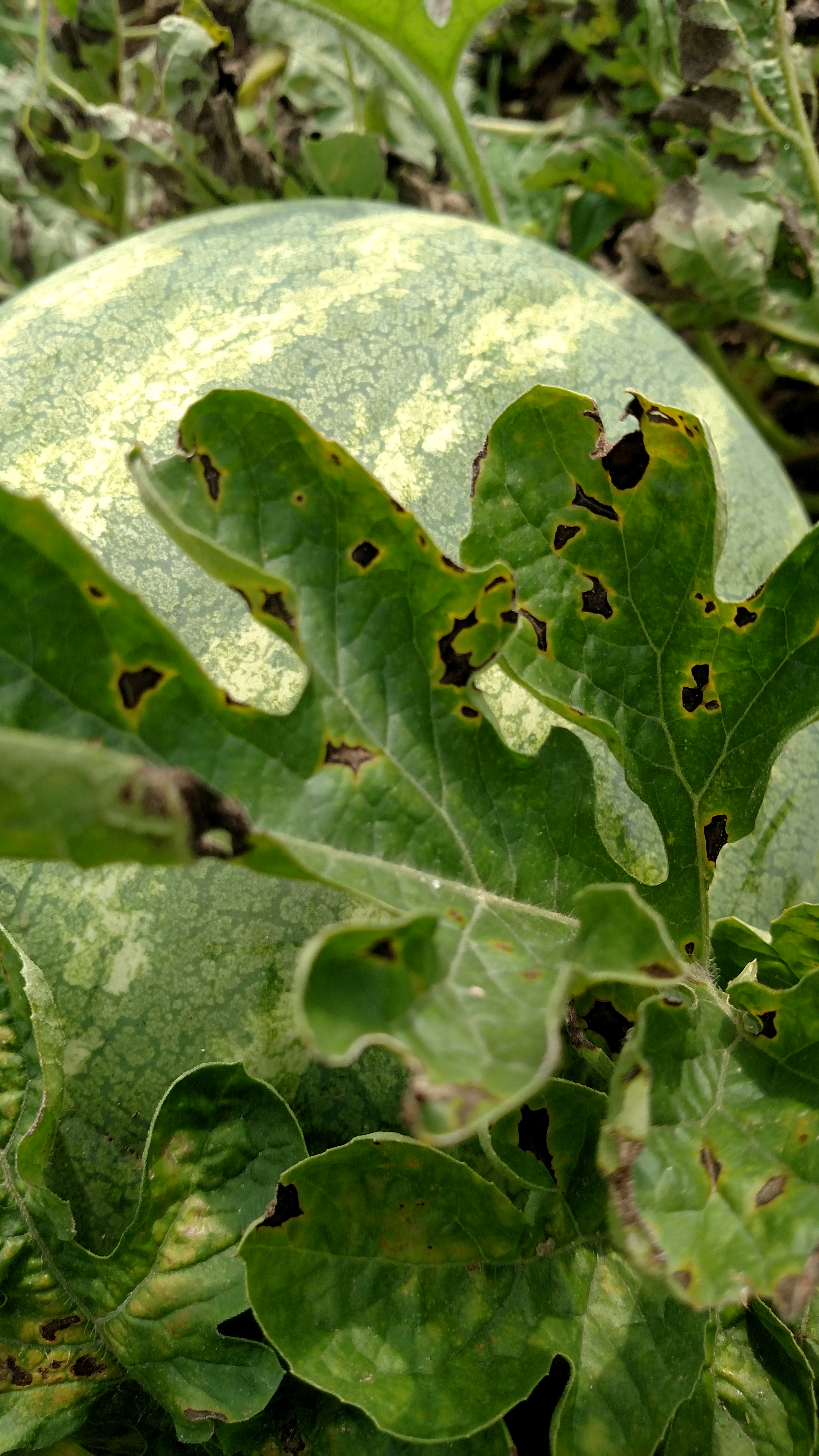 Figure 6. Another photo of anthracnose of watermelon on a leaf. Note the yellow color on the margin of the lesions.
 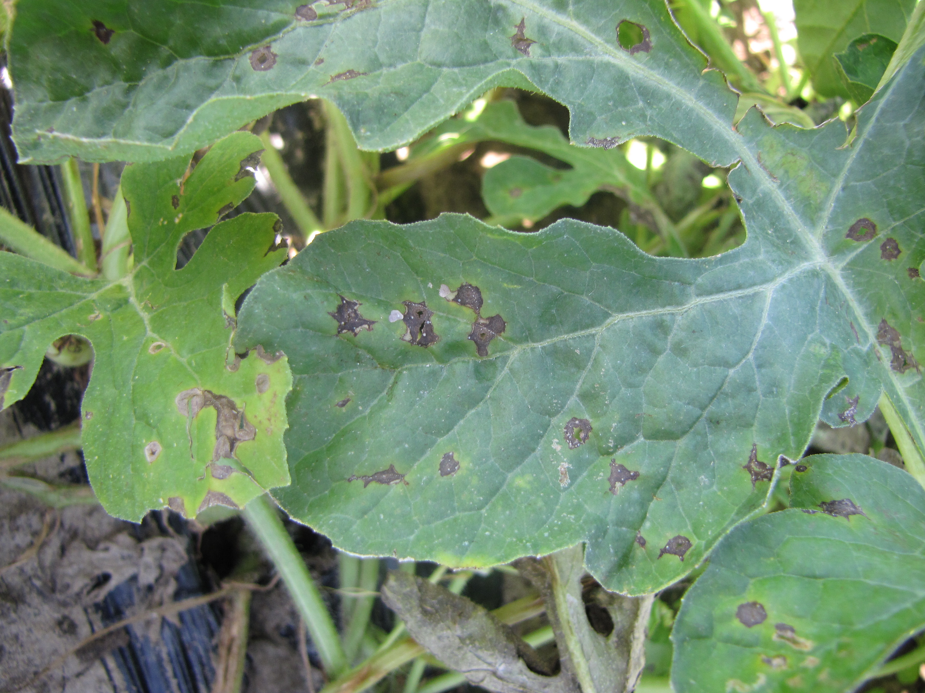 Figure 7. Several anthracnose lesions on a watermelon leaf.
 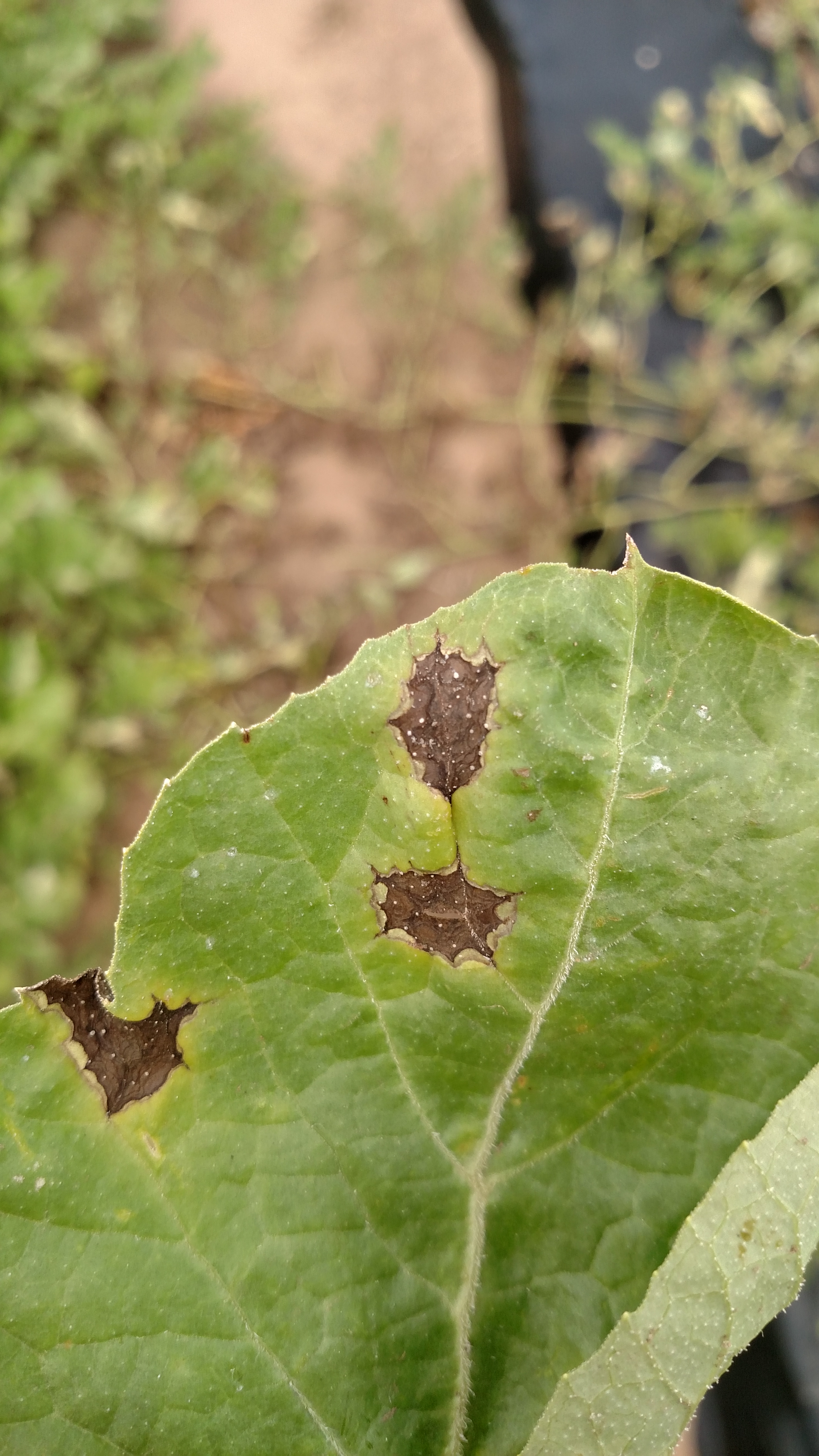 Figure 8. A close-up of a lesion of anthracnose on a watermelon leaf. Note the sharp, angular shape of the lesion.
 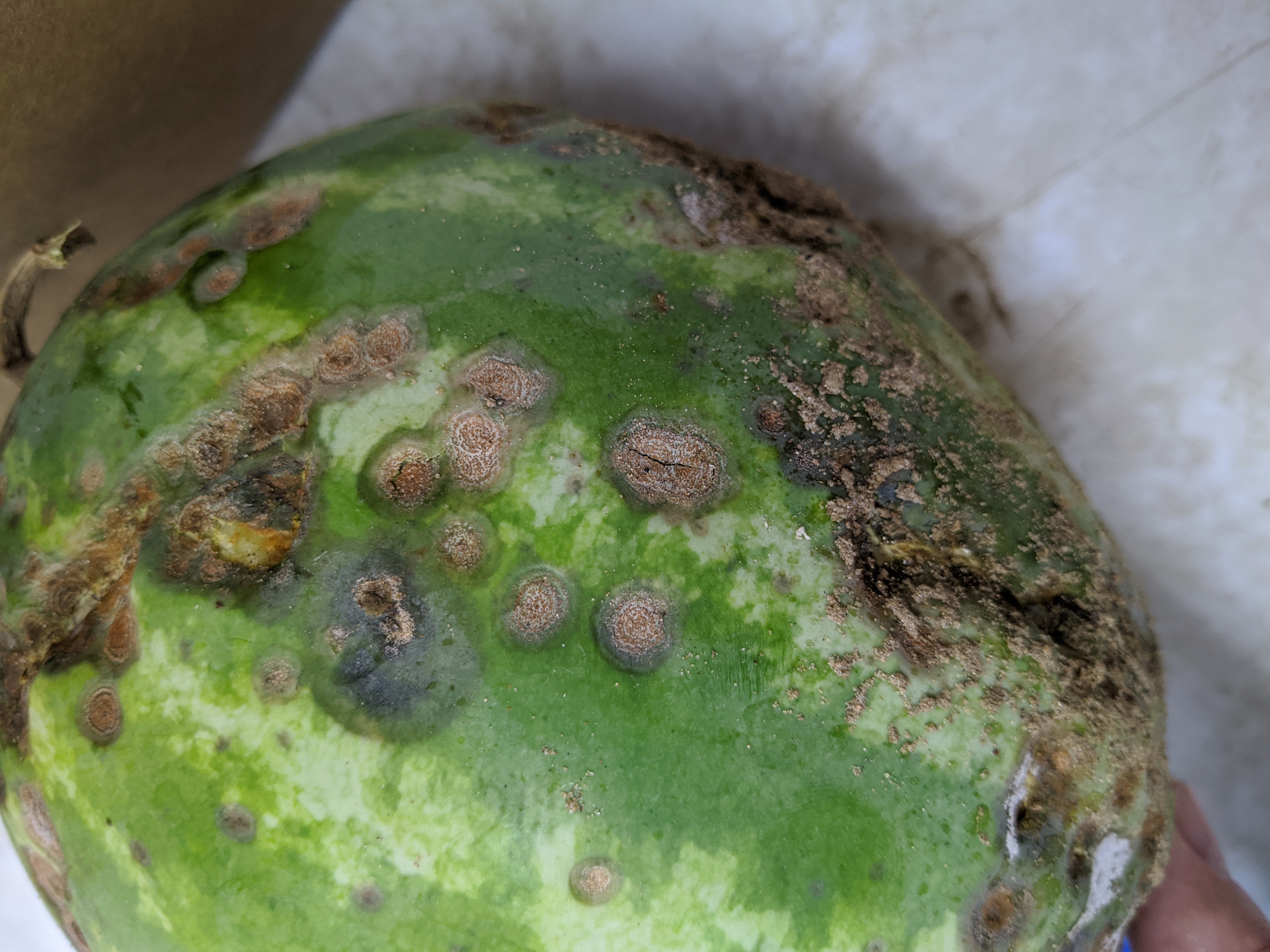 Figure 9. A watermelon fruit with pit-like lesions of anthracnose. Note the orange or salmon-like color of the lesions due to the color of the spores.
 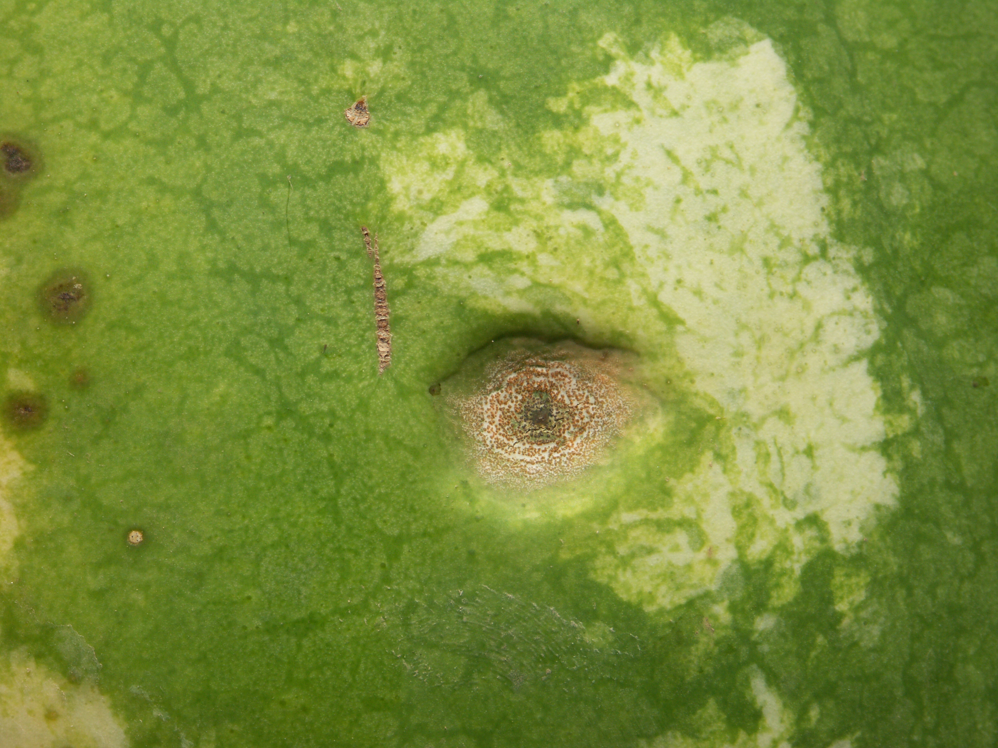 Figure 10. A close-up of a lesion of anthracnose on a watermelon fruit.
 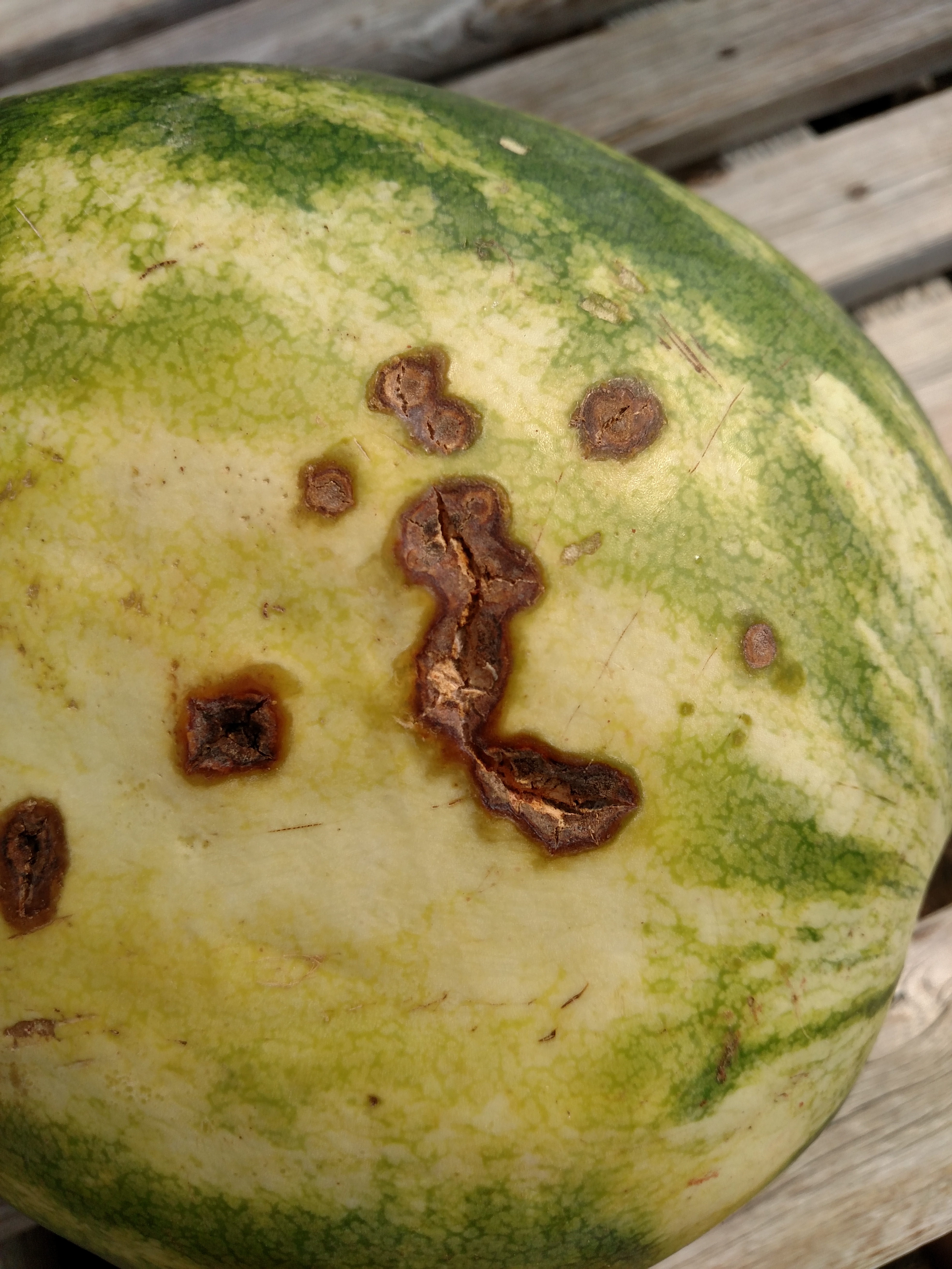 Figure 11. The anthracnose lesions on this watermelon fruit appear more as cracks than pit-like as in other photos. Note that there is still a hint of orange in some of the cracks due to the spore colors. Note also that the lesions tend to be toward the bottom of the fruit as is typical.
 Bacterial fruit blotch
Bacterial fruit blotch is easily identified by the large greasy appearing lesions on the fruit, often on the top side of the fruit. It is helpful, however, to become familiar with the necrotic lesions on leaves. While these lesions are not important economically, identification of lesions on transplants or early plantings may help one to manage the disease. Leaf lesions also provide a reservoir of inoculum for lesions on the fruit. Almost always associated with seed production.
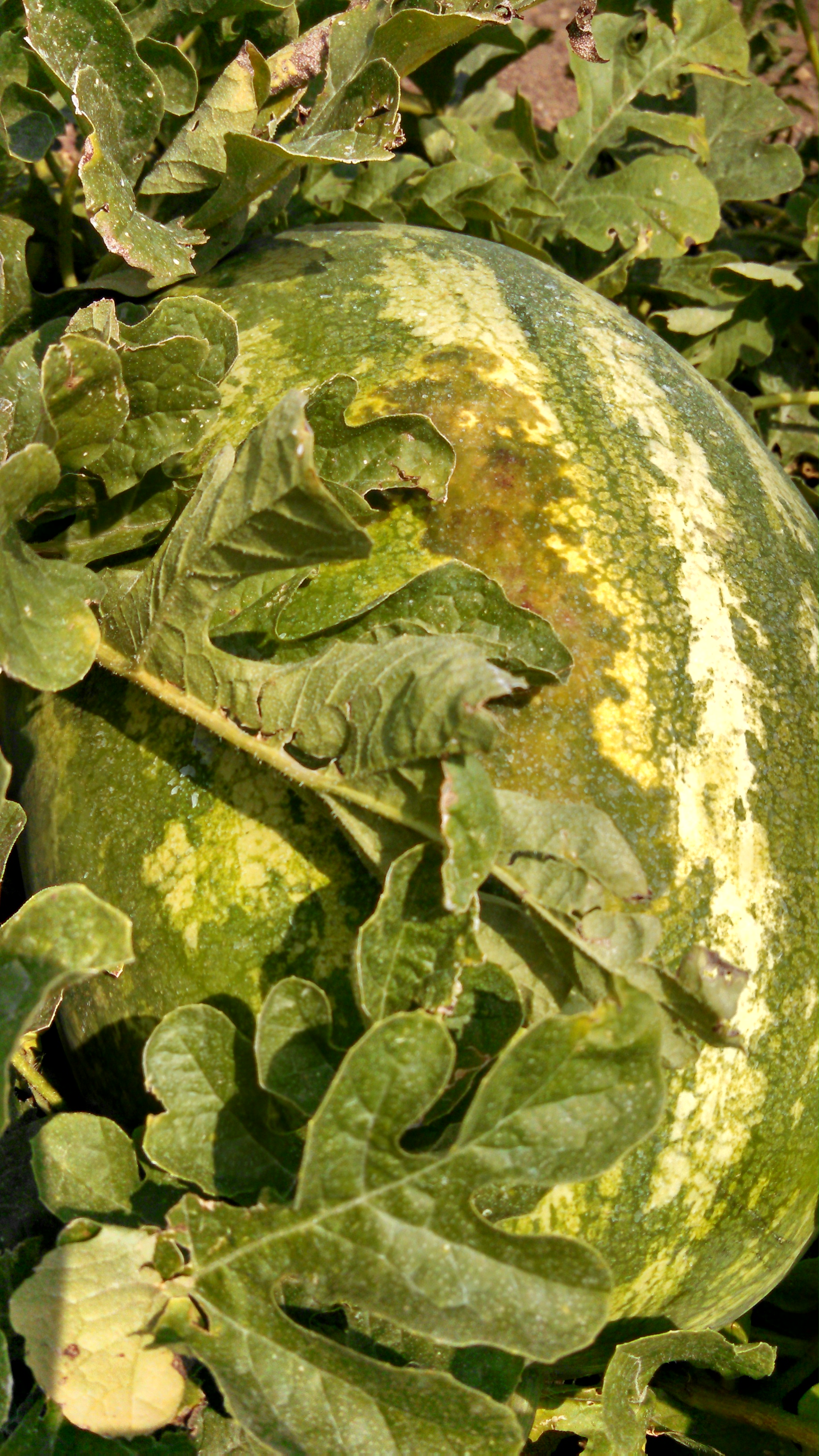 Figure 1. An irregular dark lesion can be observed on the top of this watermelon fruit caused by bacterial fruit blotch of watermelon.
 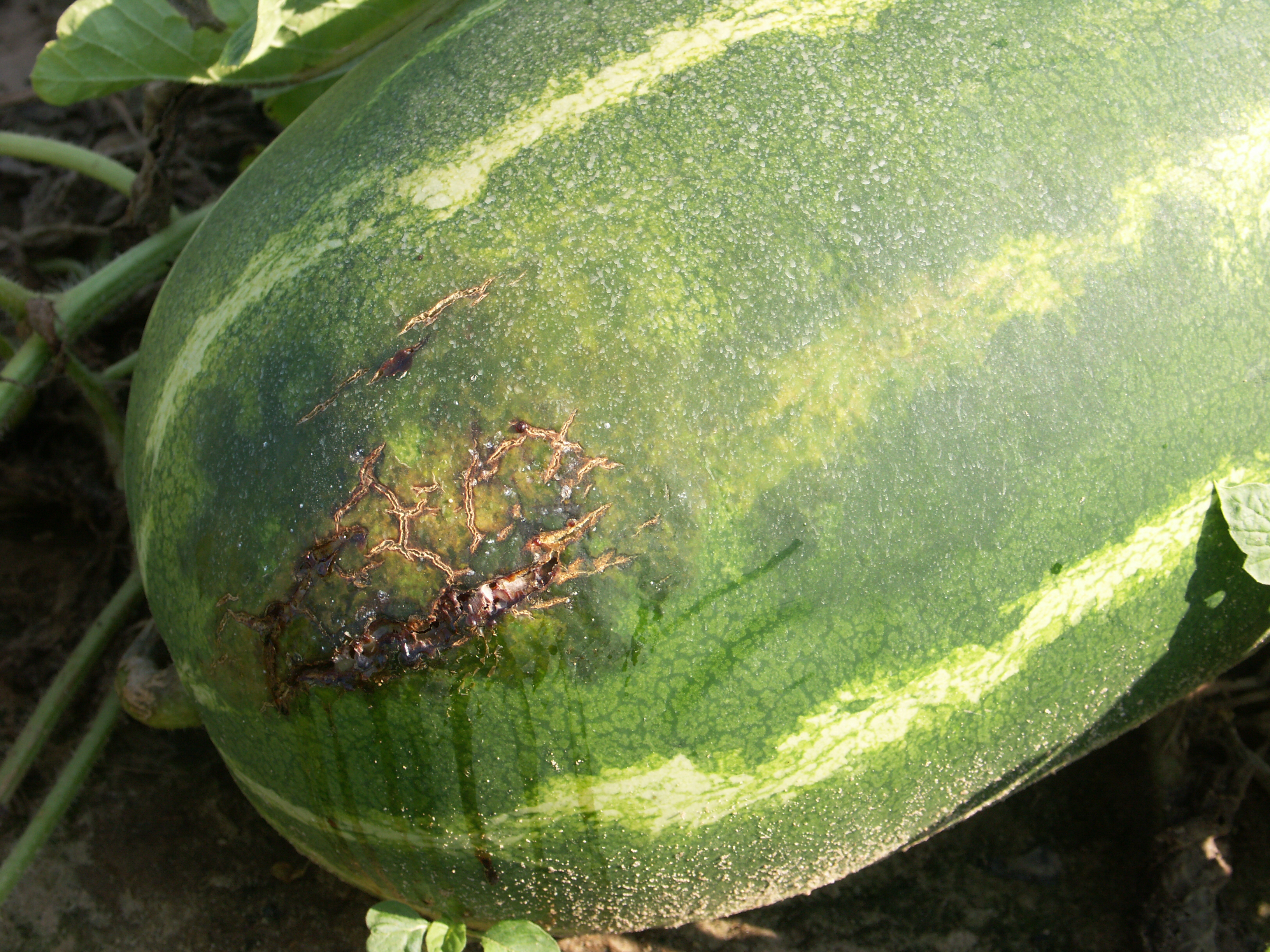 Figure 2. Watermelon is cracked probably due to secondary infection of a lesion of bacterial fruit blotch. Note leakage of fluids has dripped down side of fruit.
 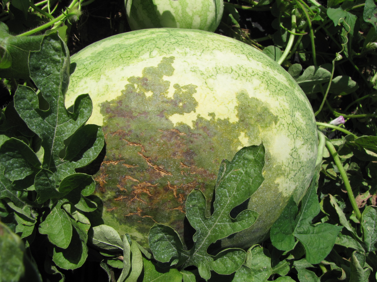 Figure 3. A large, spreading lesion due to bacterial fruit blotch is seen on the top of this watermelon. Note cracking of lesion.
  Figure 4. Leaf lesions of bacterial fruit blotch of watermelon may be irregular.
 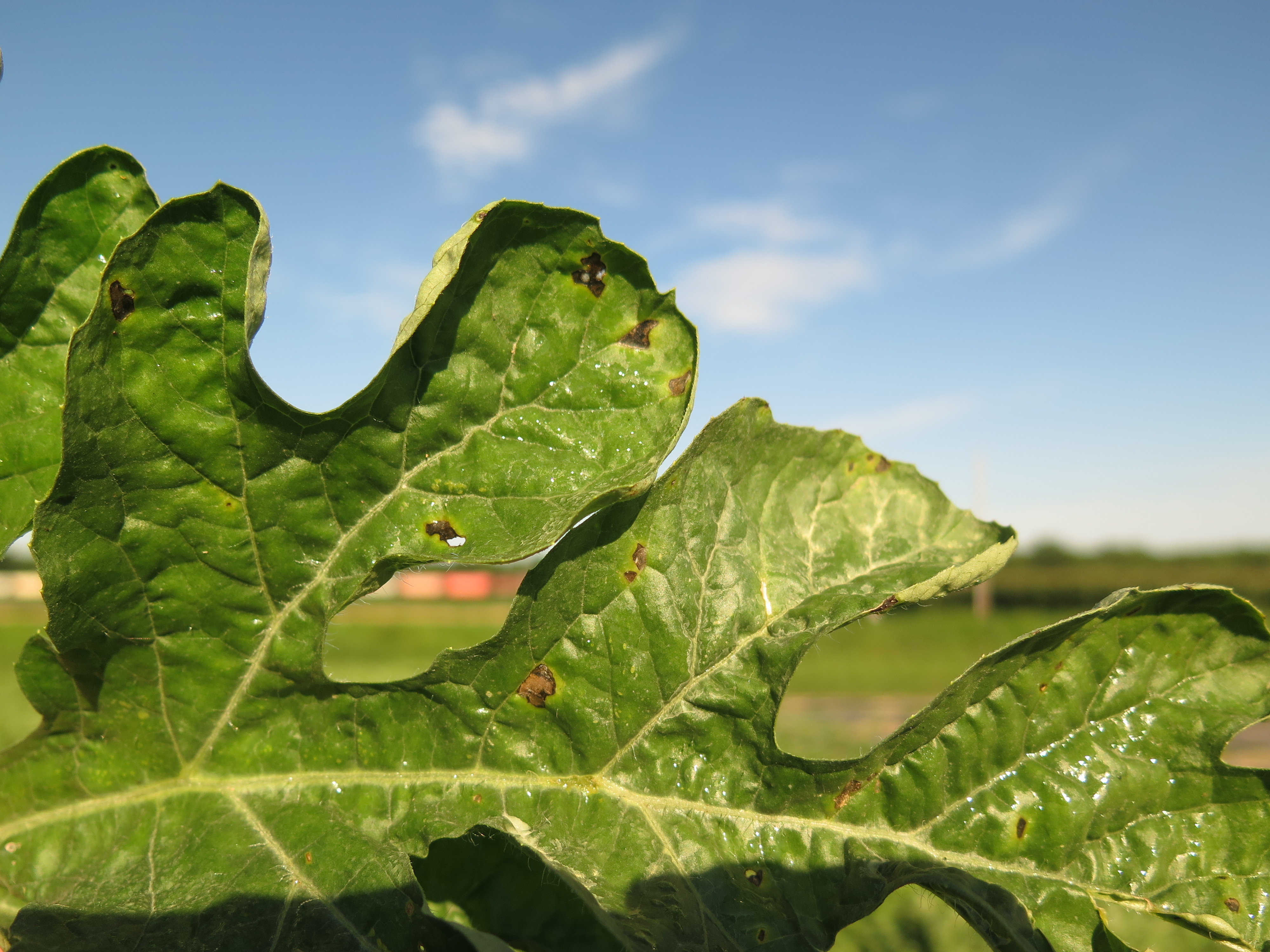 Figure 5. Leaf lesions of bacterial fruit blotch of watermelon can be irregular.
 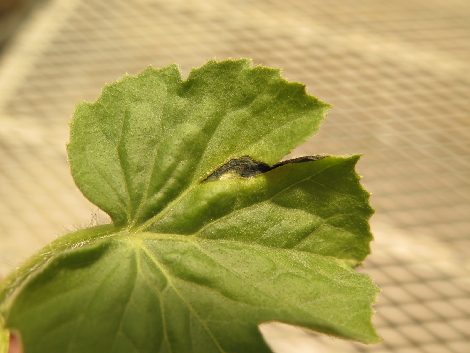 Figure 6. Lesion of bacterial fruit blotch of watermelon seedling.
 Black root rot
Black root rot-caused by soil borne fungal organism. Not common, but can be economically important when present. Note dark, coloration on base of stem (hypocotyl).
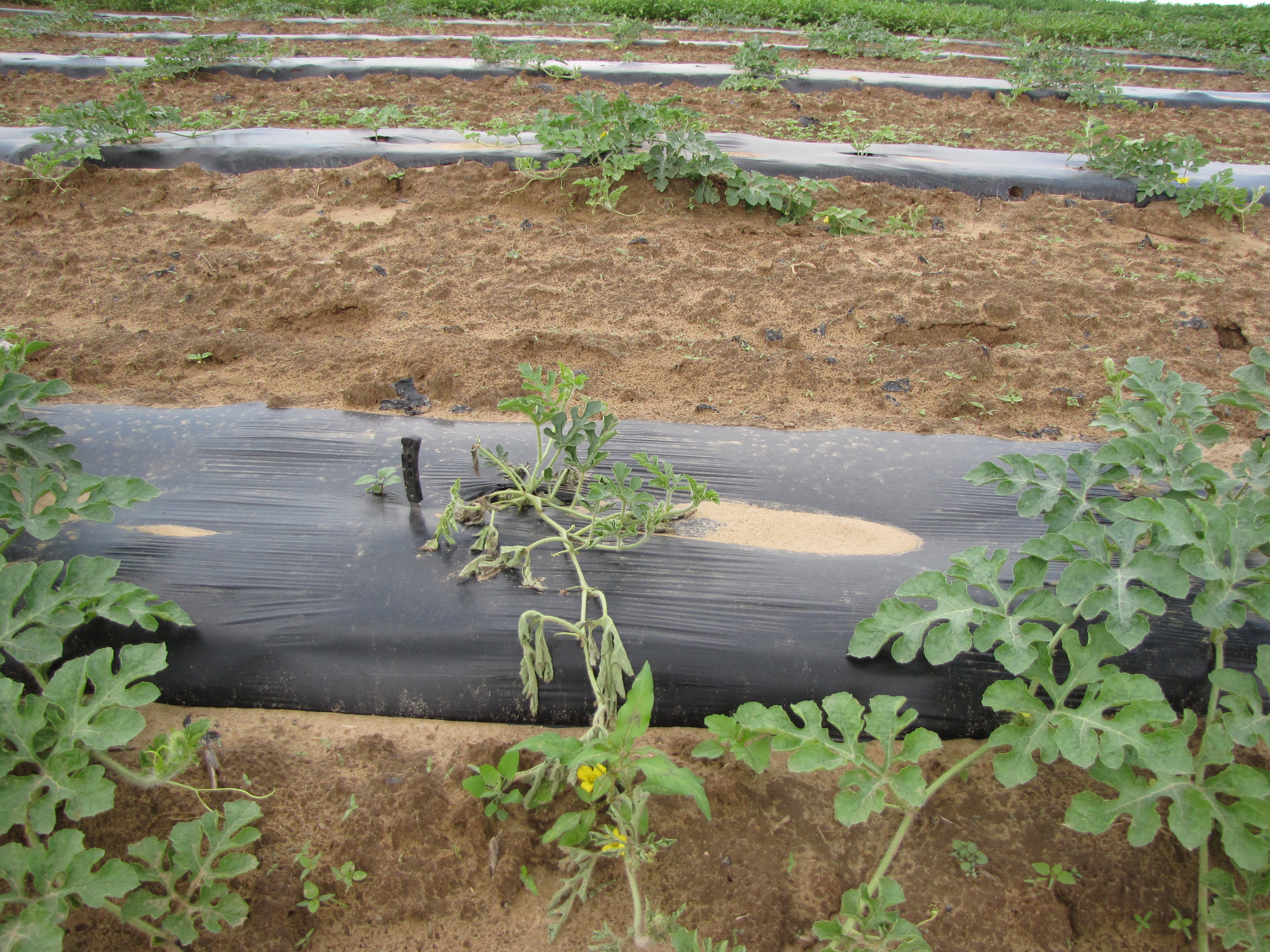 Figure 1. The symptoms of wilt in this watermelon could be from many causes. Black root rot can cause wilt such as seen here.
 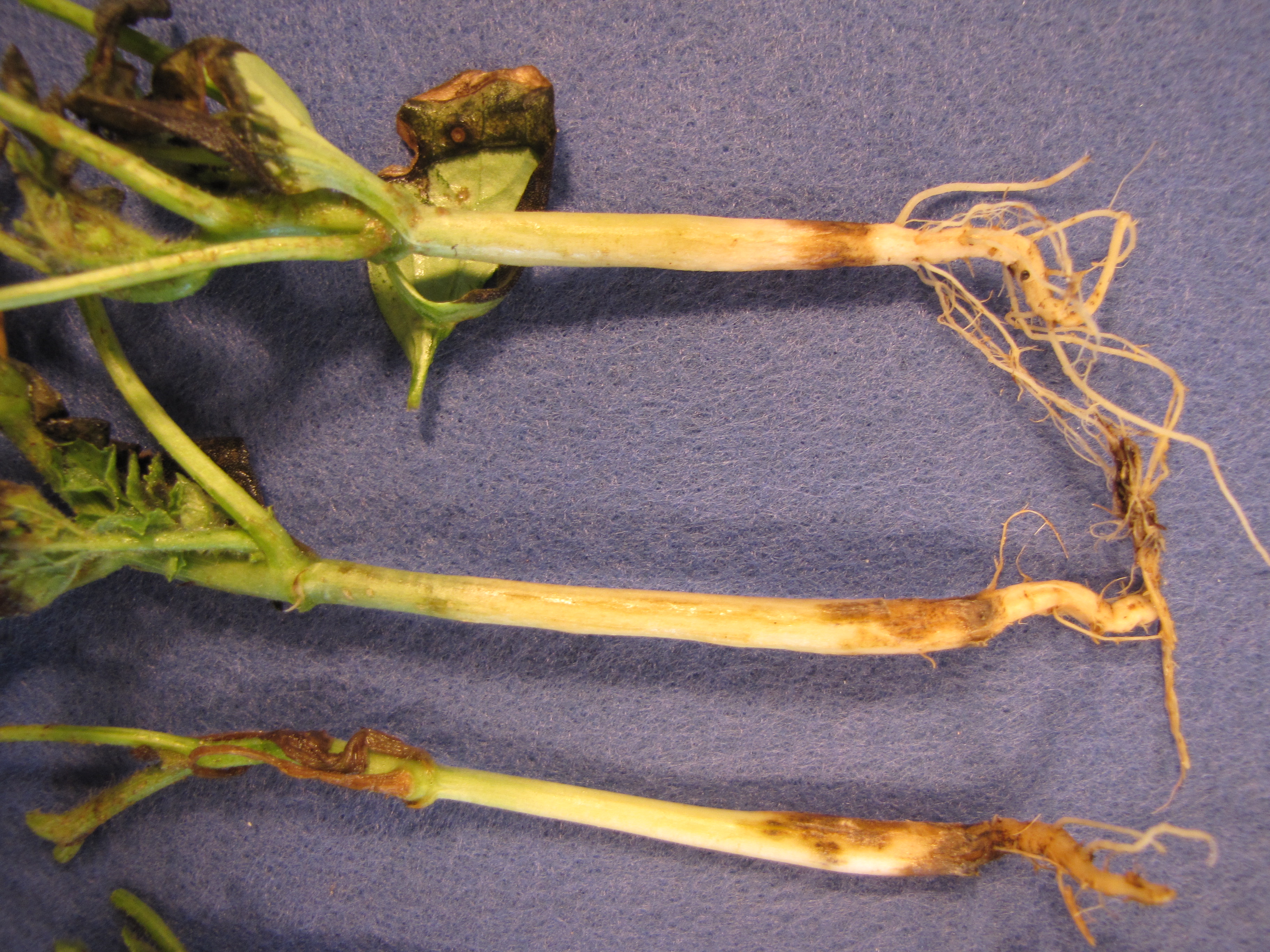 Figure 2. Dark areas on the hypocotyl of this watermelon seedling is caused by chlamydospores (resting spores) of the fungus that causes black root rot (Thielaviopsis basicola).
 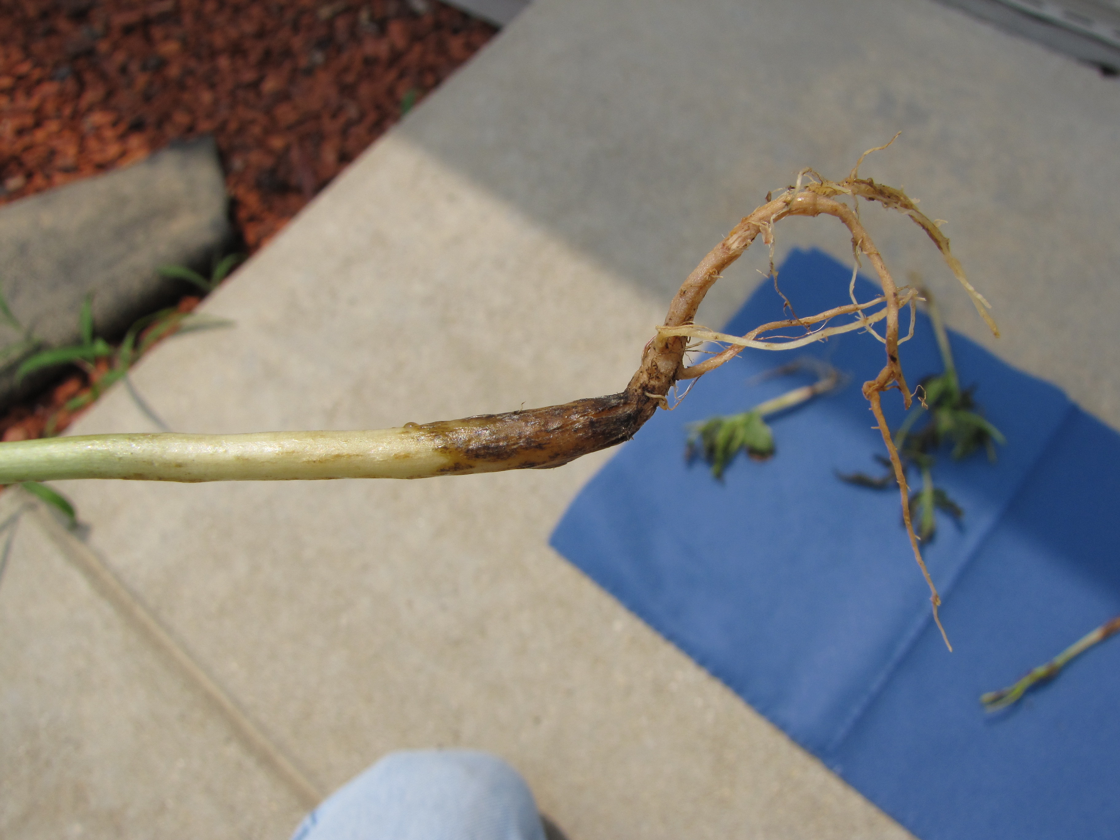 Figure 3. Dark discoloration and poor root system are as a result of black root rot of watermelon.
 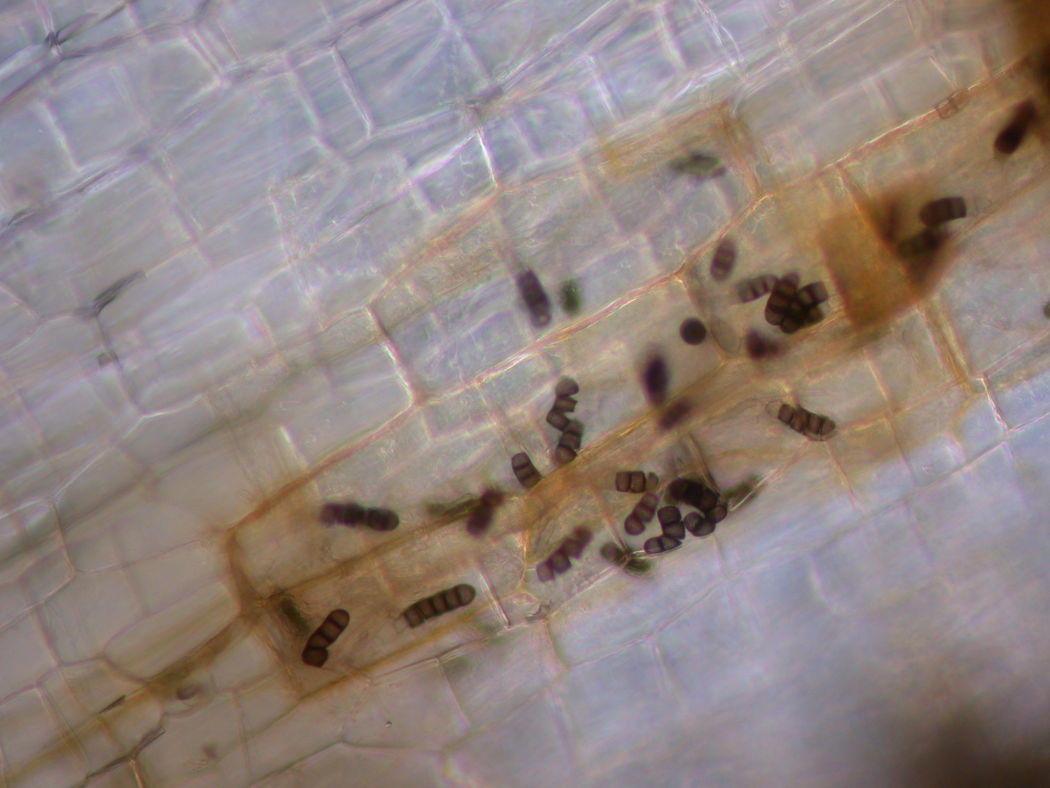 Figure 4. The structures seen here in root tissue are specialized spores known as chlamydospores. These spores are resilient resting spores that help the fungus survive for long periods in the soil. In addition, these spores impart a dark appearance to tissue.
 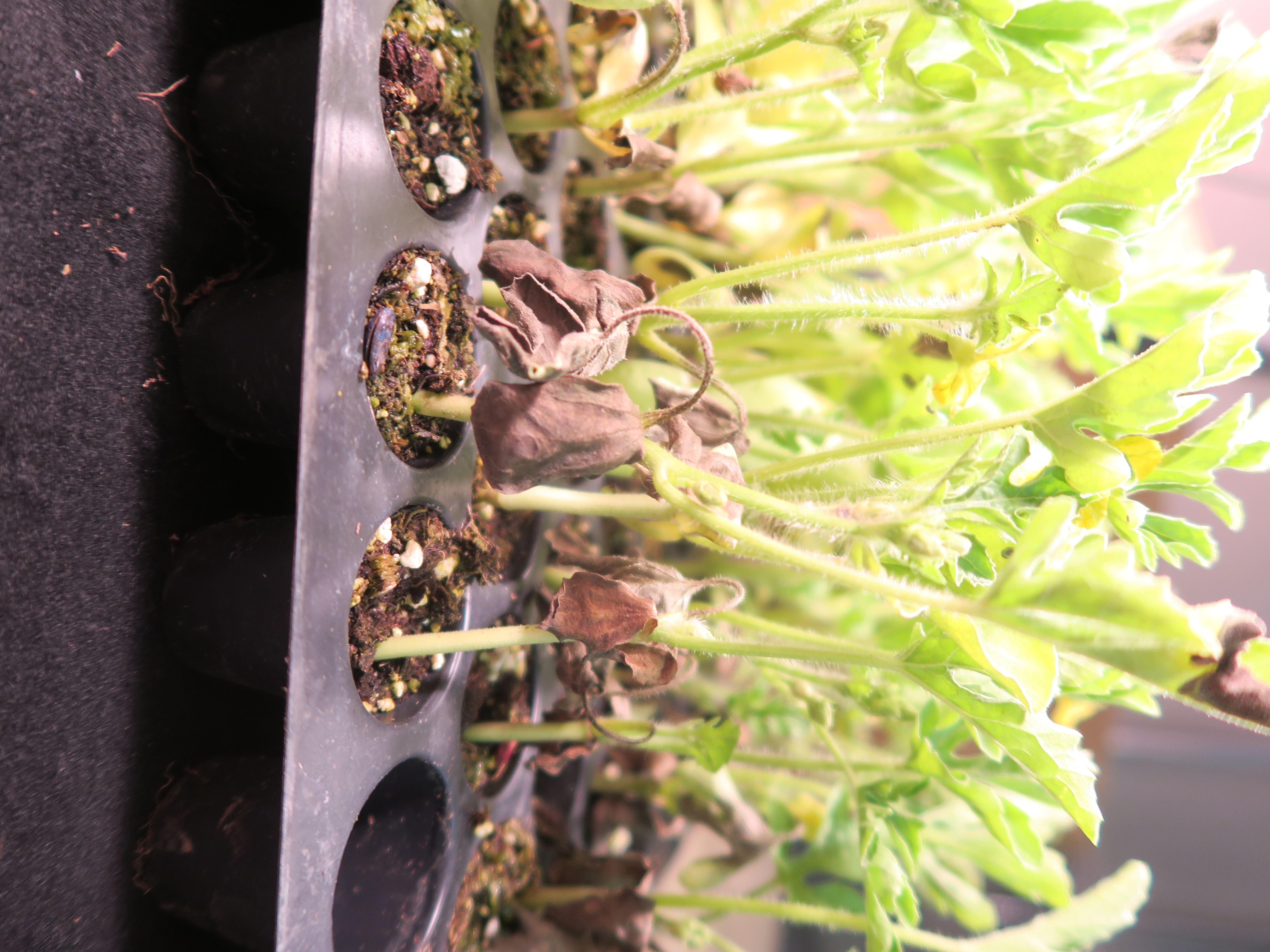 Figure 5. The watermelon seedlings in this transplant tray are declining due to black root rot.
 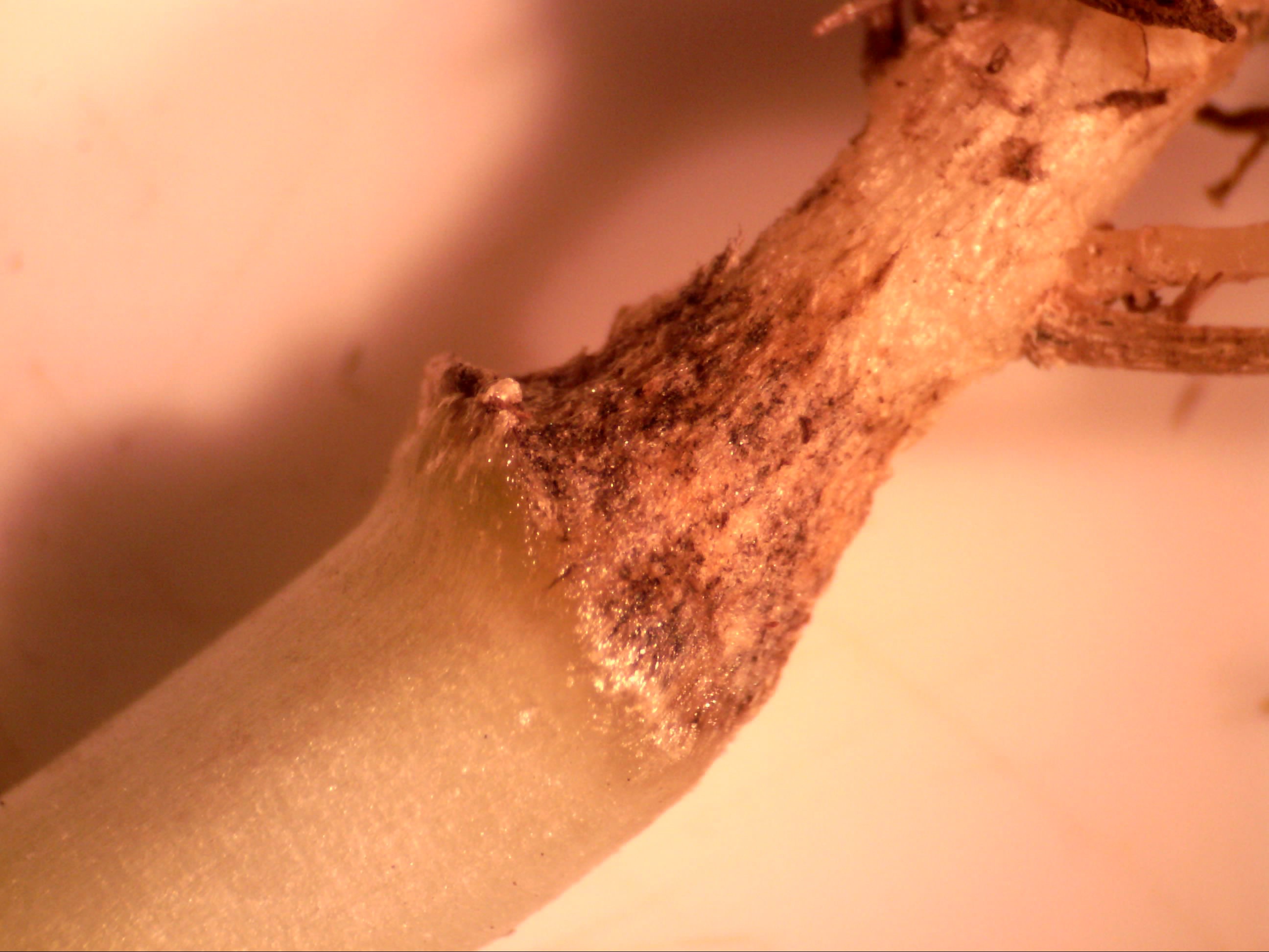 Figure 6. The watermelon transplants in the tray above are declining due black root rot which can be seen causing dark symptoms here on the hypocotyl.
 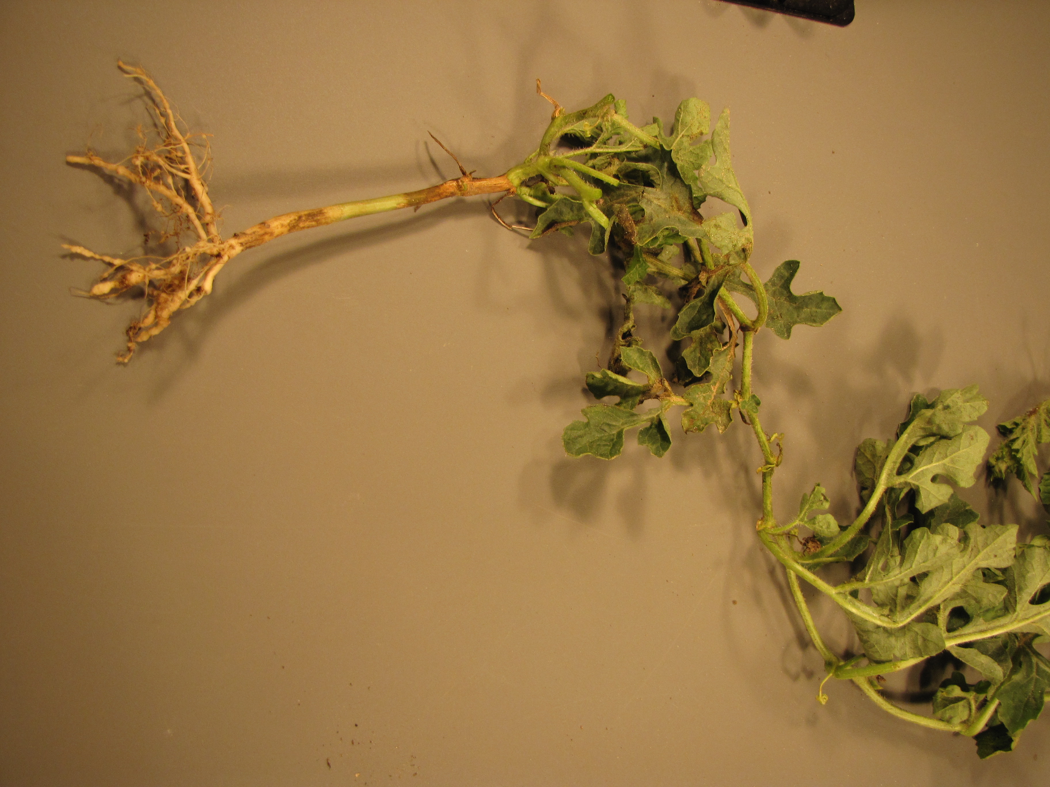 Figure 7. This watermelon is affected by gummy stem blight immediately below and above the cotyledon, black root rot at the lower hypocotyl and root knot nematodes on the roots.
 Cercospora spot
Cercospora leaf spot-Not a common or economically significant disease. Lesions are light brown and mostly round, with dark brown margins.
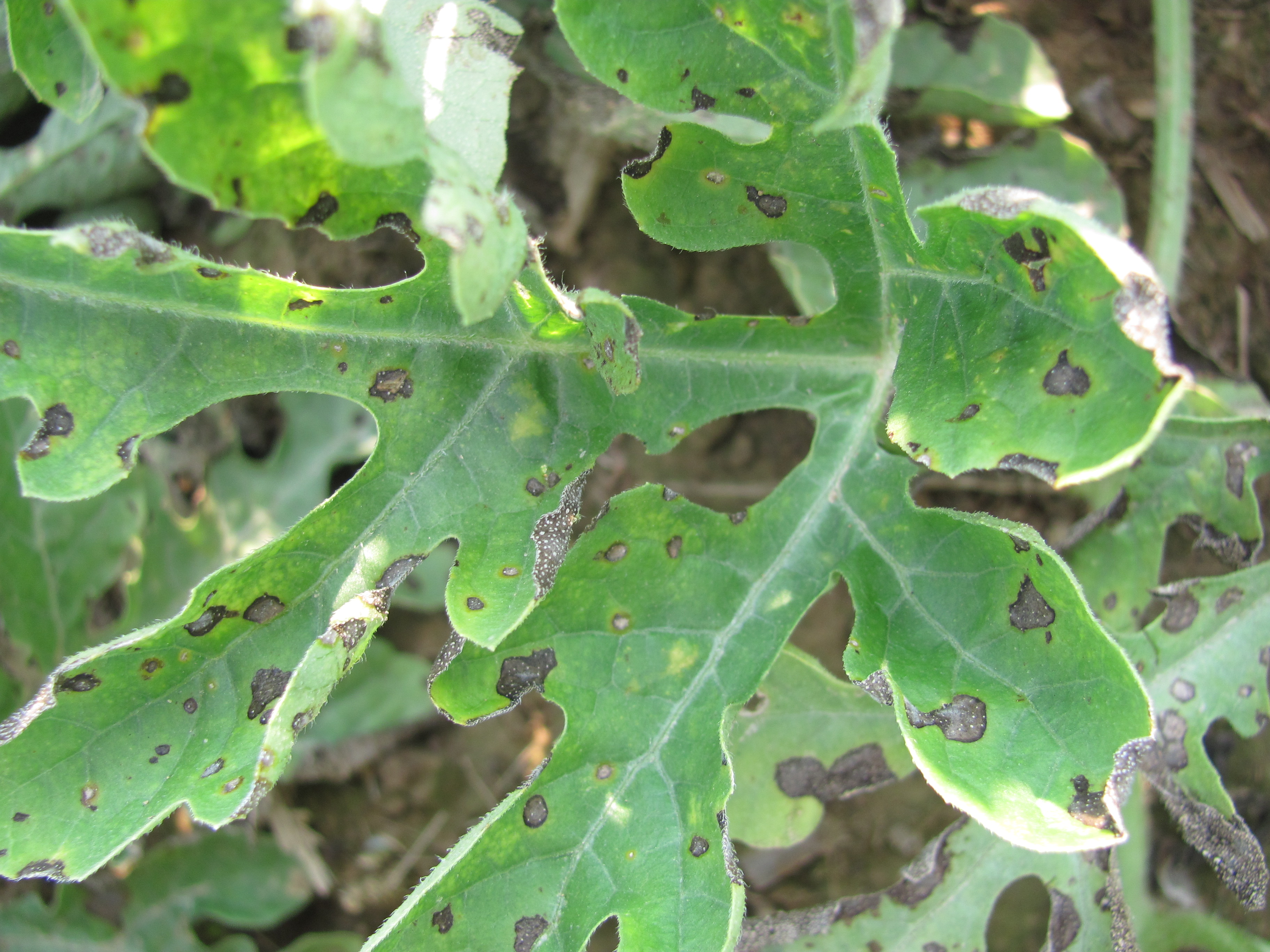 Figure 1. Cercospora leaf spot on watermelon.
 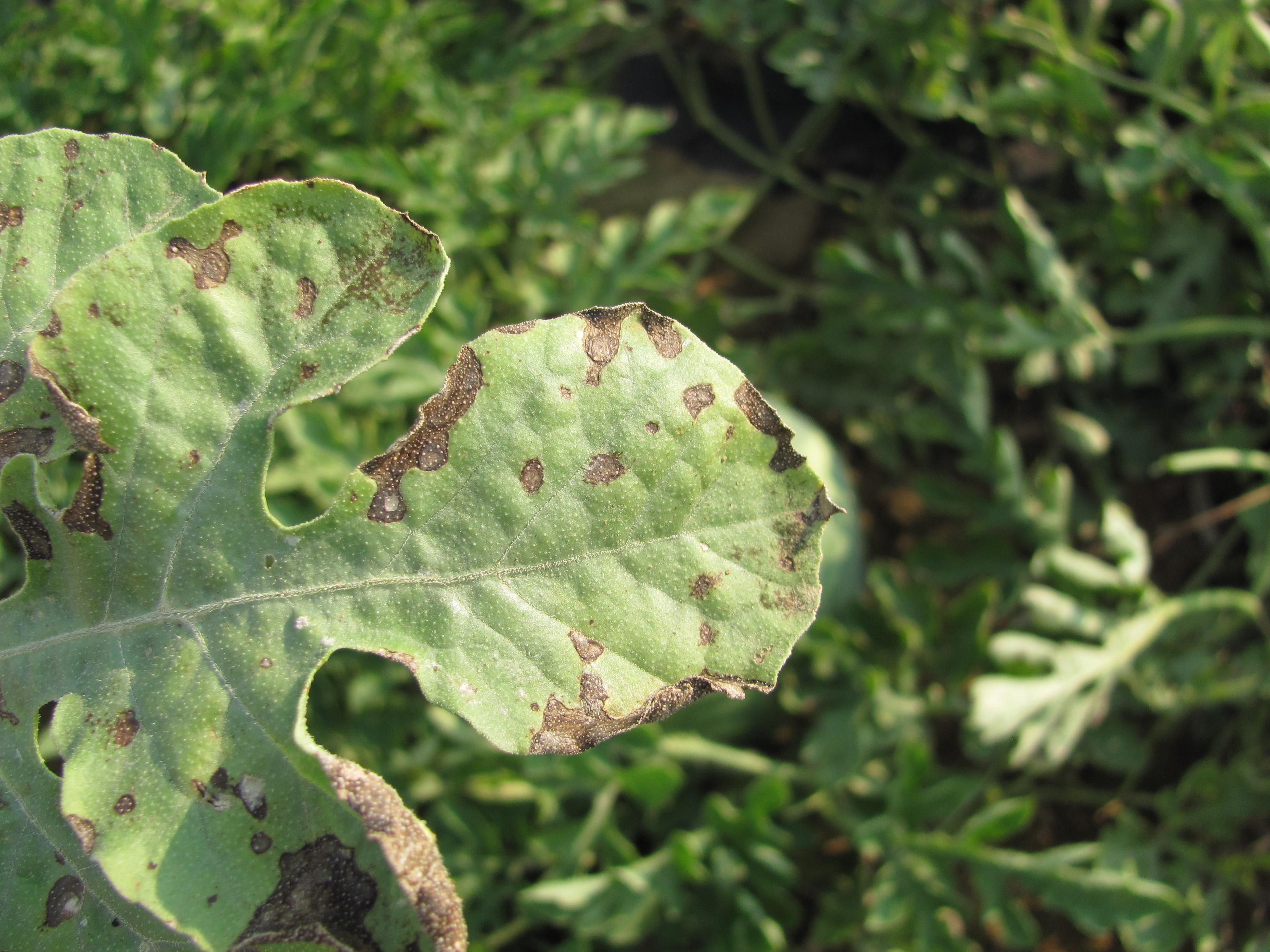 Figure 2. Closeup of Cercospora leaf spot on watermelon.
 Chimera
Chimera-also known as a somatic mutation, affected leaves often lack pigment in mottled patterns. Since the mutation occurs in portions of the plant after germination, the patterns may affect only a portion of the plant. This is not an infectious problem and rarely causes any economic loss. Somatic mutations also occur in other crops.
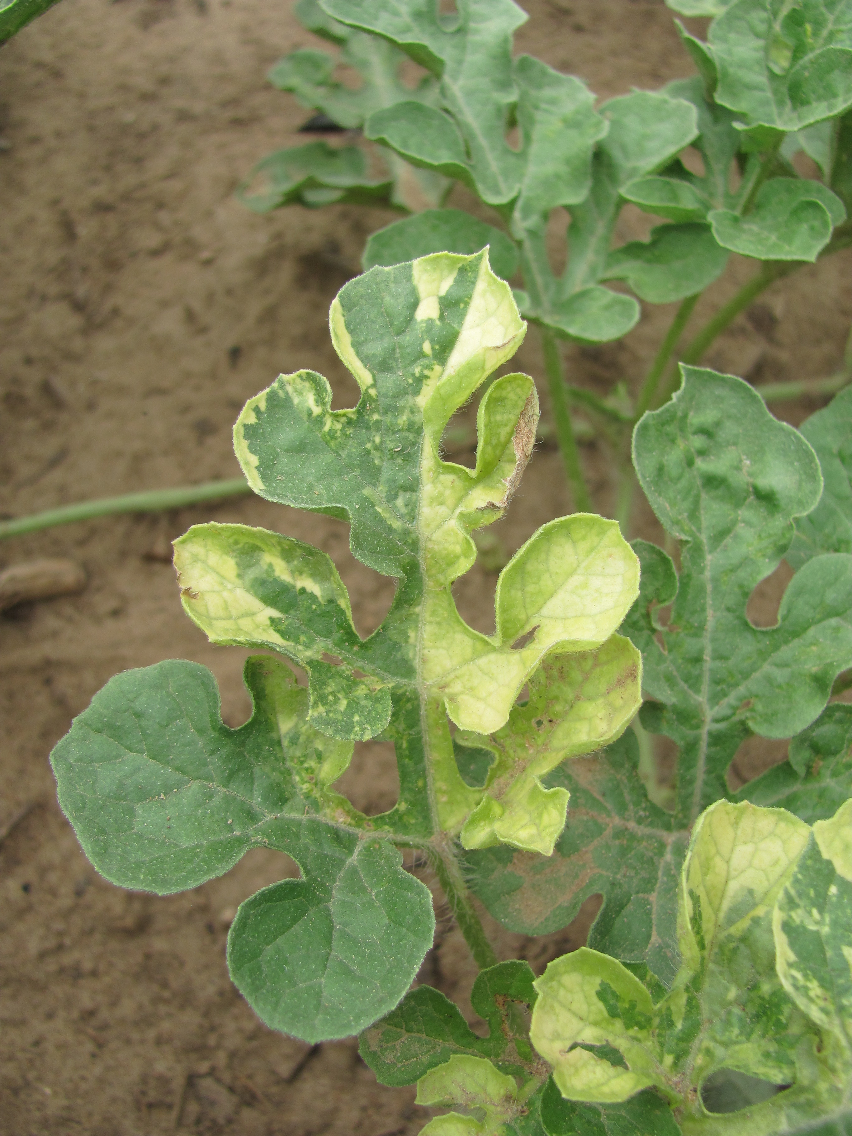 Figure 1. Watermelon with irregular color patterns due to a chimera, also known as a somatic mutation.
 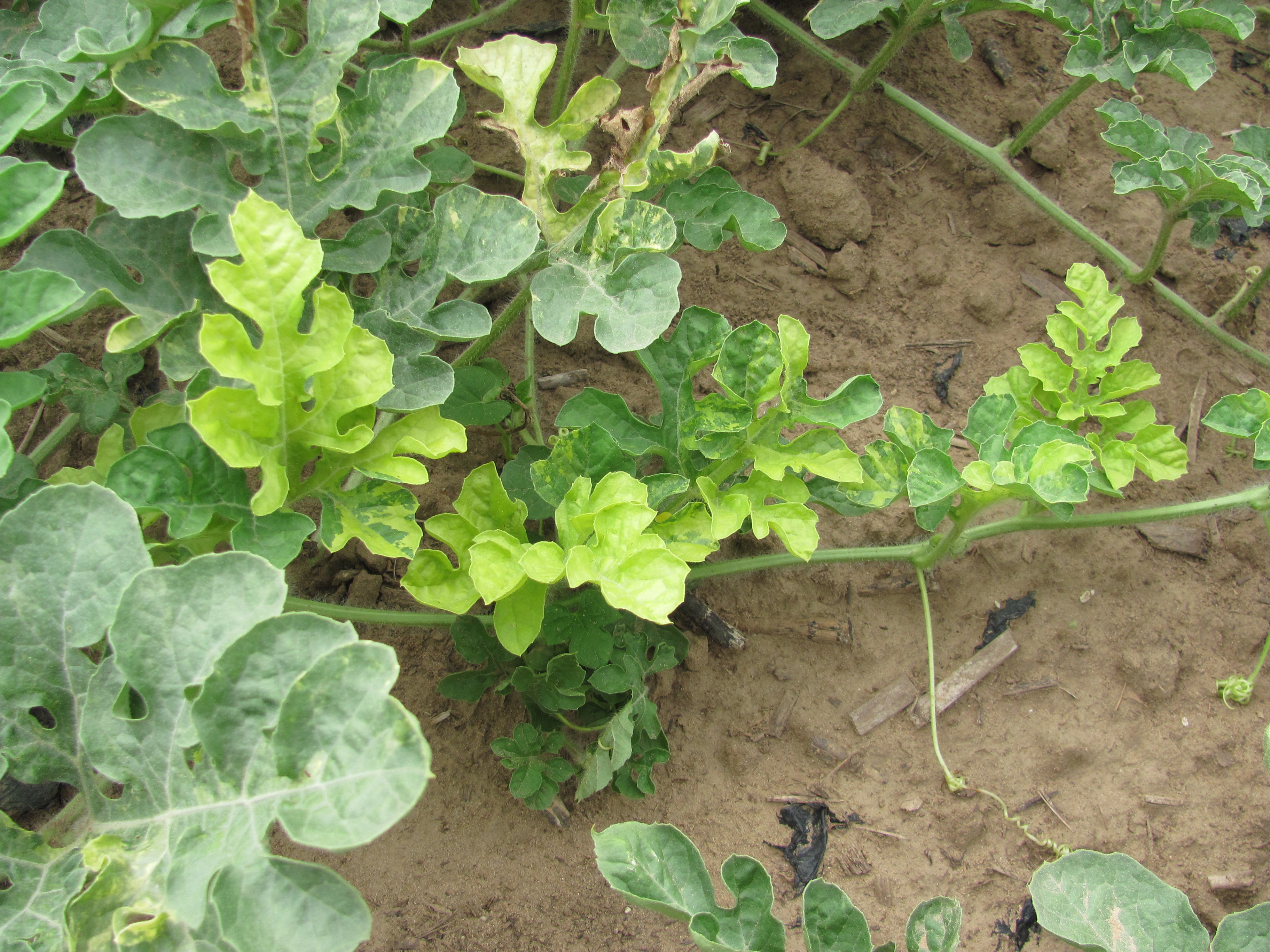 Figure 2. Chimera has caused irregular patterns on a watermelon leaves.
 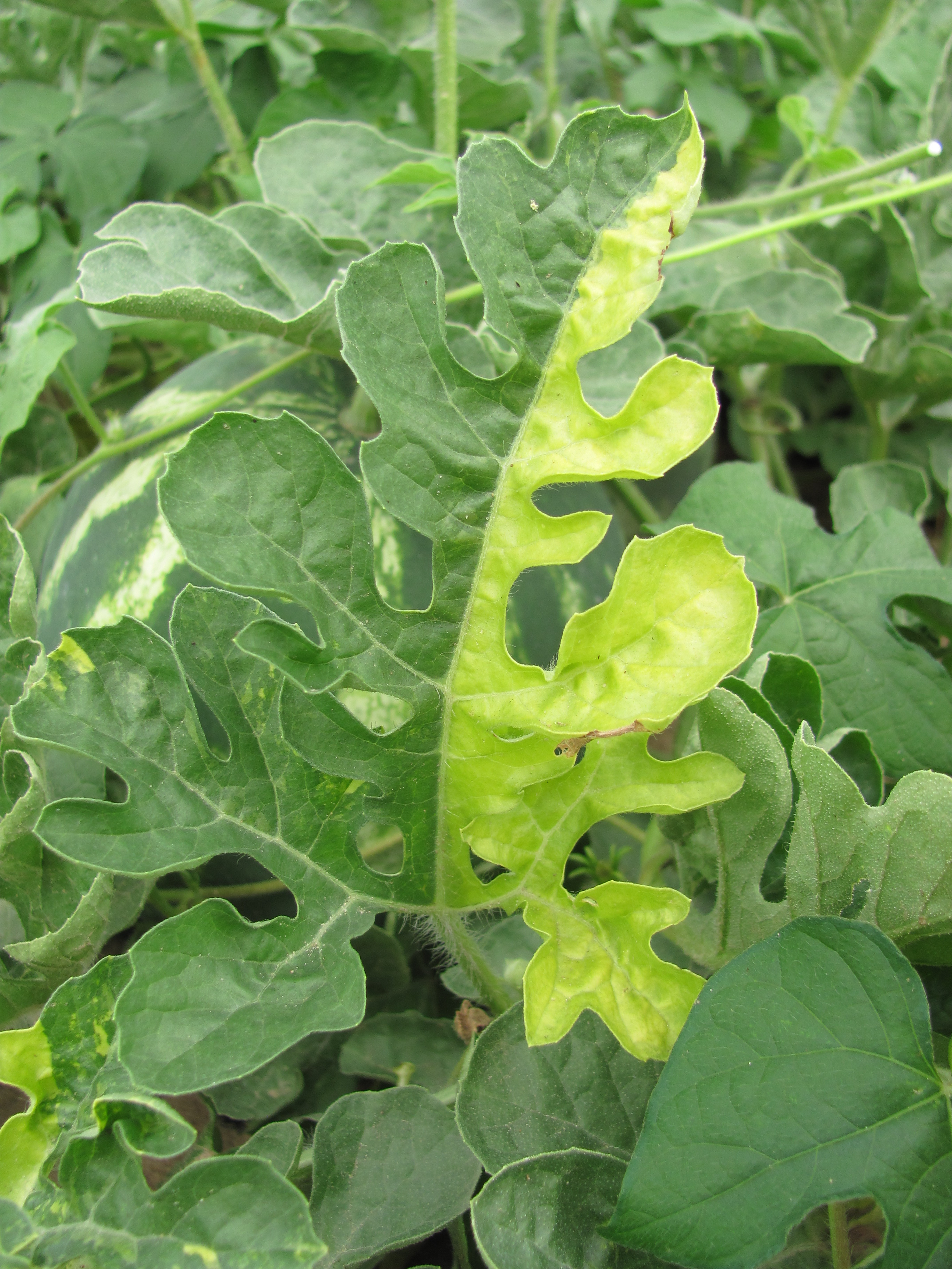 Figure 3. A chimera in a watermelon leaf.
 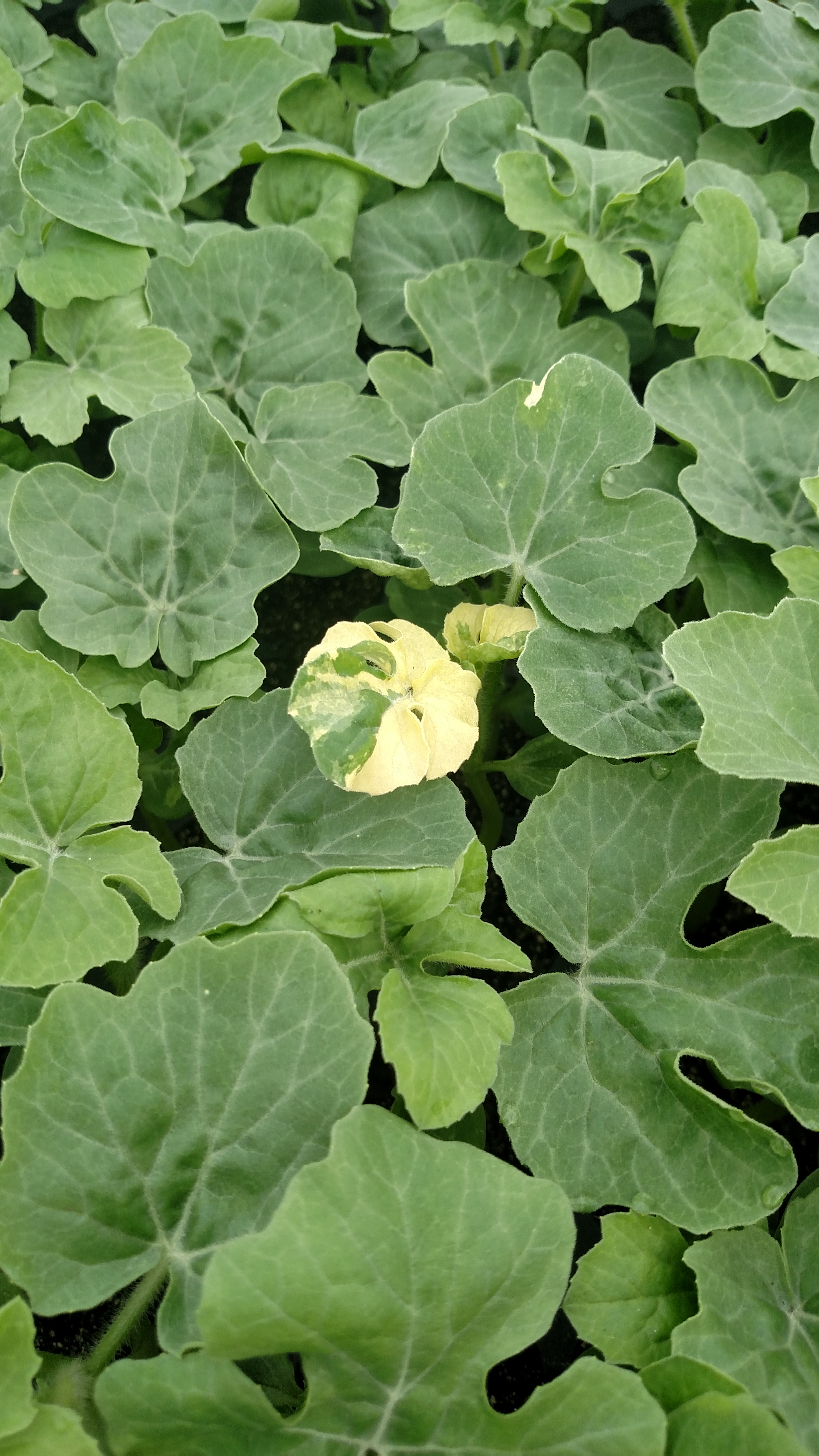 Figure 4. Chimera on a watermelon transplant.
 Cross-stitch
Cross stitch-This is an unexplained disorder. It is characterized by crack-like lesions that run perpendicular to the sutures of watermelon. This disorder is not a common one and is usually not economically important. It is apparently non-infectious.
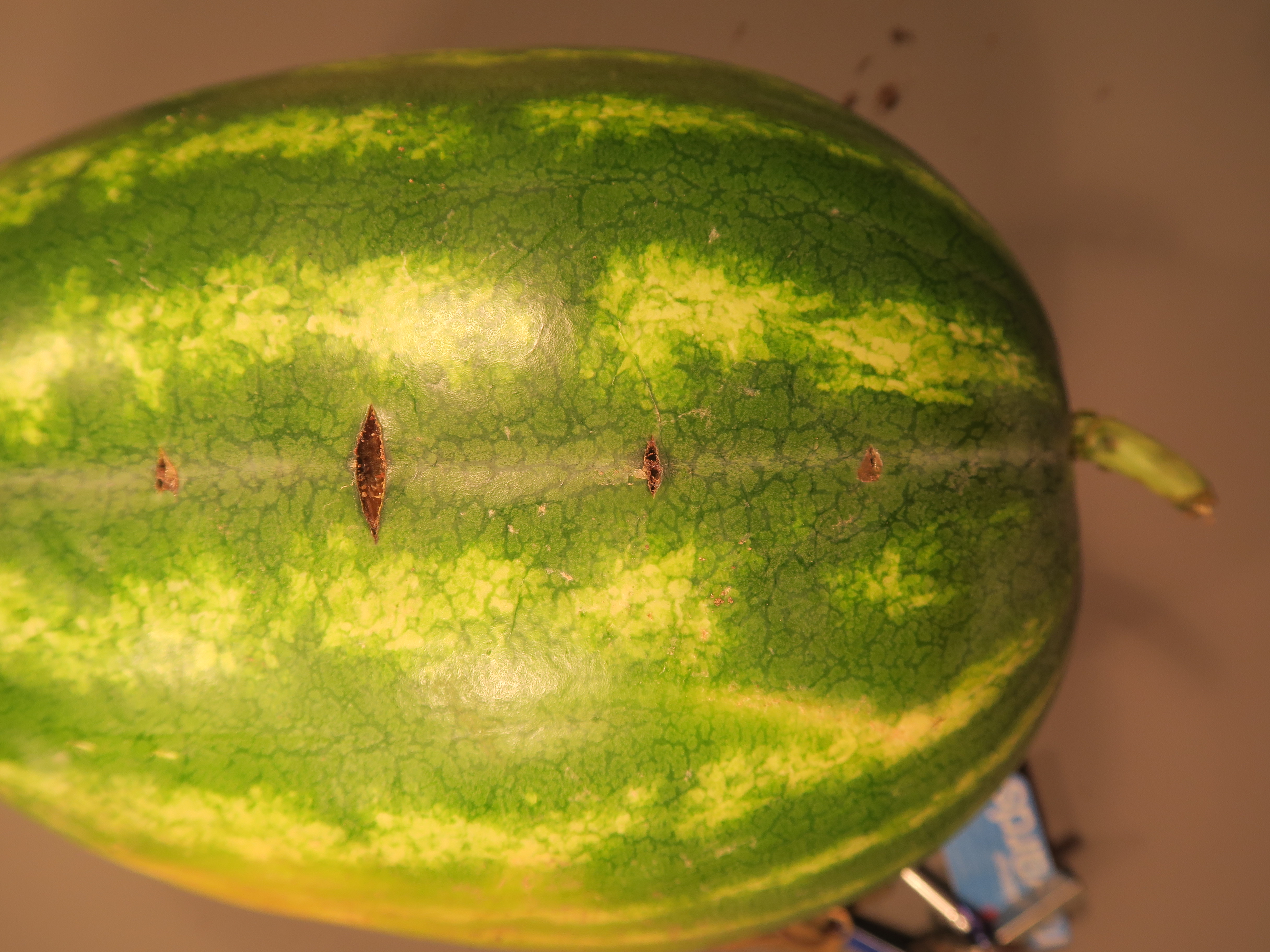 Figure 1. Cross stitch on watermelon.
 Damping-off
Damping-off of watermelon-symptoms of damping-off include the collapse and wilt of affected seedlings. A brown necrosis can often be observed at the soil level.
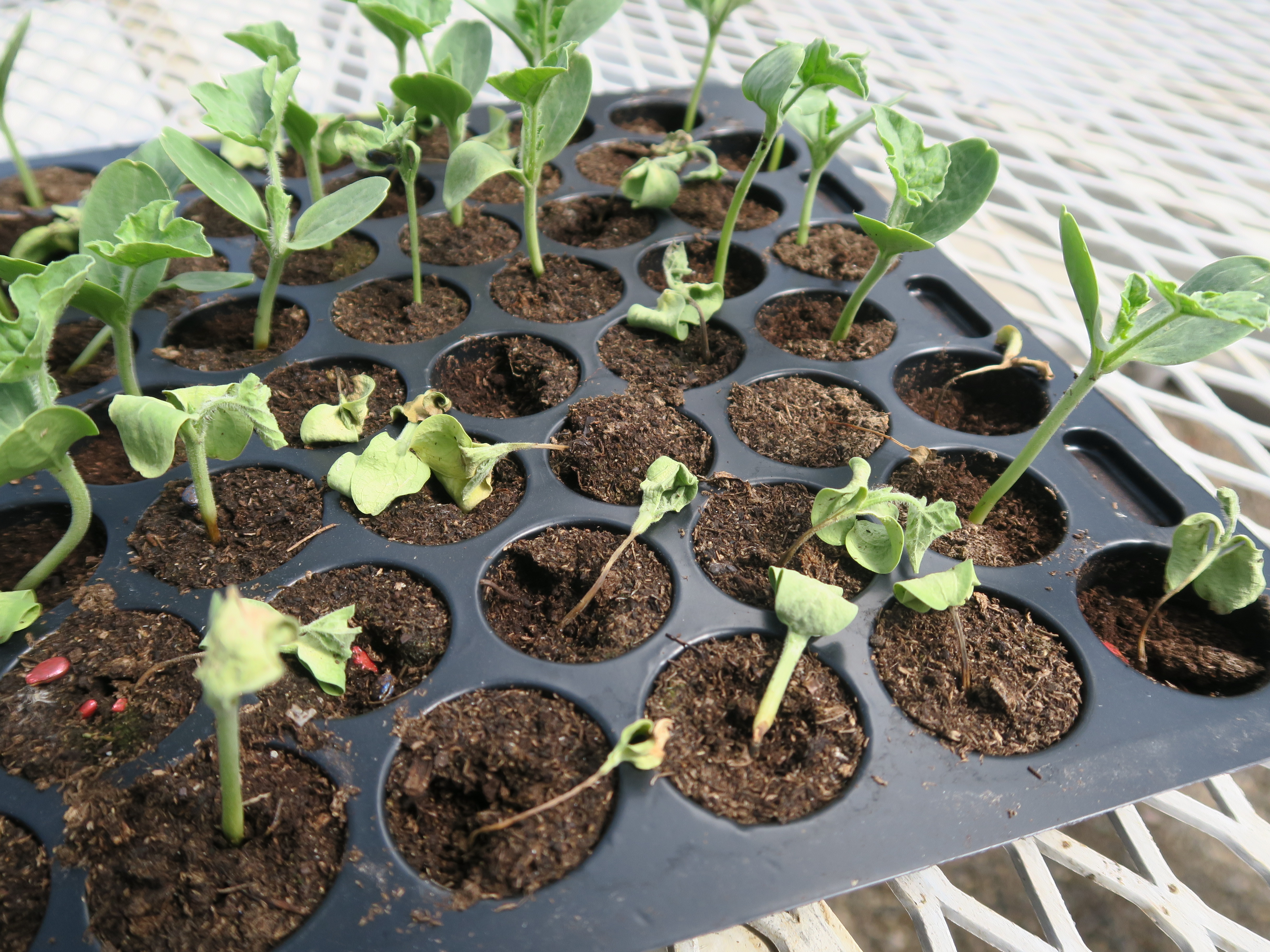 Figure 1. Damping off of watermelon.
 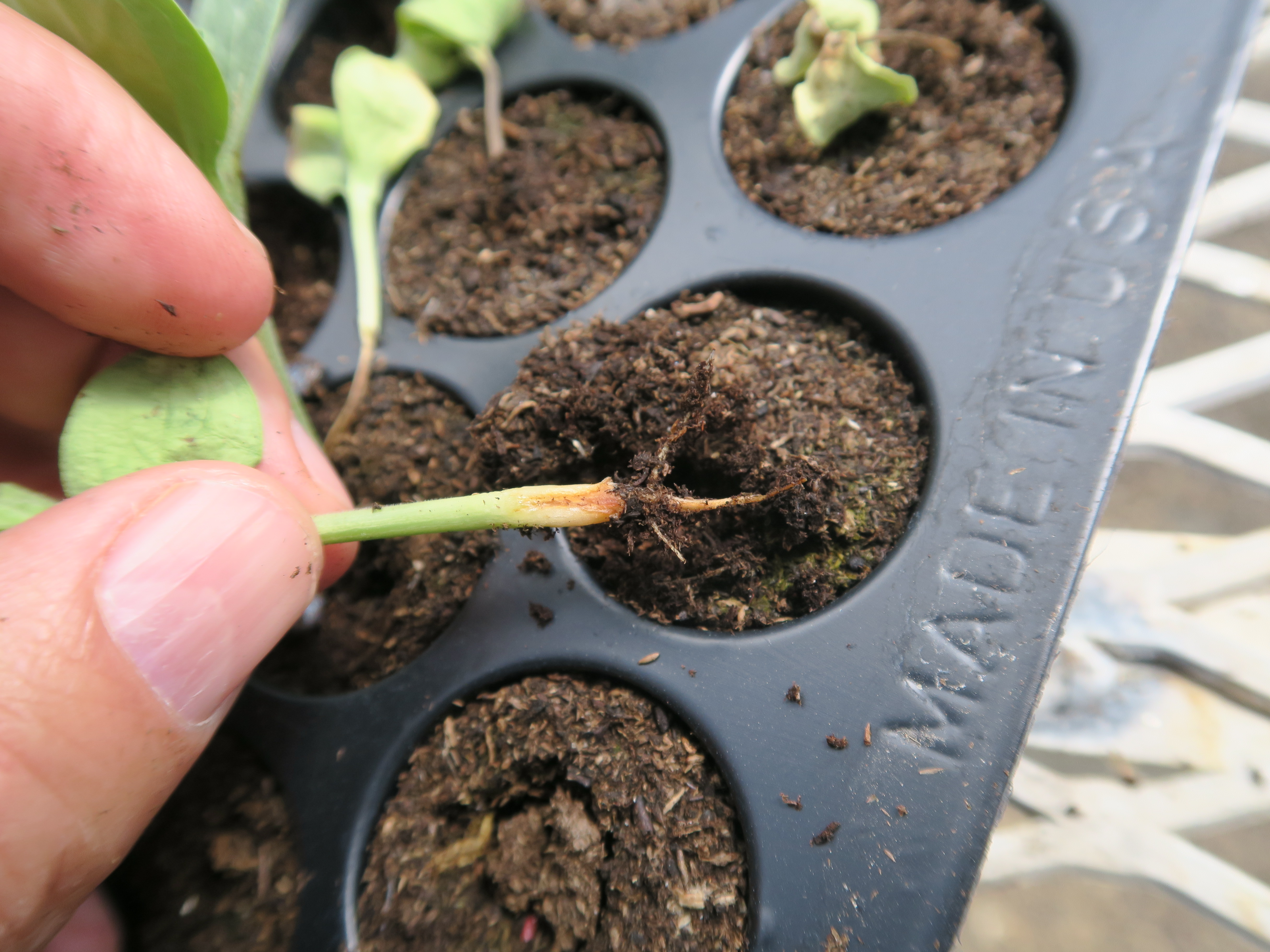 Figure 2. Damping off of watermelon.
 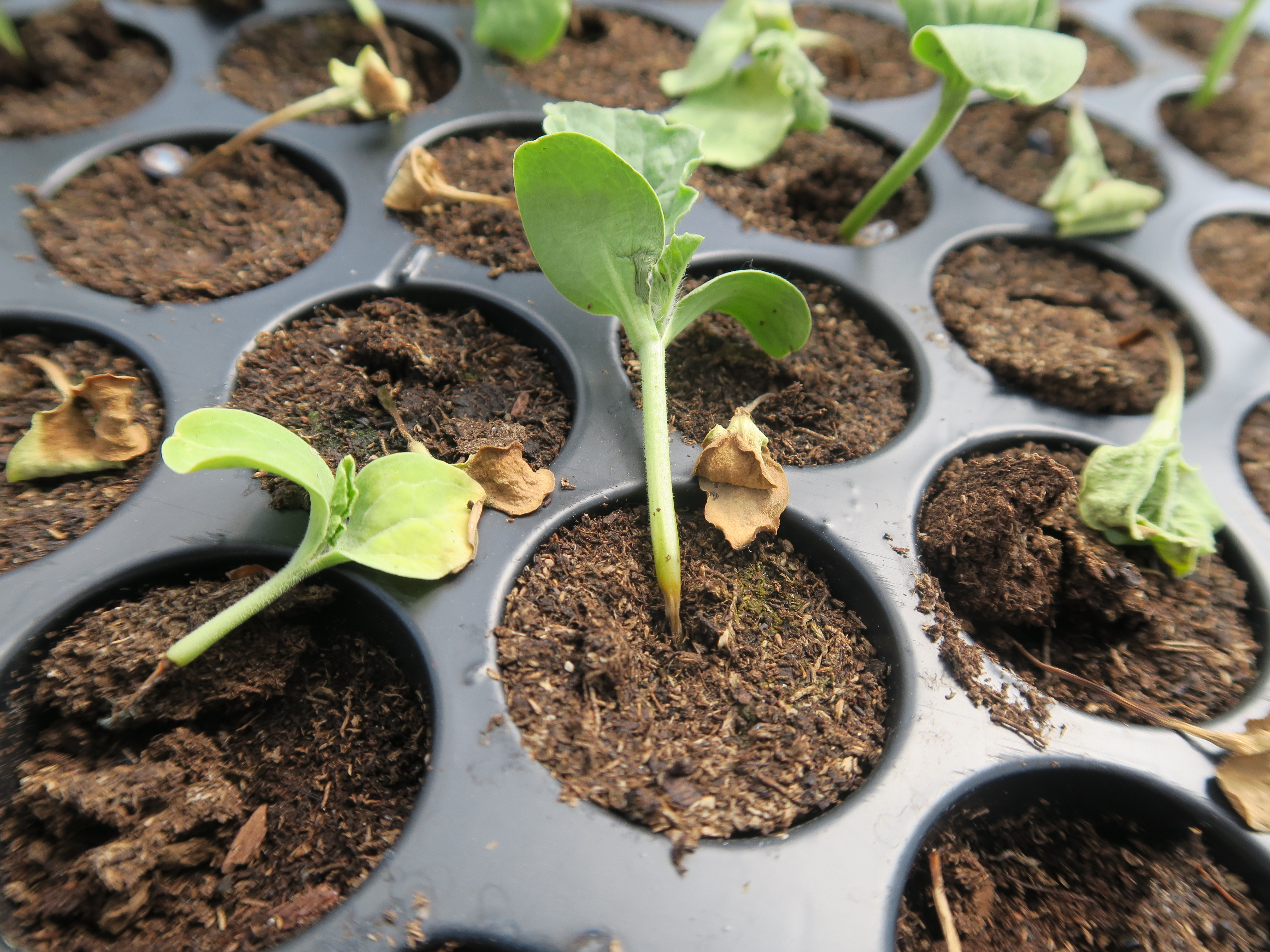 Figure 3. Damping off of watermelon.
 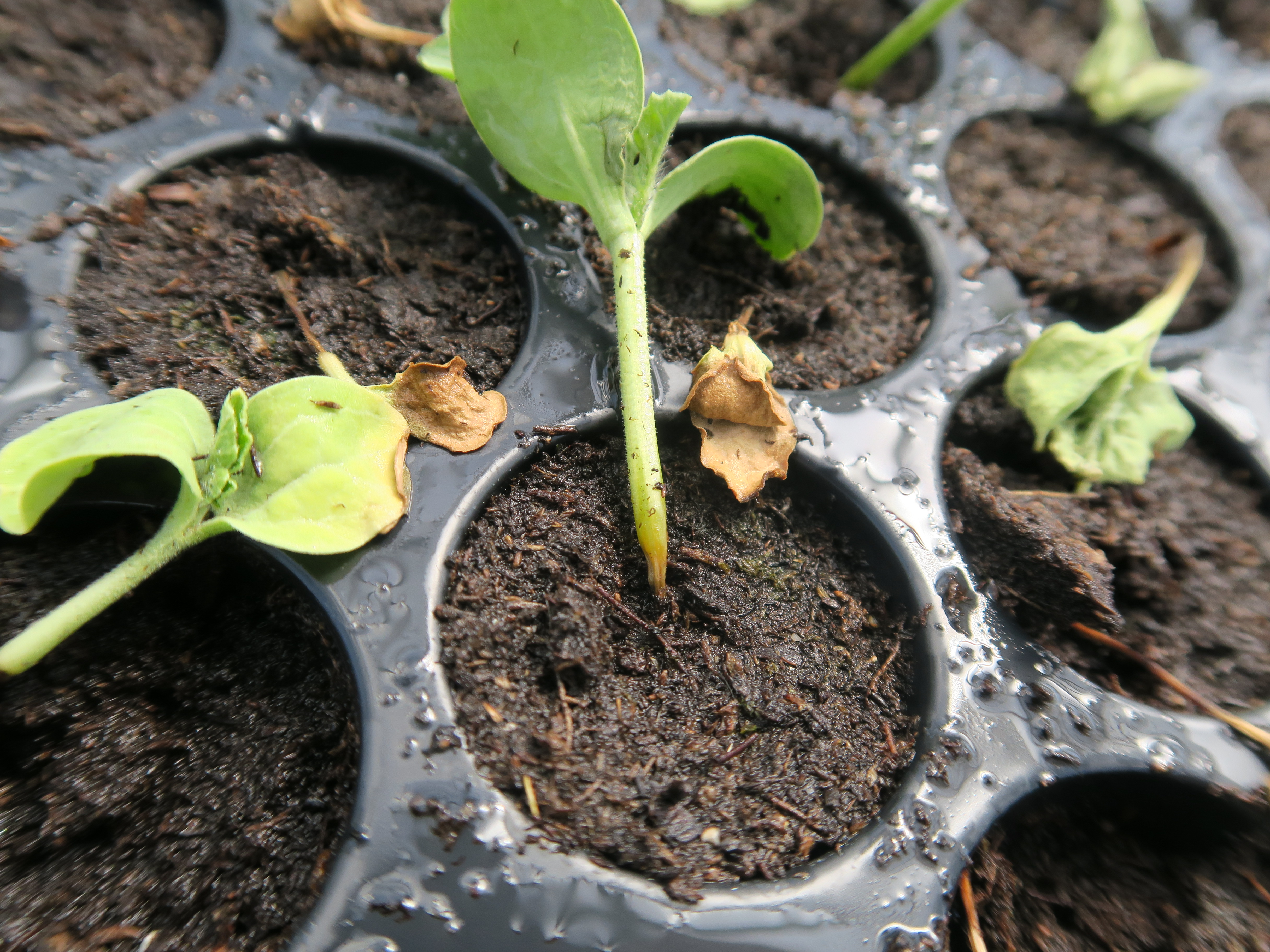 Figure 4. Damping off of watermelon.
 Dodder
Dodder-dodder is a parasitic plant that can affect watermelon as well as many other plants. The plant lacks chlorophyll and thus appears yellowish. Not common or important in Indiana.
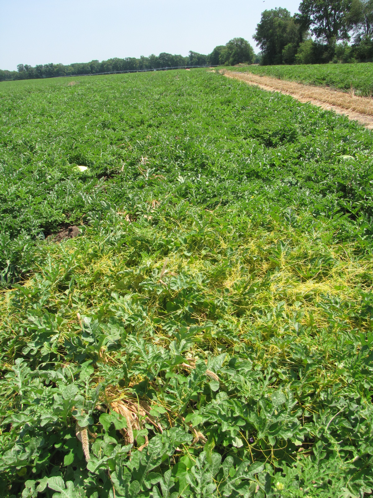 Figure 1. Dodder on watermelon.
 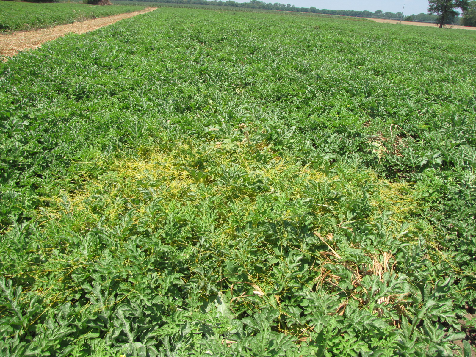 Figure 2. Dodder on watermelon.
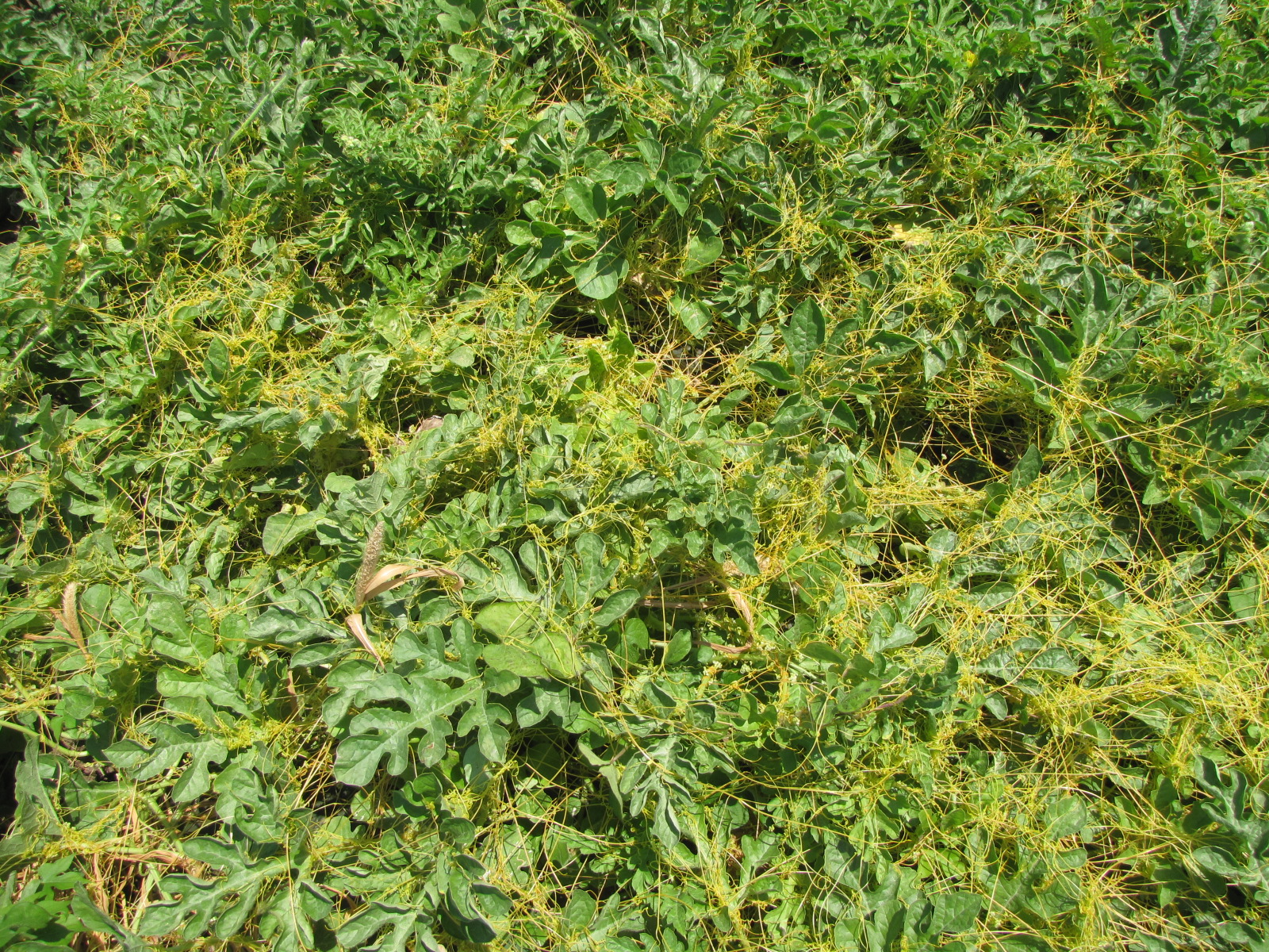 Figure 3. Dodder on watermelon.
 Downy mildew
Downy mildew-This disease does not occur every year in Indiana and watermelon may be out of production by the time the fungus-like-organisms arrives in Indiana. Initial symptoms on leaves are often light chlorotic lesions with diffuse margins. Lesions on the underside of leaves may have a black or purple sporulation under moist conditions. The center of lesions may turn necrotic with time. Stems and fruit are not directly affected.
 Figure 1. Chlorotic lesions of downy mildew on a watermelon leaf. Note that a few of the lesions are starting to become necrotic.
 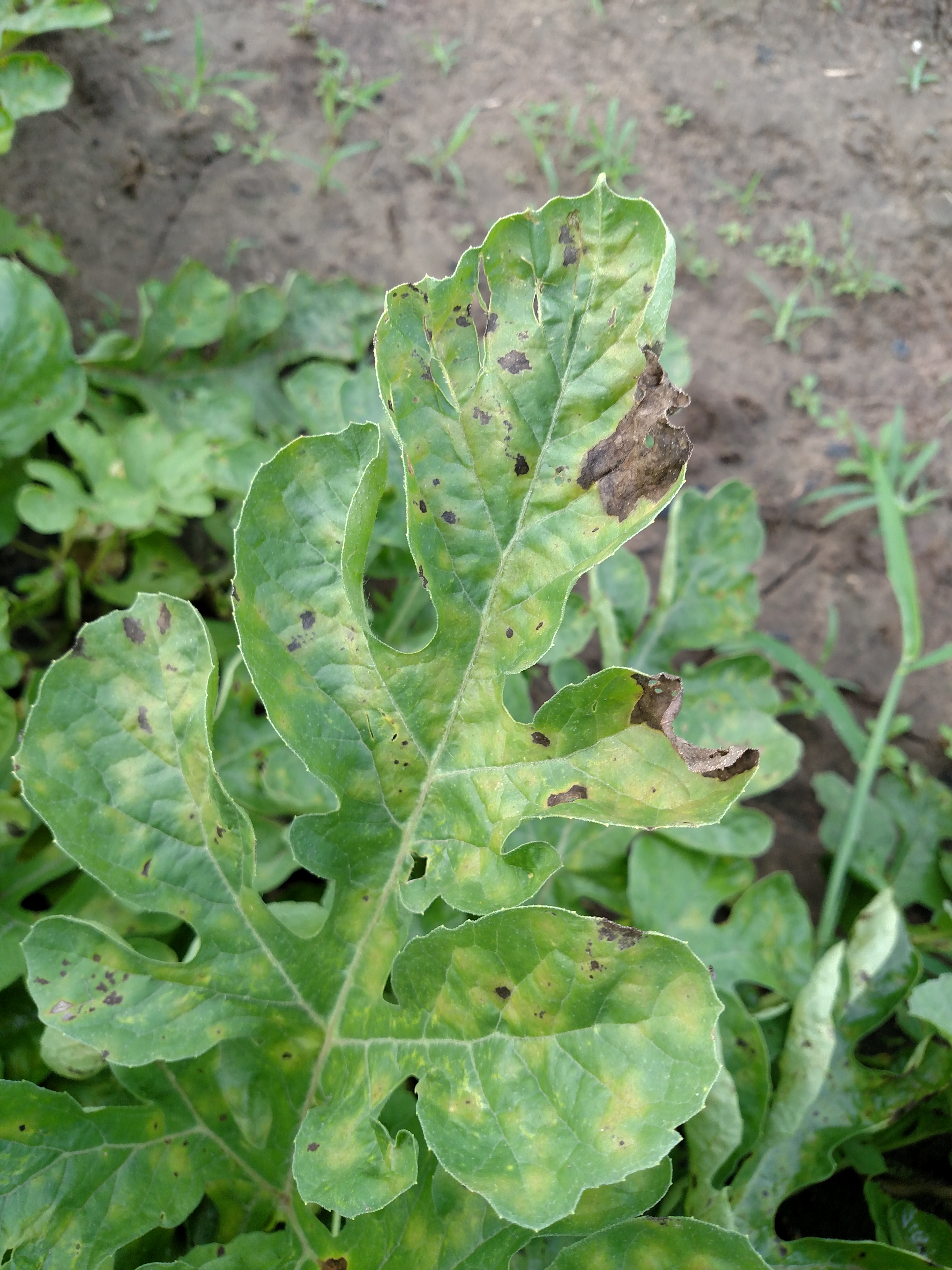 Figure 2. Chlorotic and necrotic lesions on watermelon leaf.
 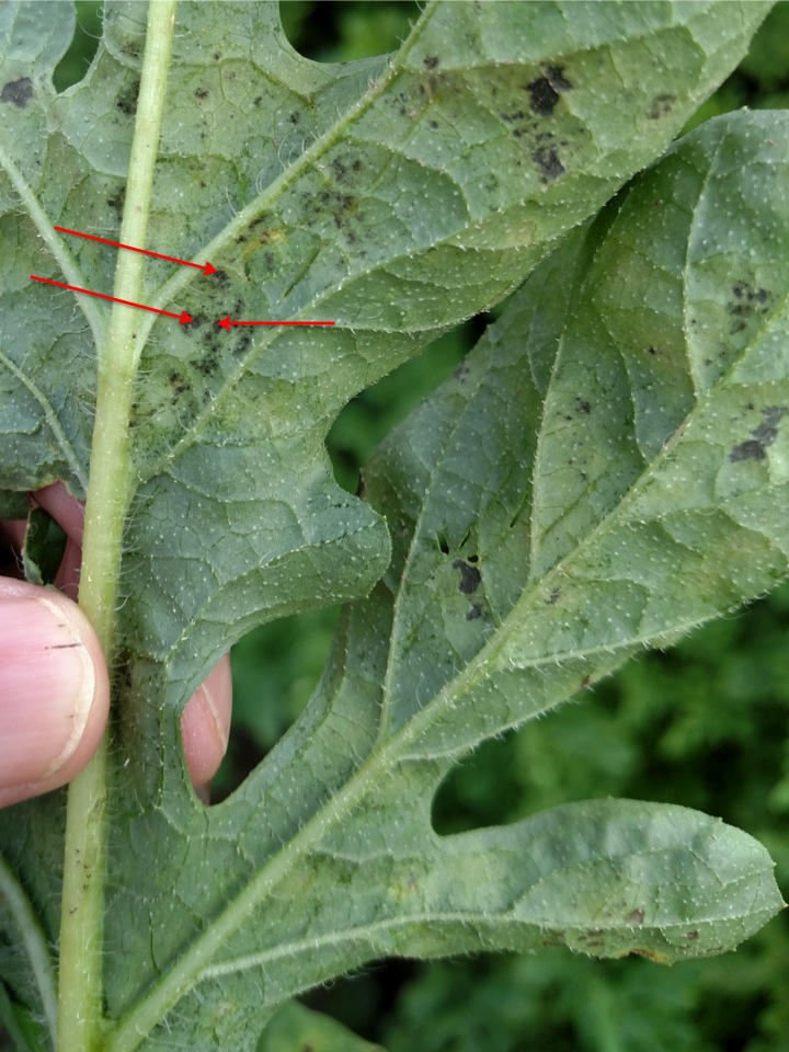 Figure 3. Underside of watermelon leaf with downy mildew. Note sporulation of downy mildew-see red arrows.
  Figure 4. Severe outbreak of downy mildew on watermelon. Note that stem and fruit of watermelon are not directly affected.
 Fusarium wilt
Fusarium wilt-An important disease and limiting factor in watermelon production in Indiana. Often the initial symptom that is observed is a one-sided wilt. Similarly, a portion of the vascular system may exhibit discoloration. Symptomatic plants are often clustered in the field.
 Figure 1. Fusarium wilt of watermelon often causes one vine to wilt while the rest of the plant appears unaffected. Symptoms of this disease often begin when plants are just starting to vine.
 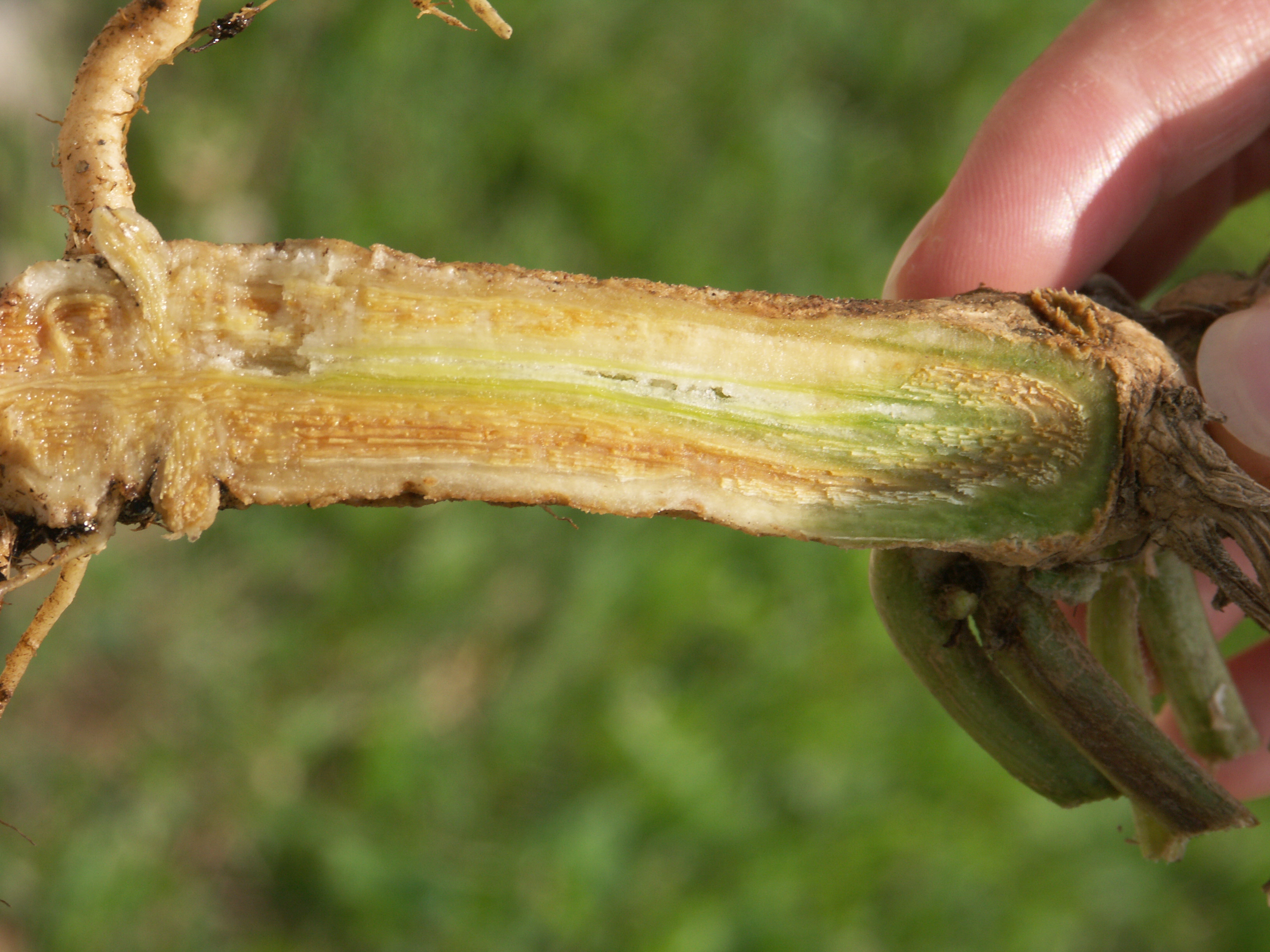 Figure 2. Vascular discoloration present in lower stem may be a symptom of Fusarium wilt of watermelon. Note that one-sided vascular discoloration in the stem may correspond to one-sided wilt in plant.
 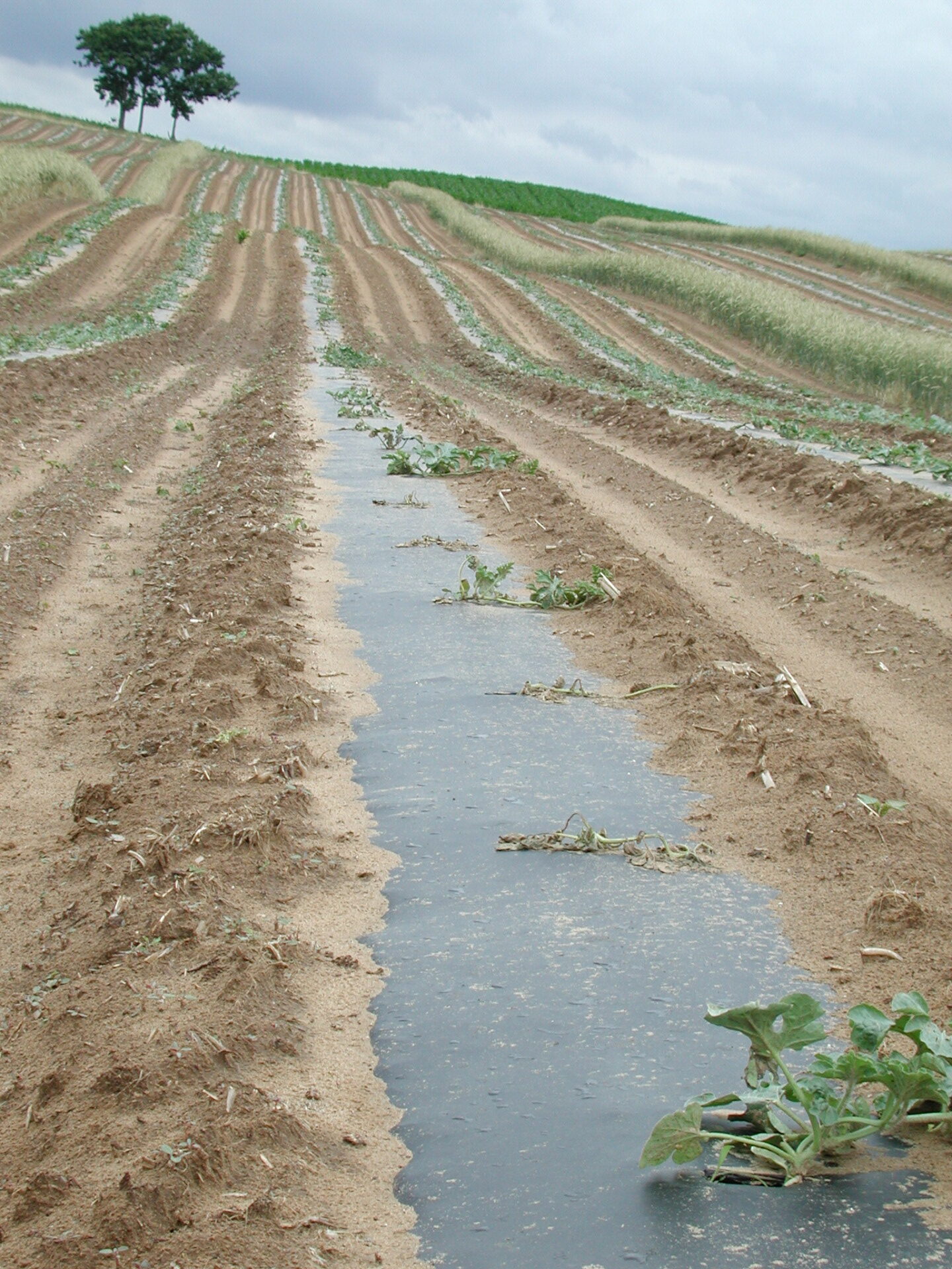 Figure 3. The distribution of Fusarium wilt of watermelon in the field is often clustered.
 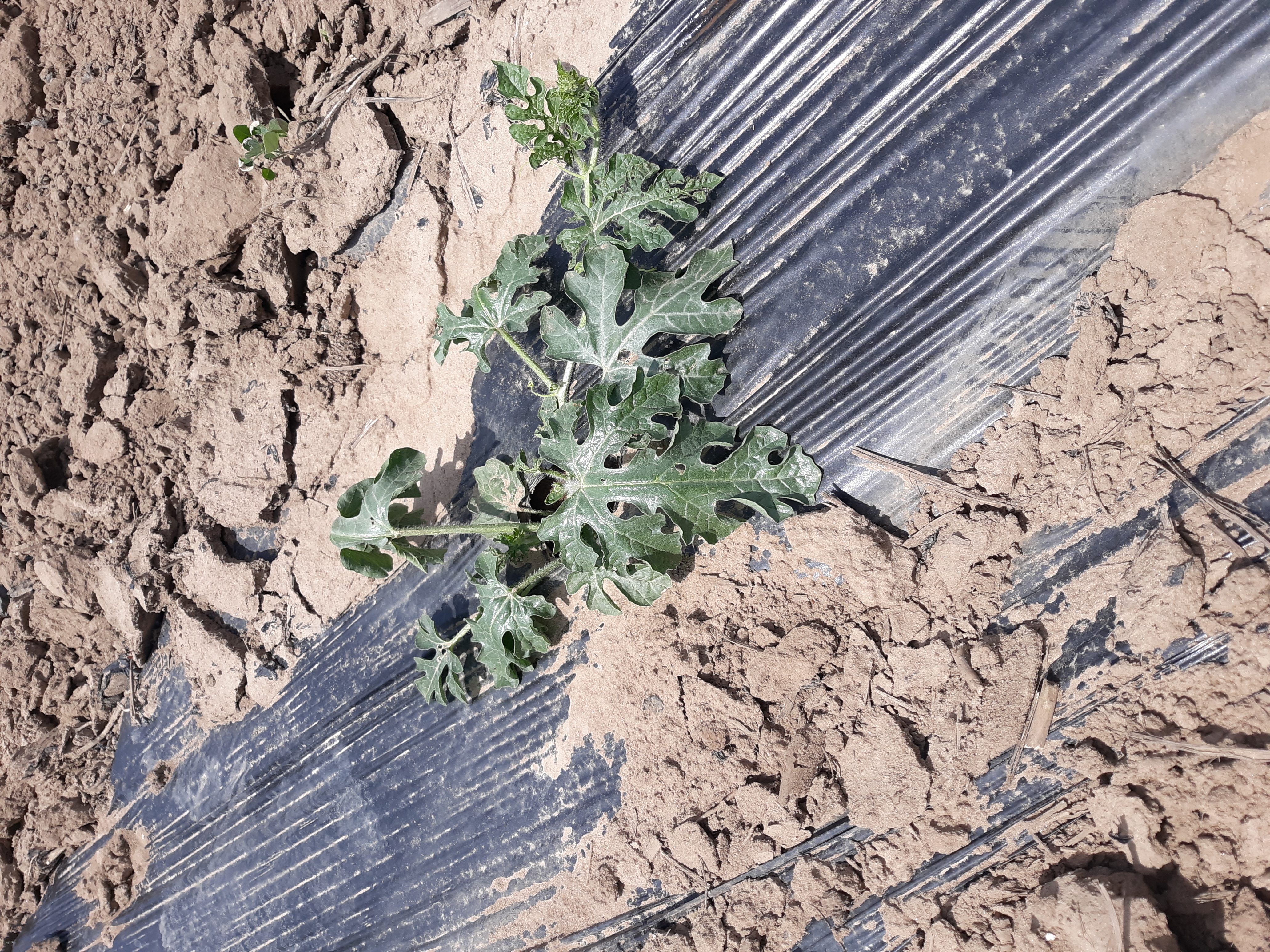 Figure 4. Early symptoms of Fusarium wilt in watermelon include wilt of almost all leaves. Note coloration of wilted leaves.
 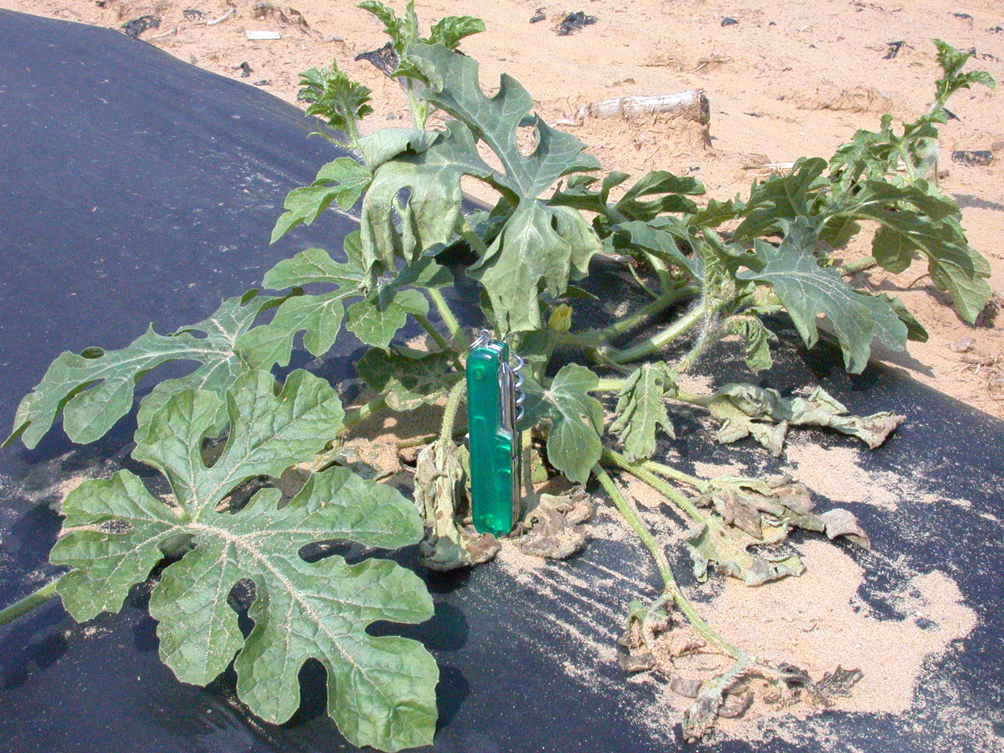 Figure 5. One sided wilt of watermelon leaf. Note also that older leaves usually wilt before young leaves.
 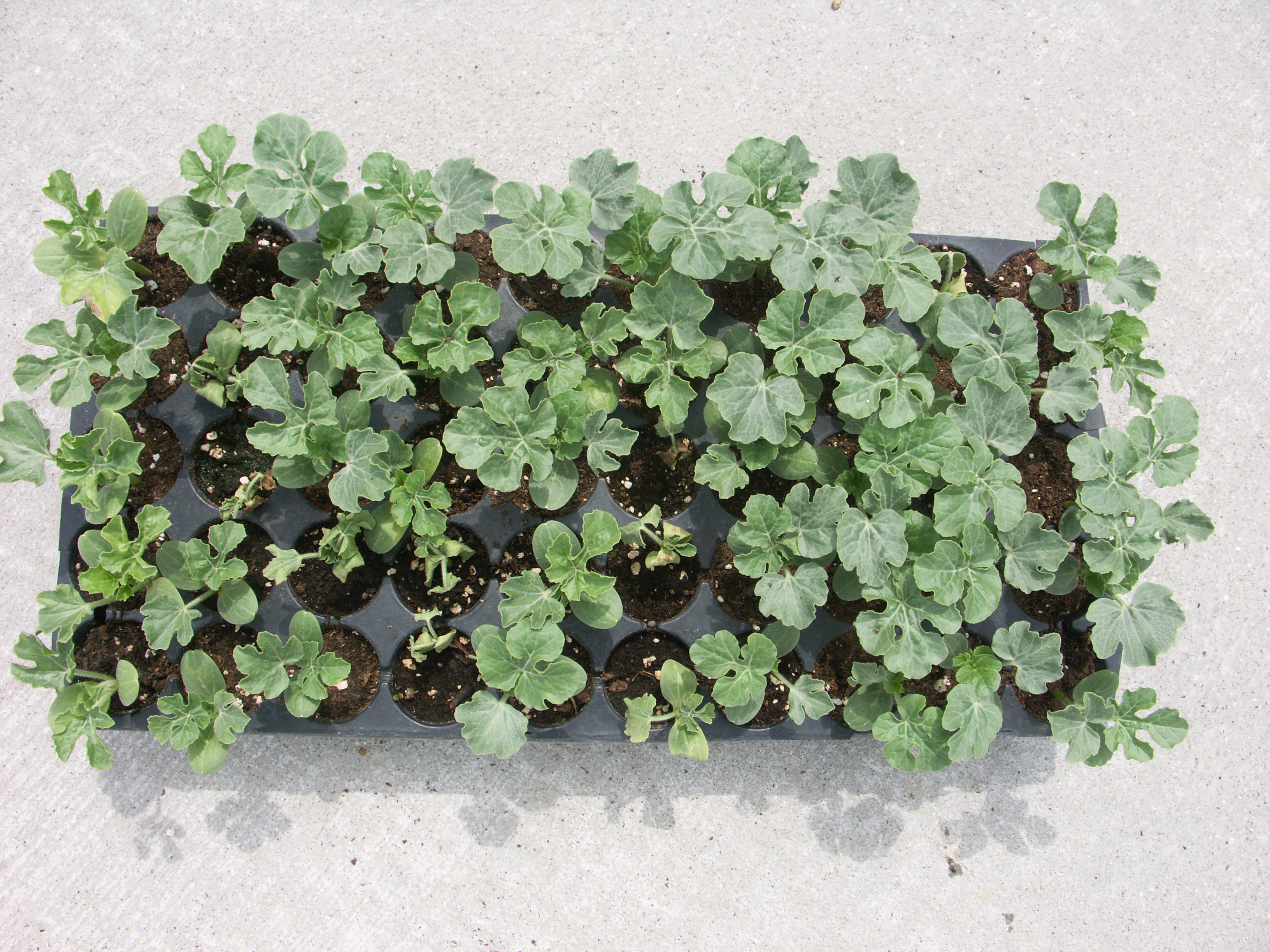 Figure 6. Seedling distribution of Fusarium wilt in watermelon may be randomly distributed in transplant trays.
 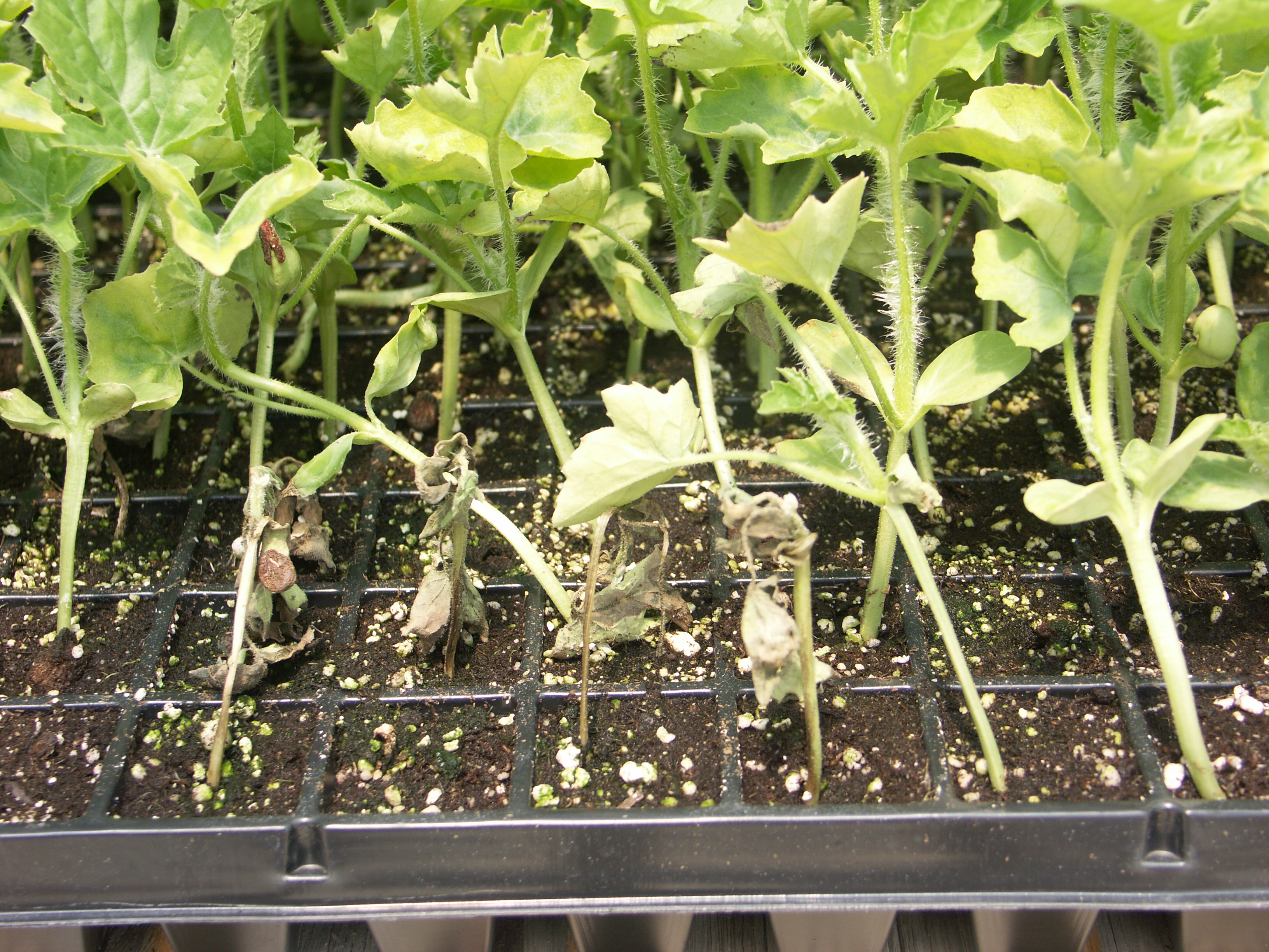 Figure 7. Distribution of Fusarium wilt of watermelon in transplant trays may be clustered under some circumstances.
 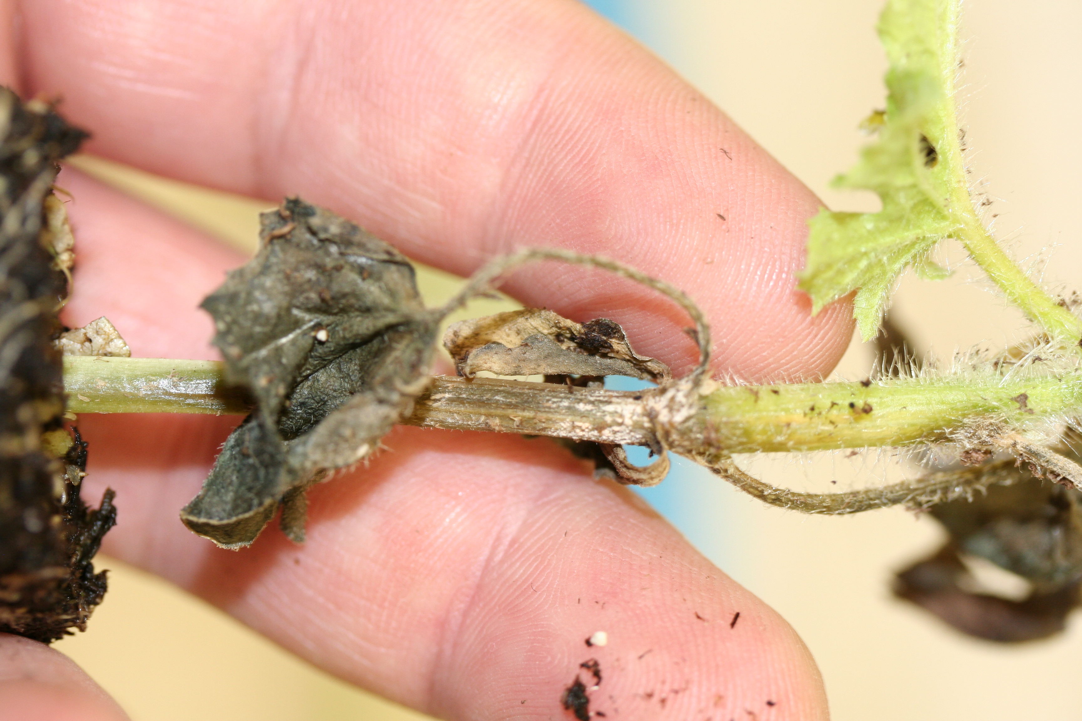 Figure 8. Hypocotyl has collapsed and become necrotic due to Fusarium wilt in watermelon at the transplant stage. Note pink-like sporulation of Fusarium fungus at top of hypocotyl.
 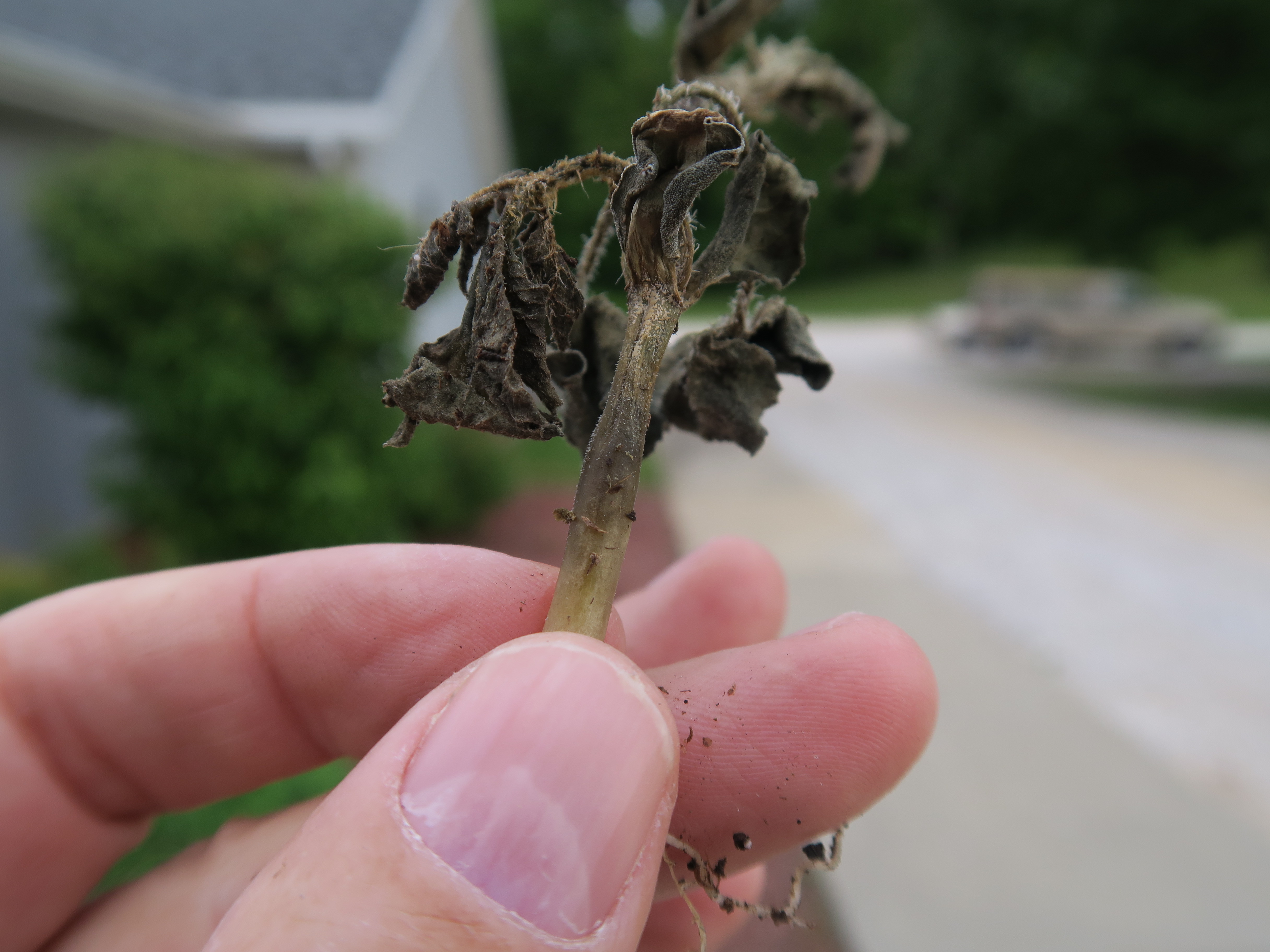 Figure 9. Close-up of hypocotyl of watermelon seedling infected with Fusarium wilt.
 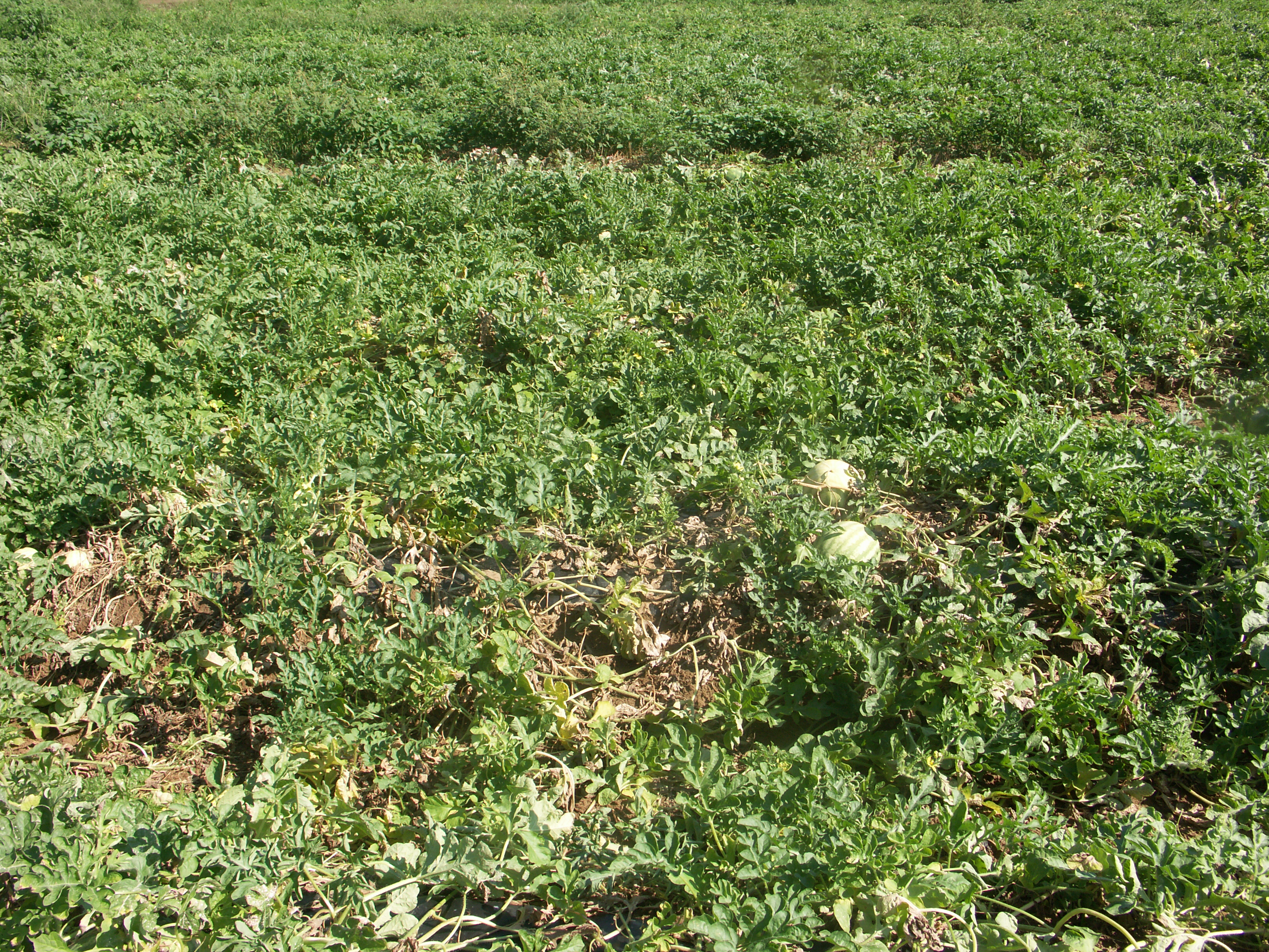 Figure 10. When Fusarium wilt in watermelon occurs in late season, wilt and collapse of vines such as seen here in the foreground may occur.
 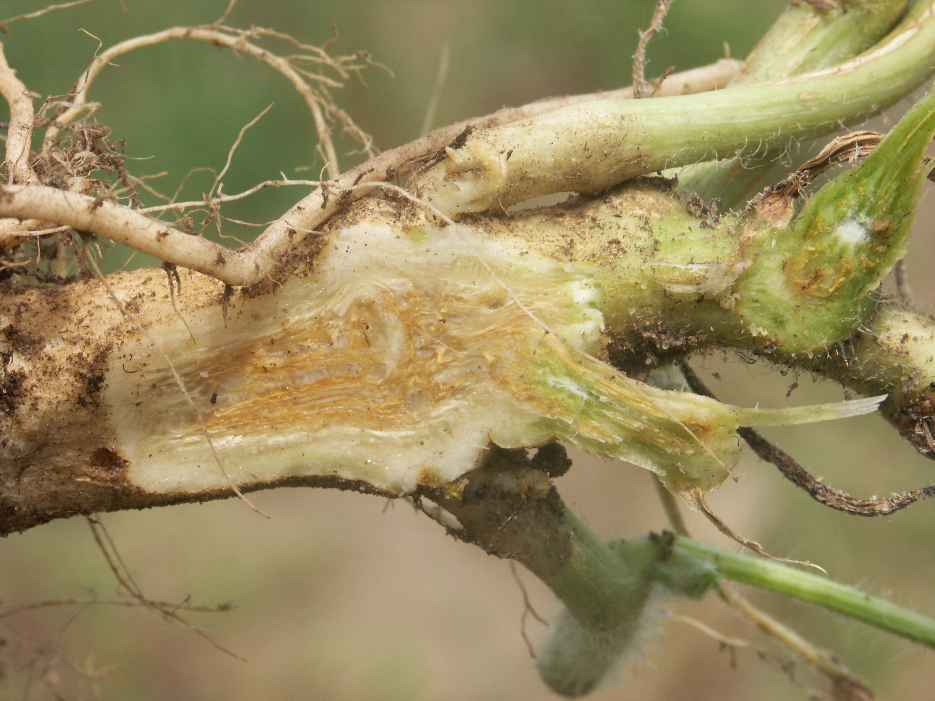 Figure 11. Vascular discoloration in Fusarium wilt of watermelon in a late season production.
 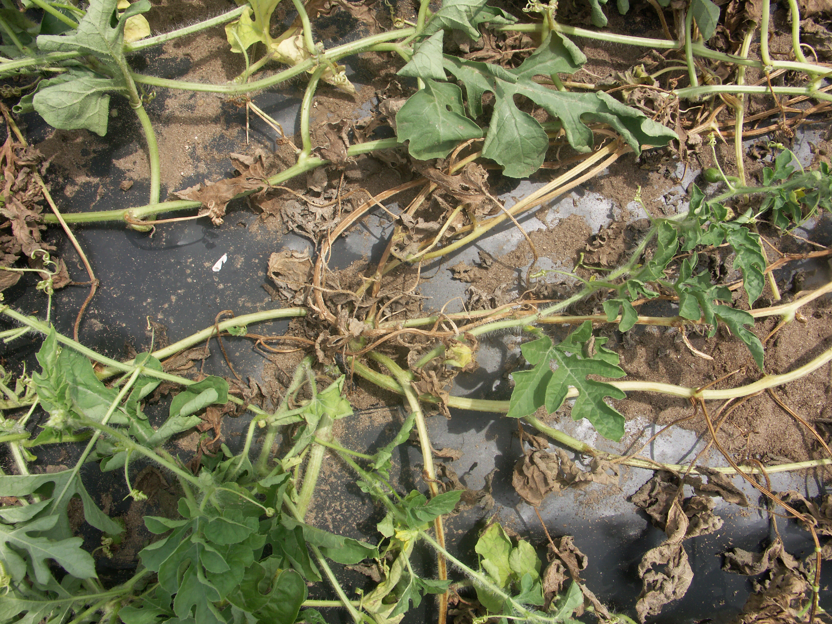 Figure 12. Fusarium wilt of watermelon in this late season field has caused some of the vines to become necrotic while some vines remain apparently healthy.
 Gummy stem blight
Gummy stem blight-One of the most common foliar diseases of watermelon in Indiana. Lesions on leaves are amorphous, dark and may appear water-soaked. Dark fungal bodies (pycnidia) may be observed in lesions on leaves or stems. I have never observed fruit infection of watermelon in Indiana.
 Figure 1. Gummy stem blight causes dark, amorphous, necrotic lesions on watermelon leaves.
 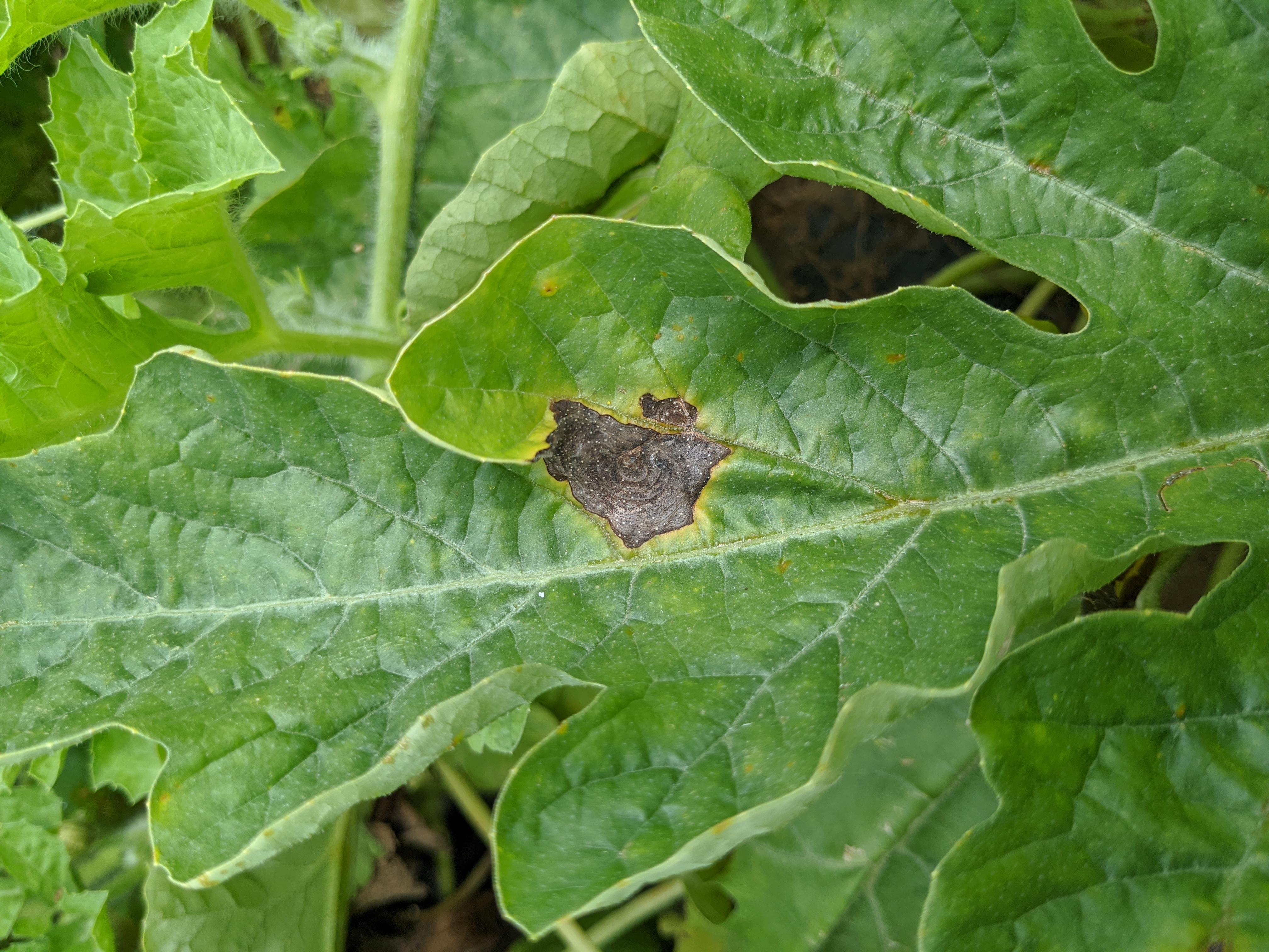 Figure 2. Gummy stem blight lesions on watermelon leaves sometimes have a ring-like structure.
 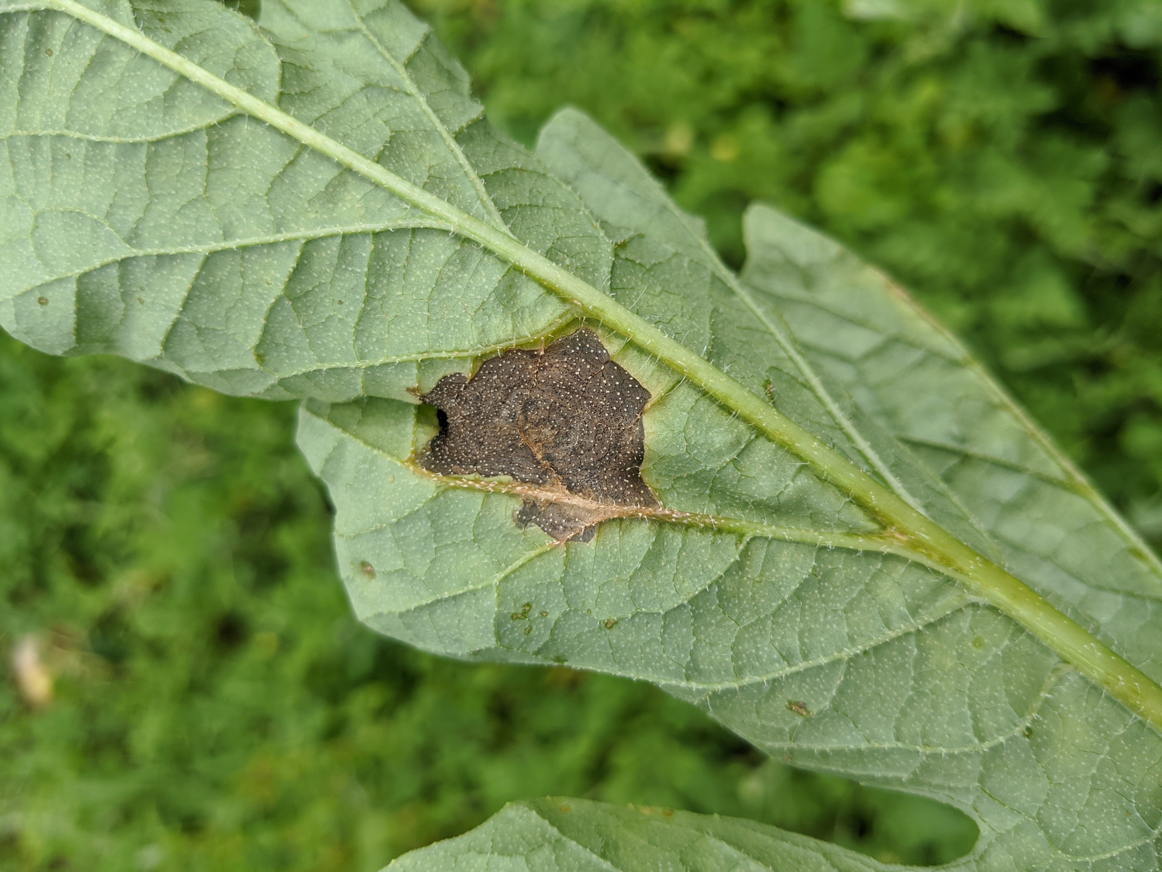 Figure 3. Underside of a watermelon leaf with a gummy stem blight lesion.
 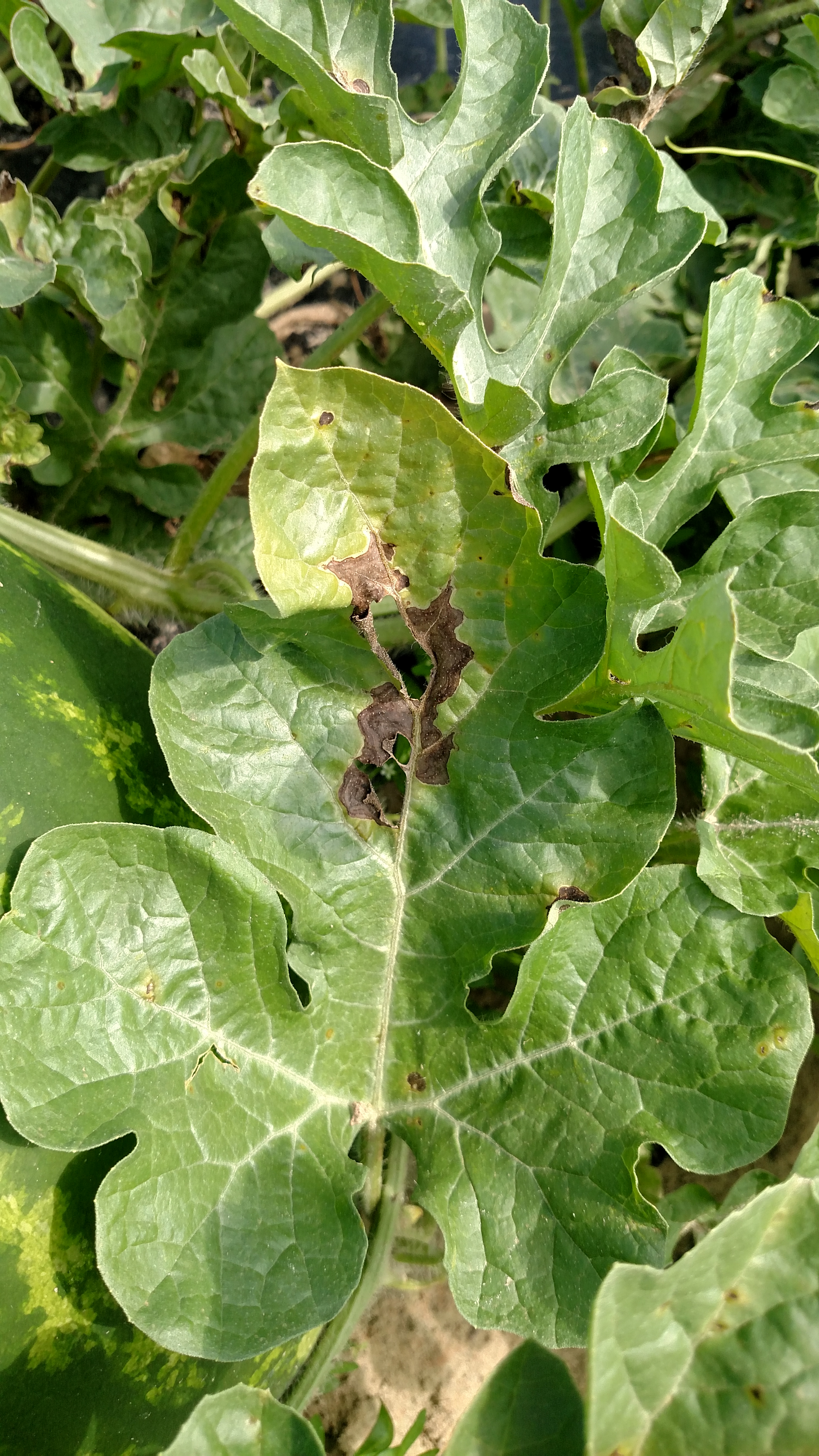 Figure 4. Large gummy stem blight lesion along the midrib of a watermelon leaf
 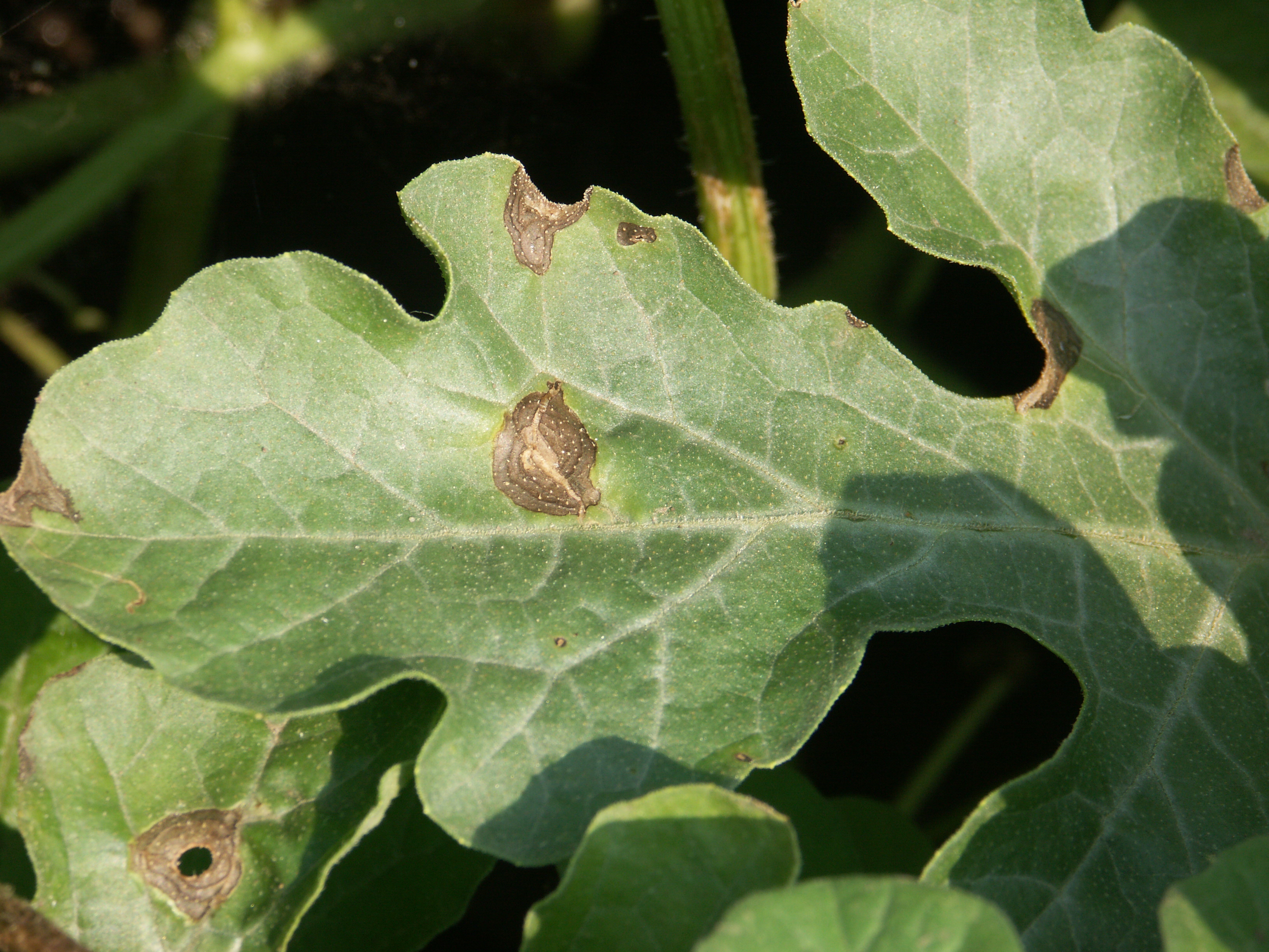 Figure 5. Close up of gummy stem blight lesions on a watermelon leaf, one lesion with a shot-hole.
 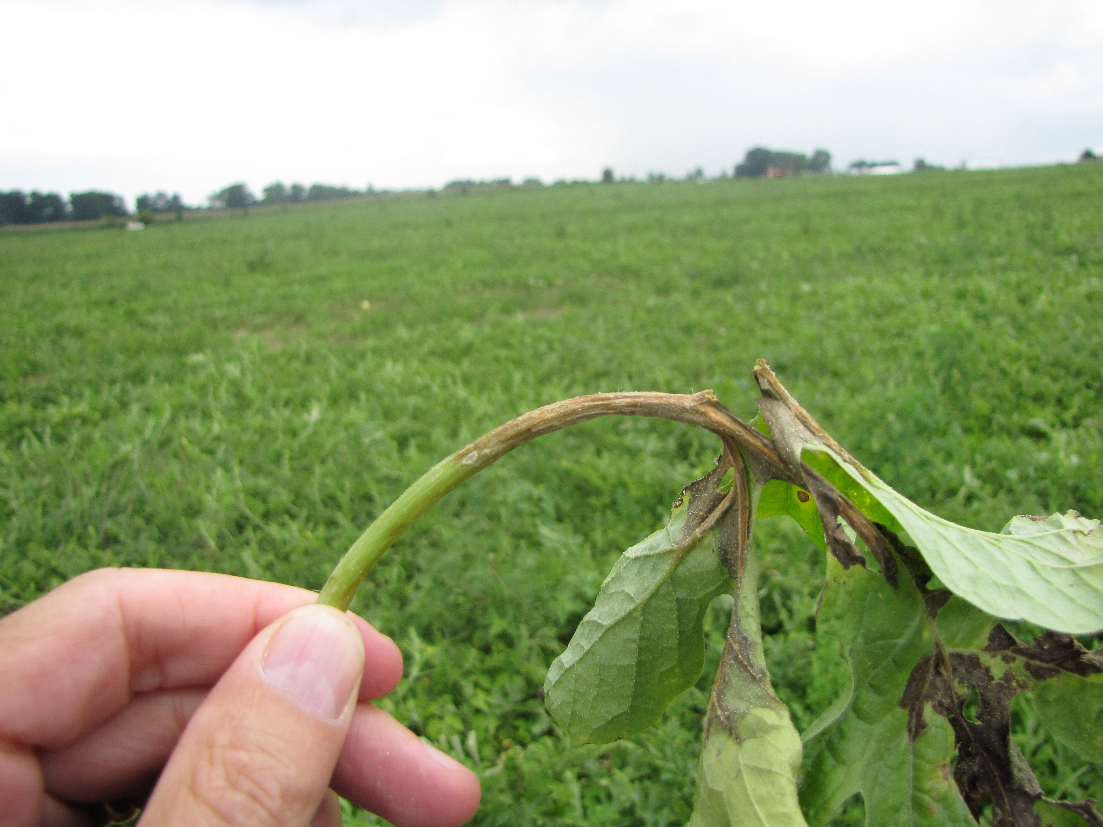 Figure 6. Lesion of gummy stem blight on a watermelon leaf petiole.
 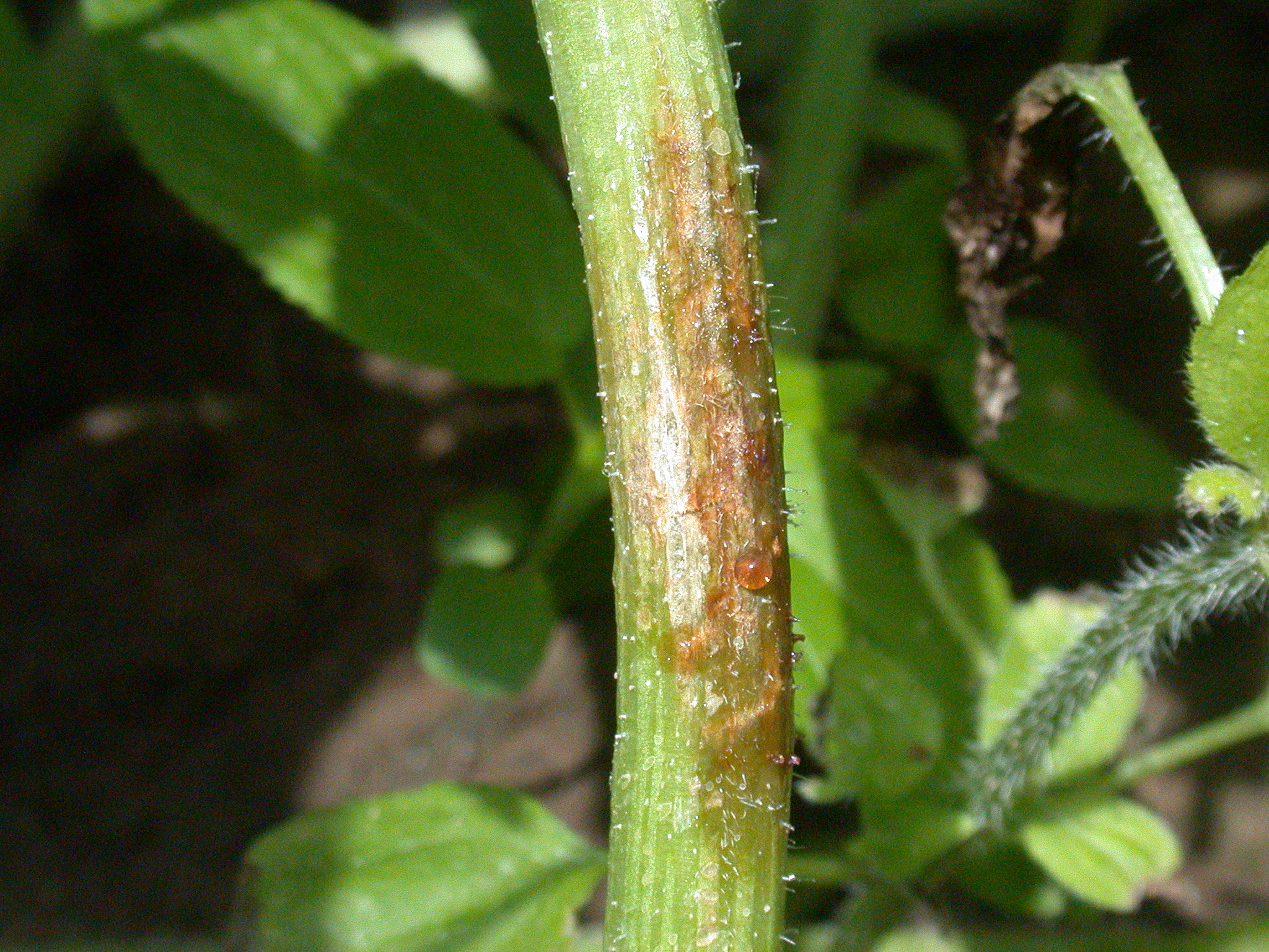 Figure 7. Lesion of gummy stem blight on a watermelon stem. The brown ooze or gum does not always occur in association with this disease and many other diseases and injuries to the stem may cause such an ooze.
 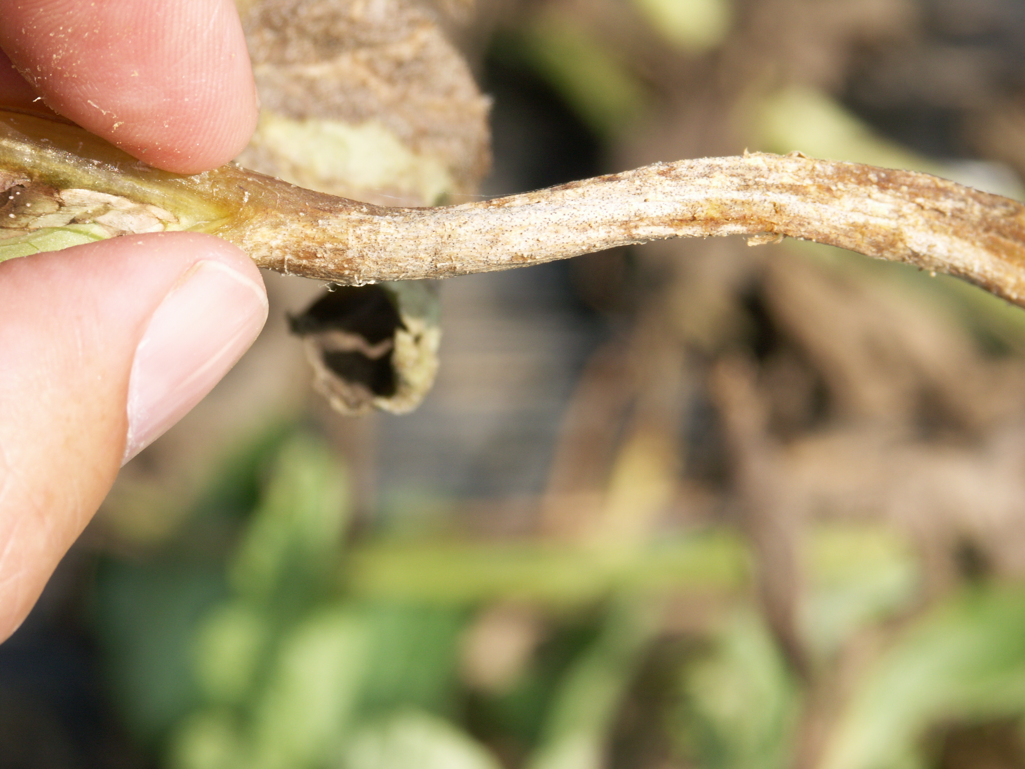 Figure 8. Fruiting bodies, pycnidia, on a watermelon leaf petiole.
 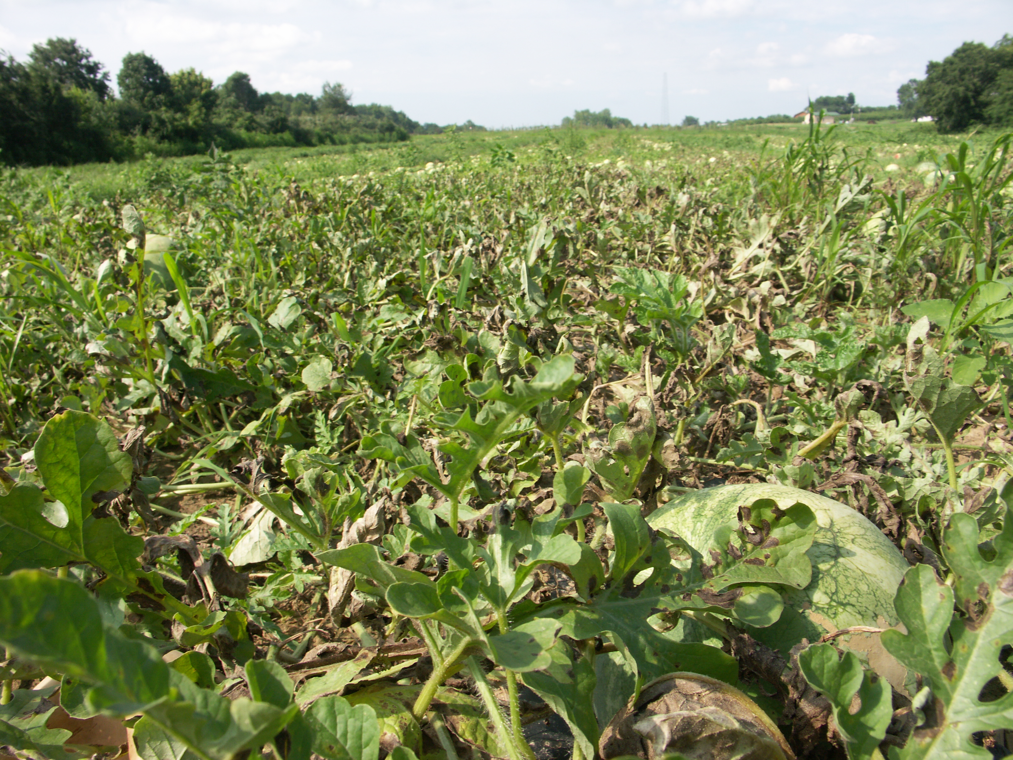 Figure 9. A field of watermelon with a severe outbreak of gummy stem blight manifested by defoliation and numerous lesions.
 Lightning Damage
Lightning damage-This is not an infectious problem and it not economically important. However, it is important to recognize lightning damage and distinguish it from other problems. Symptoms vary, but affected areas in fields may be roughly round, but do not expand over time. Affected plants may be rotted or even appear scorched. Note that nearby weeds are also affected.
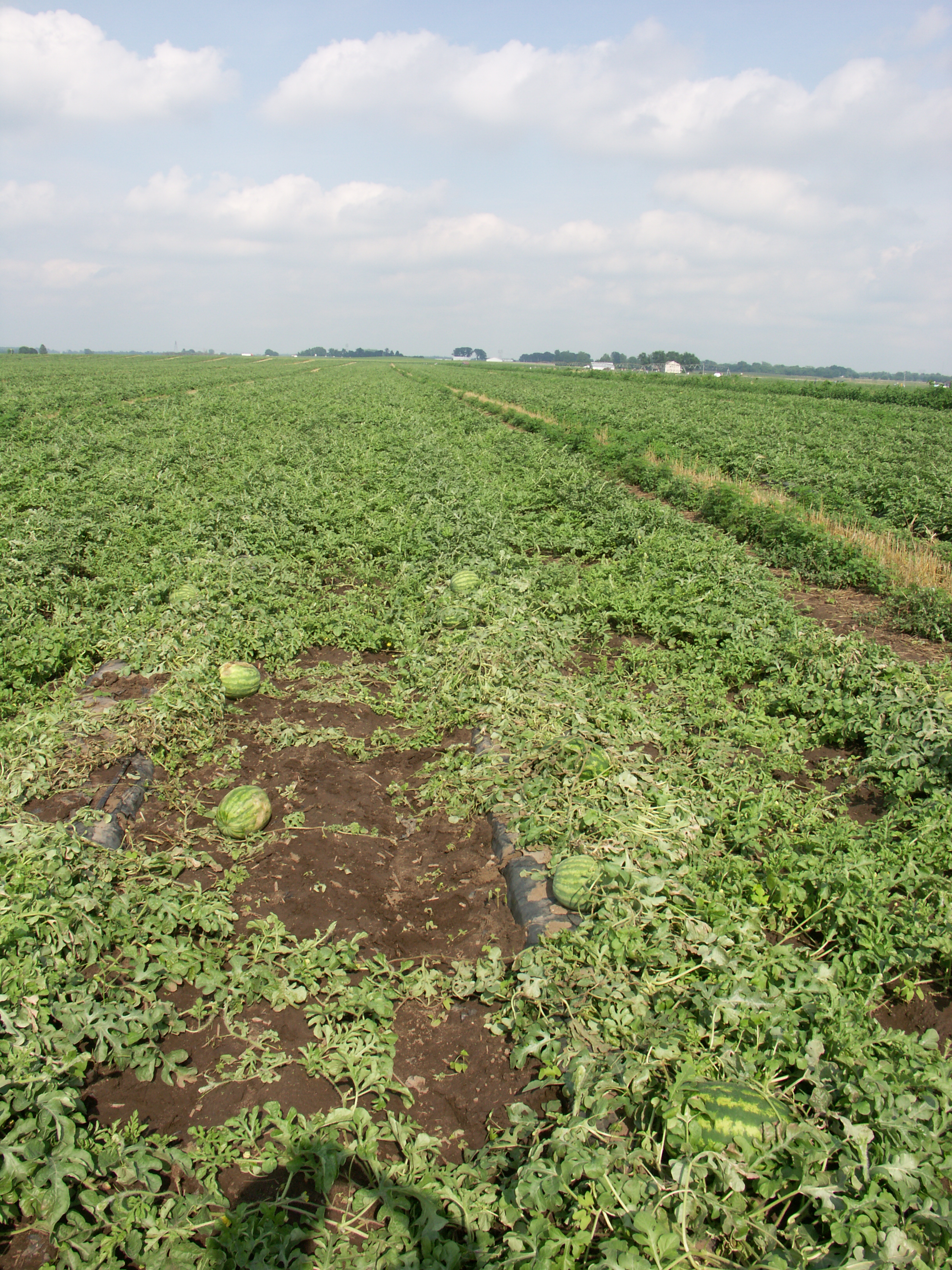 Figure 1. Lightning damage on watermelon. Note wilt and other damage.
  Figure 2. Lightning damage on watermelon. Note approximately round area of damage. This photos was taken a few days after the strike occurred.
  Figure 3. Lightening damage on watermelon. Note wilt and leaf burn.
  Figure 4. Lightning damage on watermelon. Note wilt and leaf burn.
 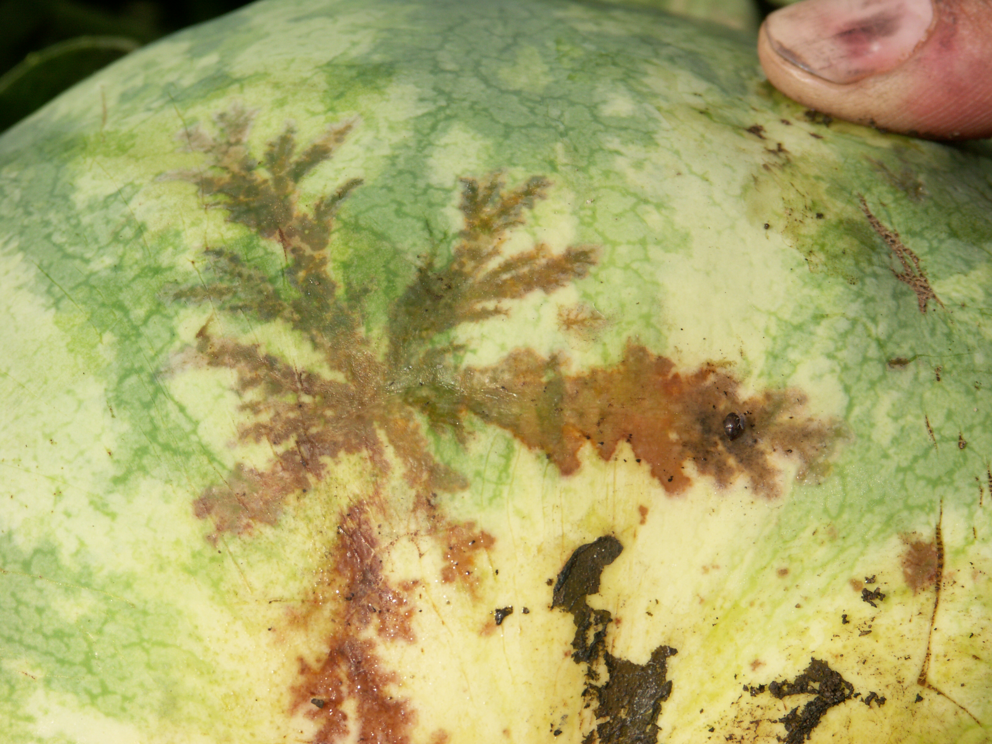 Figure 5. Lightning damage on watermelon.
  Figure 6. Lightning damage on weeds in watermelon field.
 Manganese toxicity
Manganese toxicity-This is not an infectious disorder. However, it is important to recognize the symptoms since economic loss can occur. Also, the symptoms tend to be clustered in areas of the field where the soil pH is relatively low, thus mimicking an infectious problem.
 Figure 1. Manganese toxicity on watermelon. Note symptomatic vines are clustered in areas of low soil pH.
  Figure 2. Manganese toxicity on watermelon.
 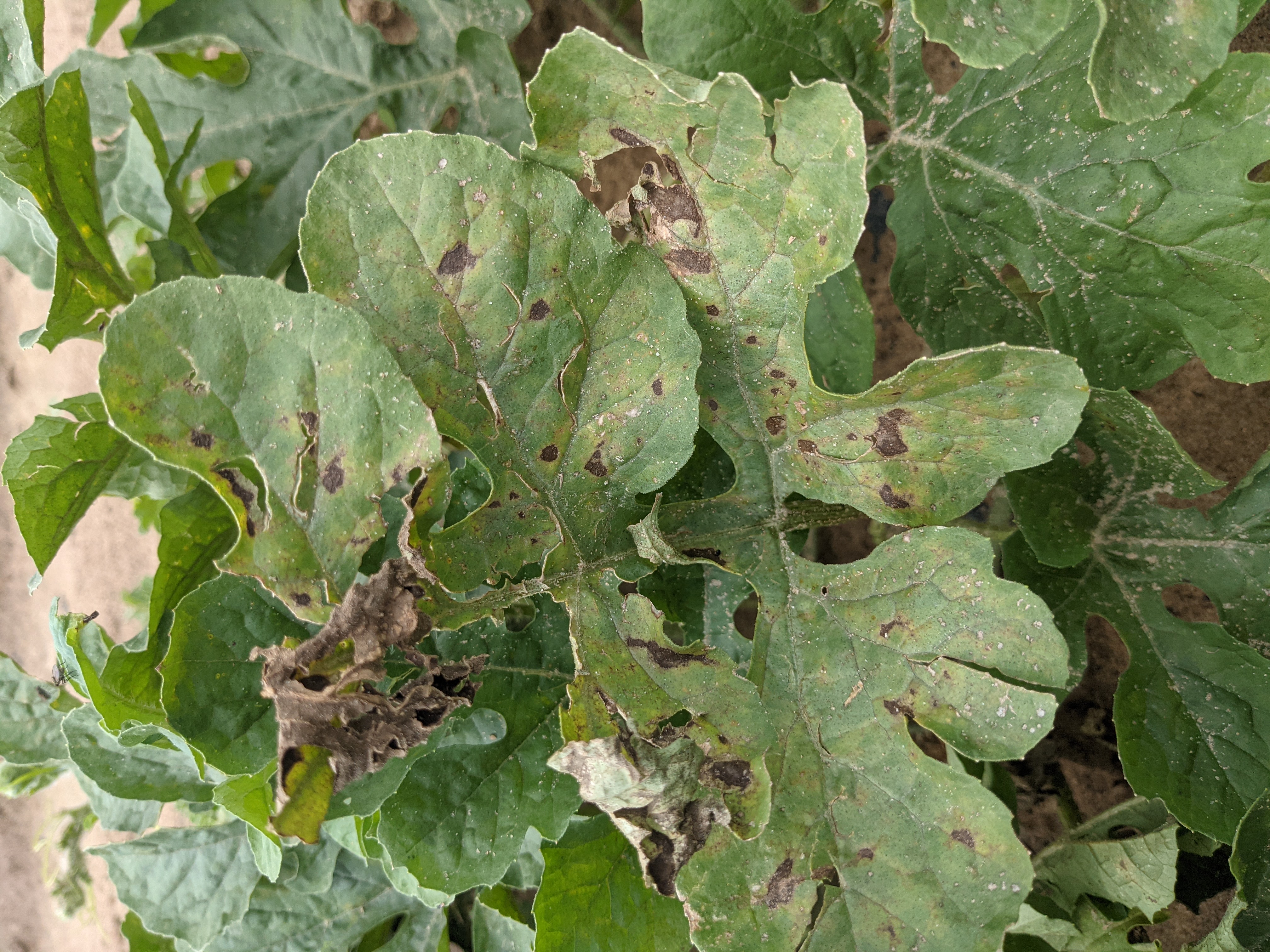 Figure 3. Manganese toxicity on watermelon.
 Phytophthora blight
Phytophthora blight-Perhaps the most important disease of watermelon in Indiana. Generally, leaves and stems are not affected (compare with pumpkin). Fruit become soft, water-soaked and a white mold will develop on the fruit.
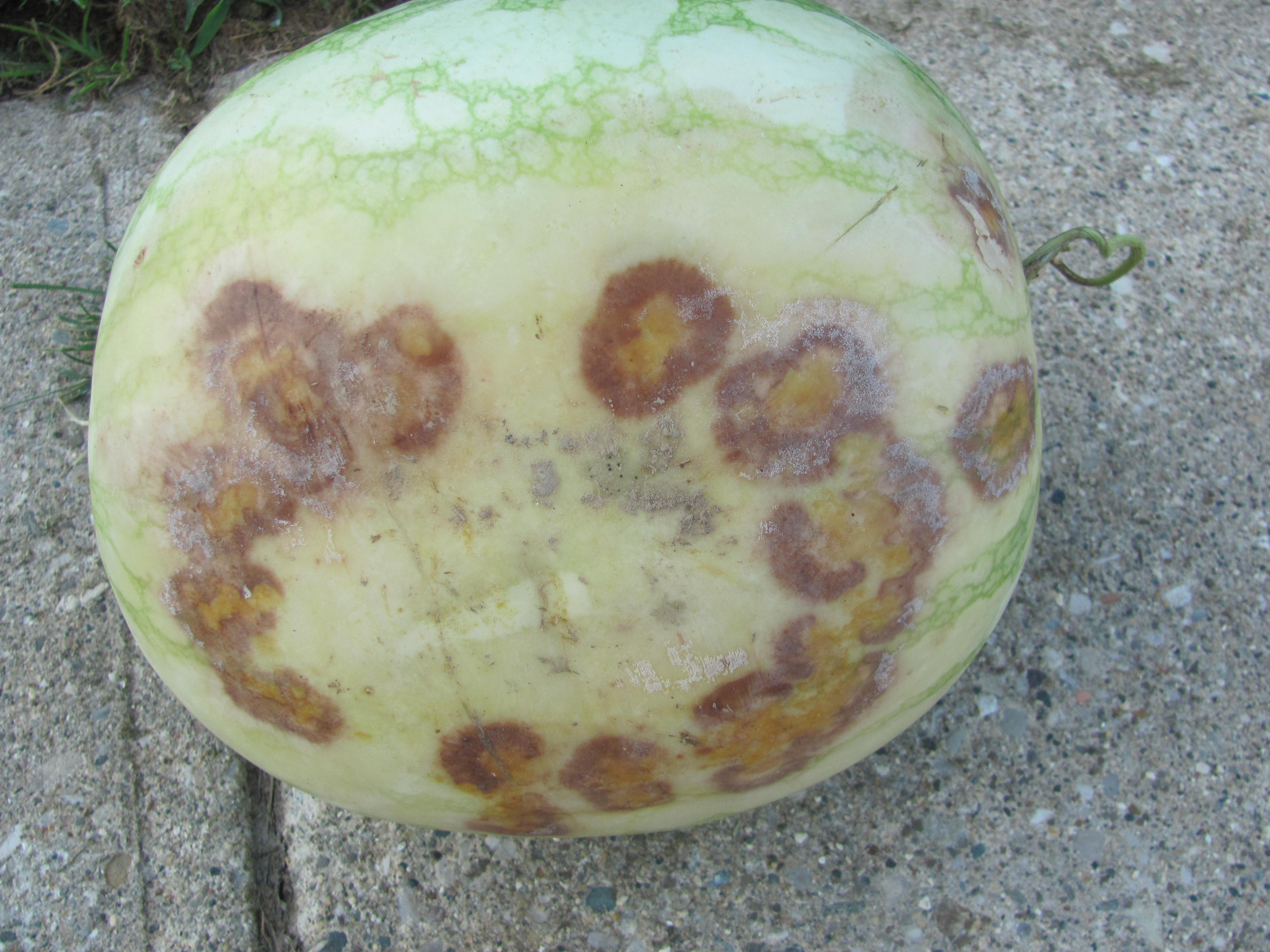 Figure 1. Phytophthora blight has caused the round bruised-like areas on the watermelon.
 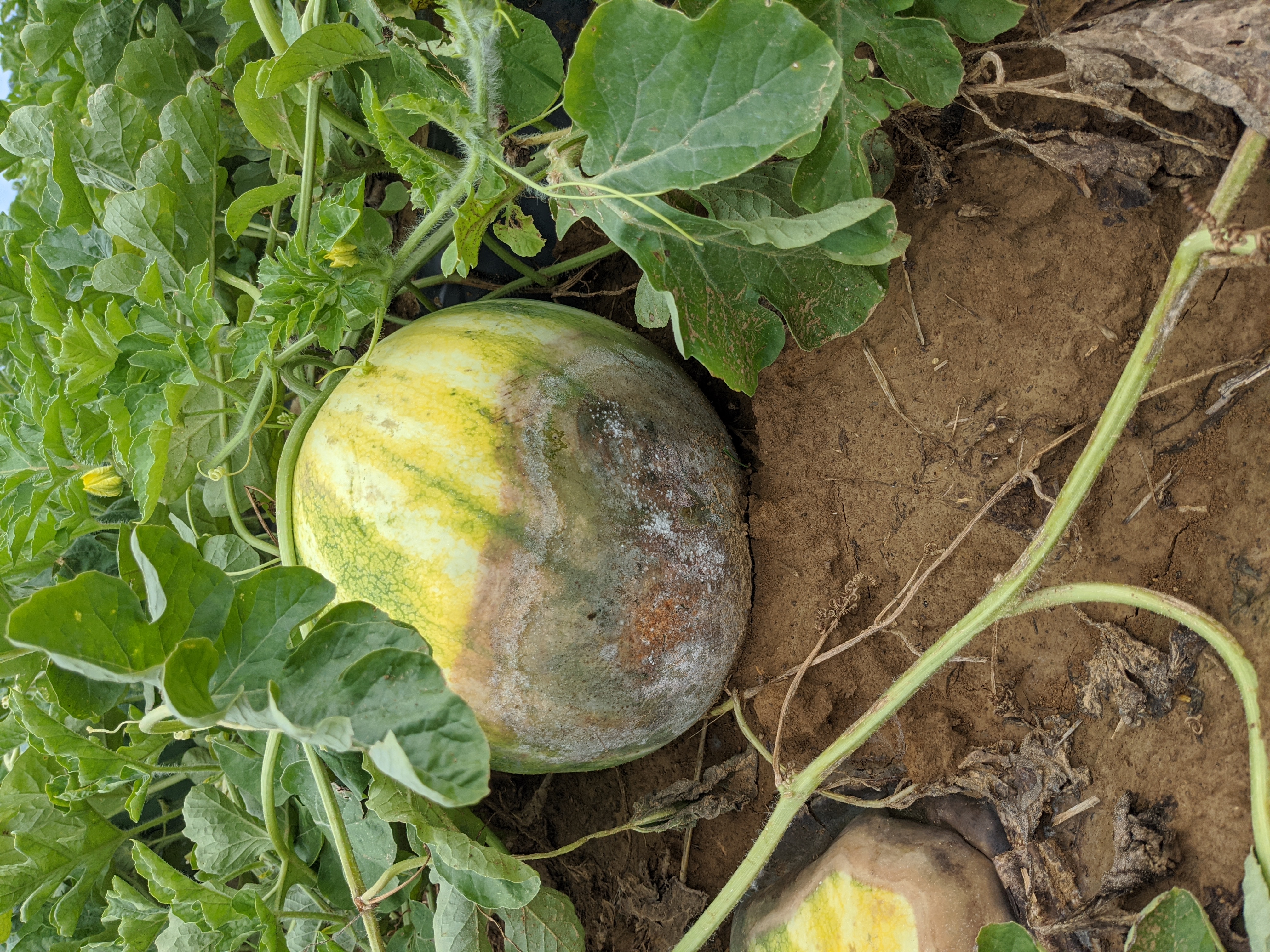 Figure 2. Phytophthora blight has caused the water-soaked symptoms on the base of this watermelon including the white sporulation of the fungus.
 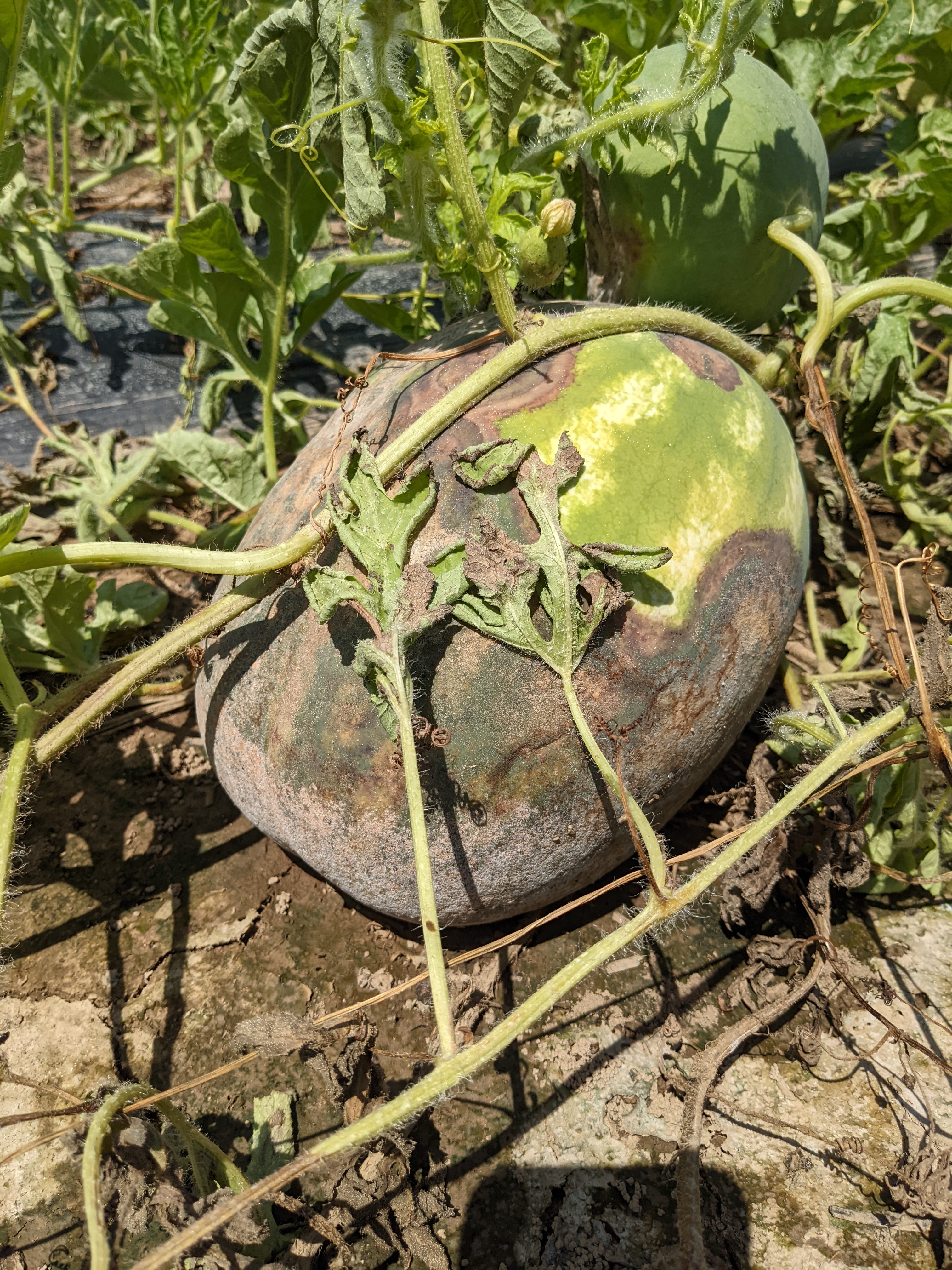 Figure 3. Phytophthora blight of watermelon has caused the large necrotic regions on this watermelon. The vines of watermelon are usually not affected by Phytophthora blight, but in extreme cases can become symptomatic.
 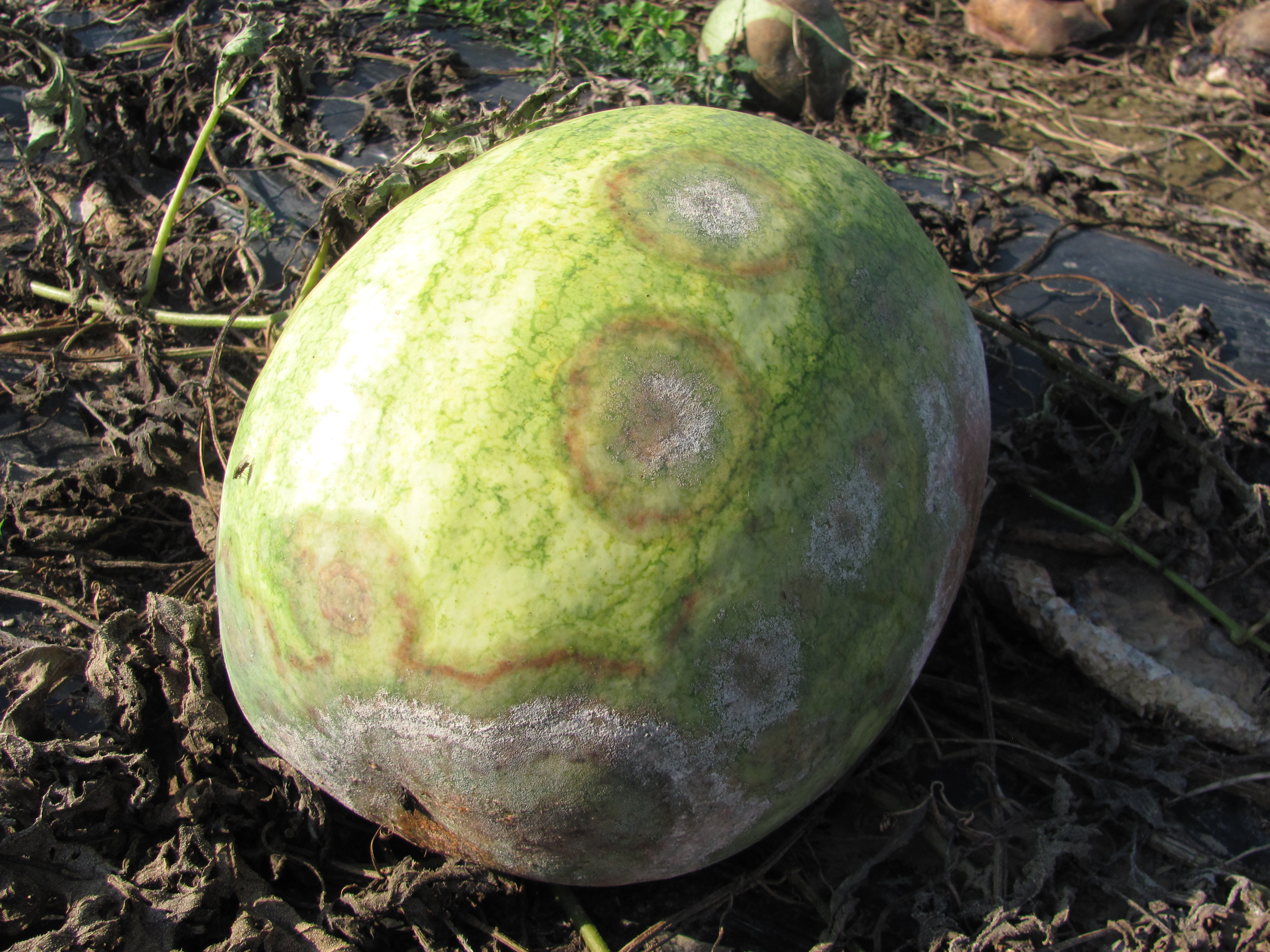 Figure 4. Phytophthora blight of watermelon has caused the lesions with gray/white sporulation. Note the bruised circular areas around each lesion.
 Powdery mildew
Powdery mildew-not as important or common a problem in Indiana as it is in the southeast US. Symptoms are easily recognizable from the white talc-like symptoms of lesions.
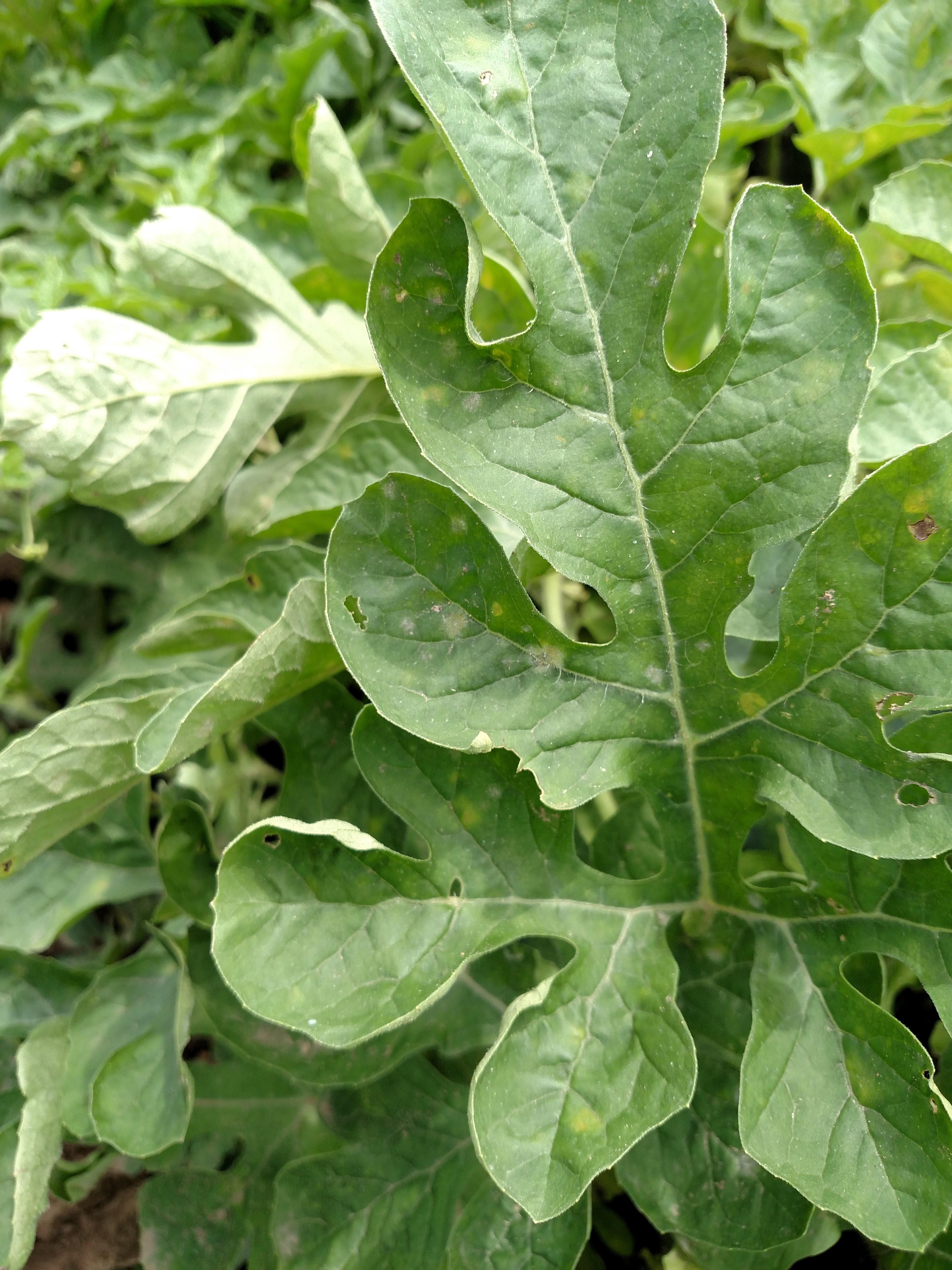 Figure 1. Leaf lesions of powdery mildew of watermelon may be white due to the sporulation of the fungus or colonies on the reverse of the leaf may show up chlorotic.
 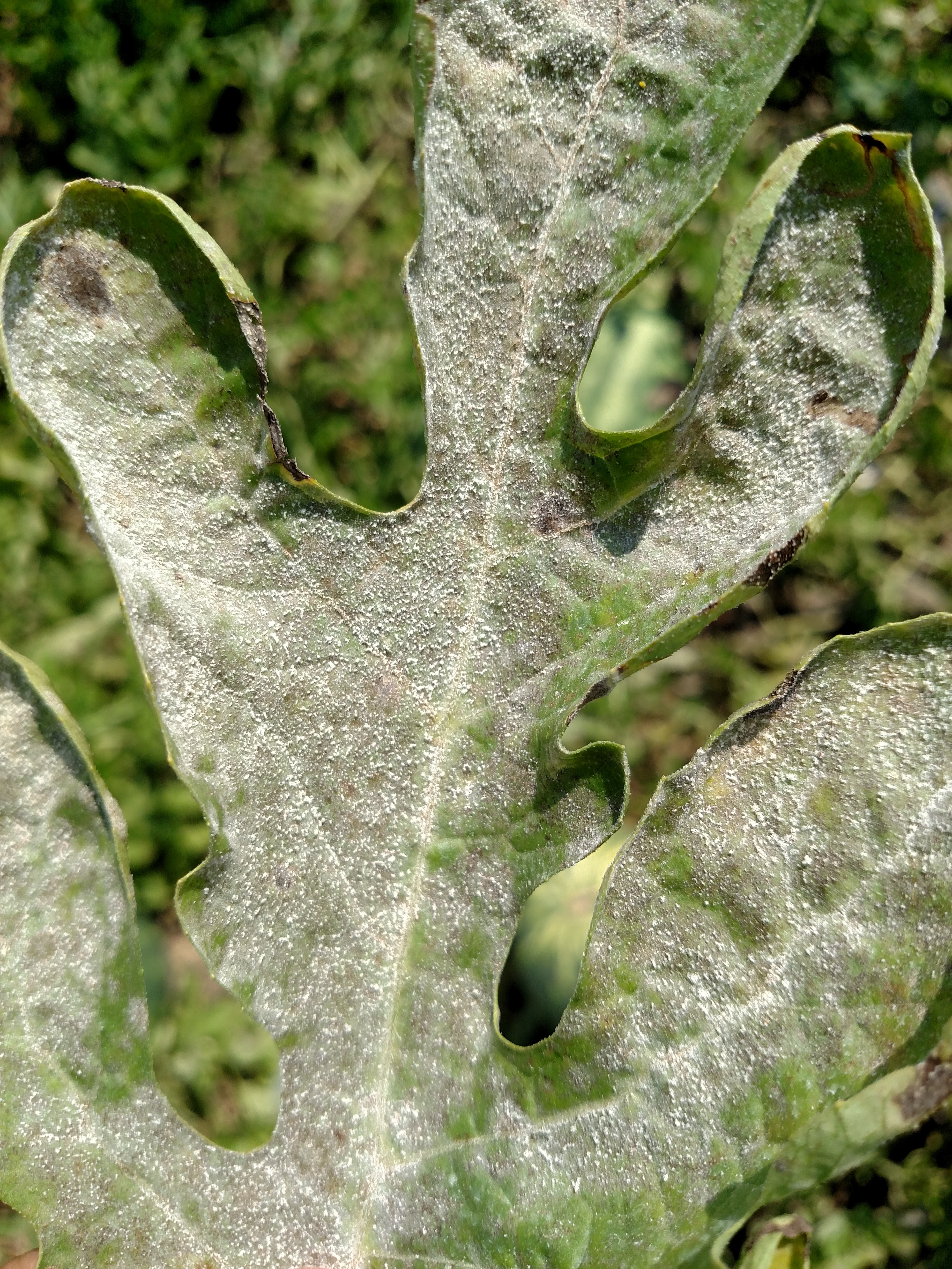 Figure 2. Leaf with sporulation of powdery mildew.
 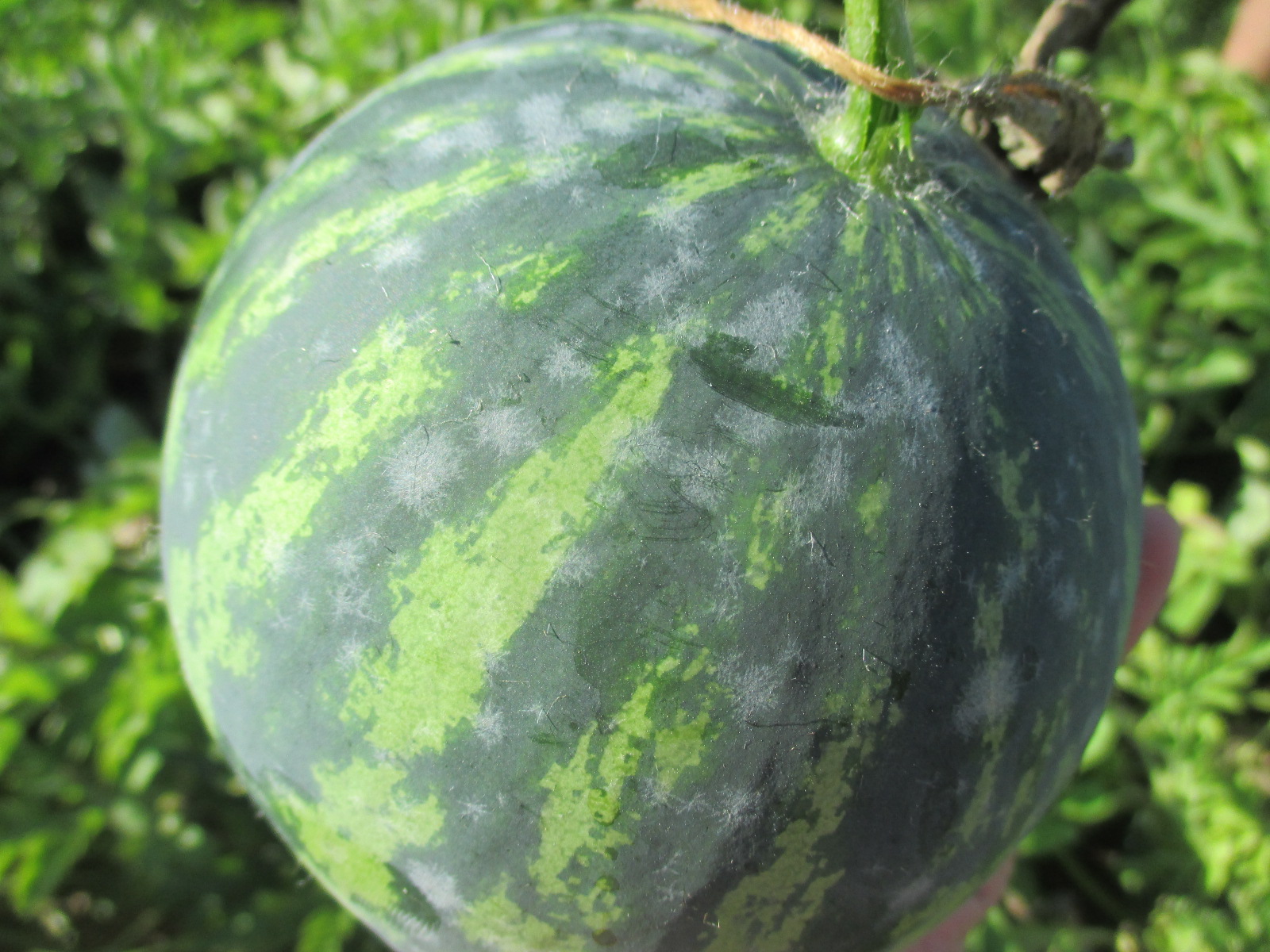 Figure 3. Powdery mildew of watermelon fruit. (Photo by Wenjing Guan.)
 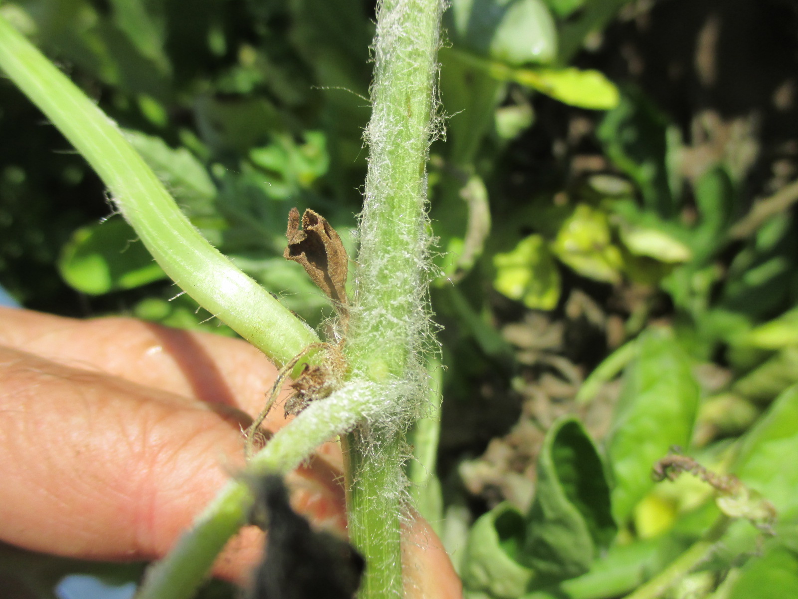 Figure 4. Powdery mildew on stem of watermelon.
 Rind Necrosis
Rind necrosis-Occurs only sporadically and is not usually an important problem. Not an infectious disease. Affected watermelon may feel ‘knobby’ on the surface before being cut. Lesions inside the rind consist of a brown necrosis and may appear rotten. May affect marketability.
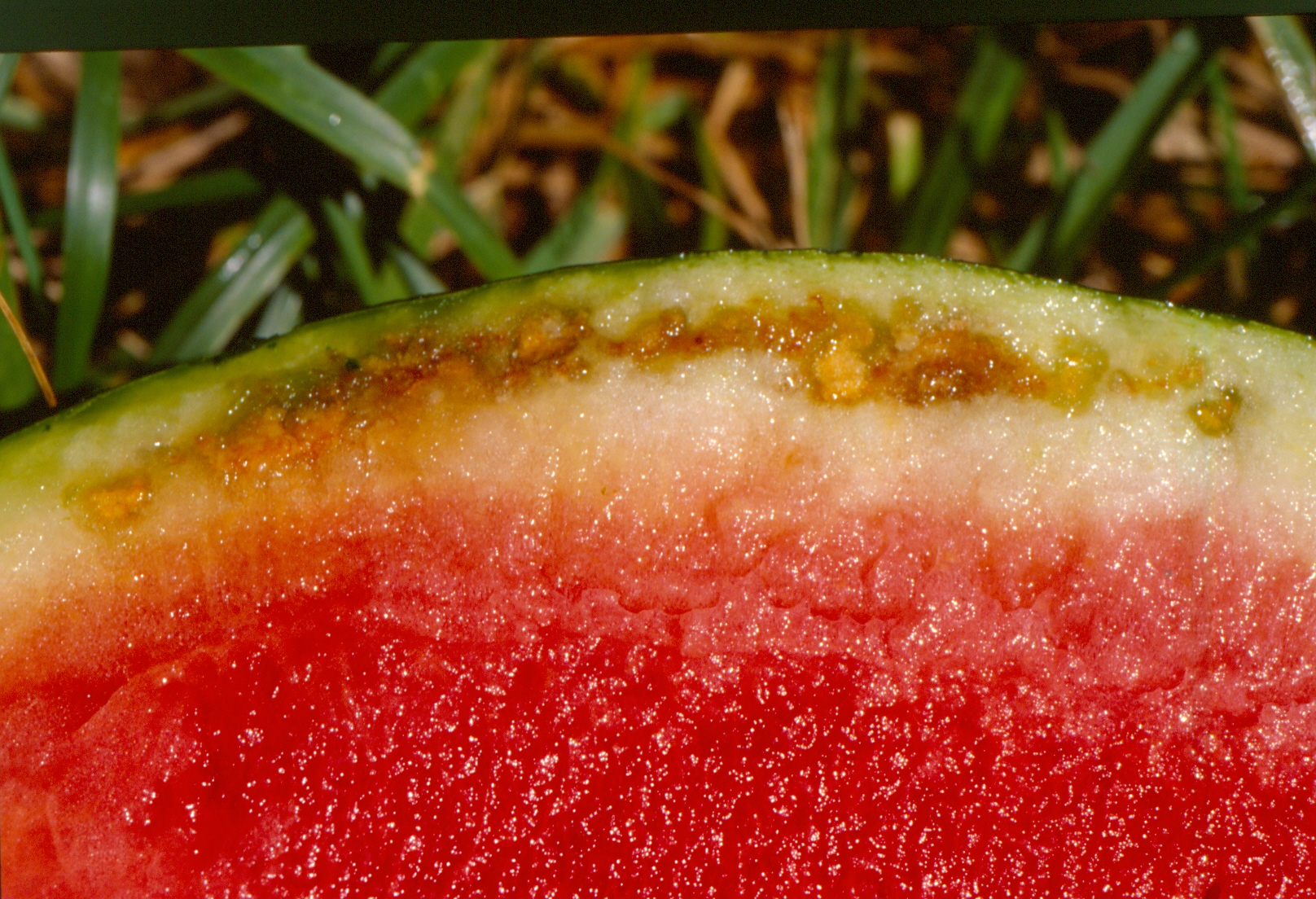 Figure 1. Rind necrosis of watermelon
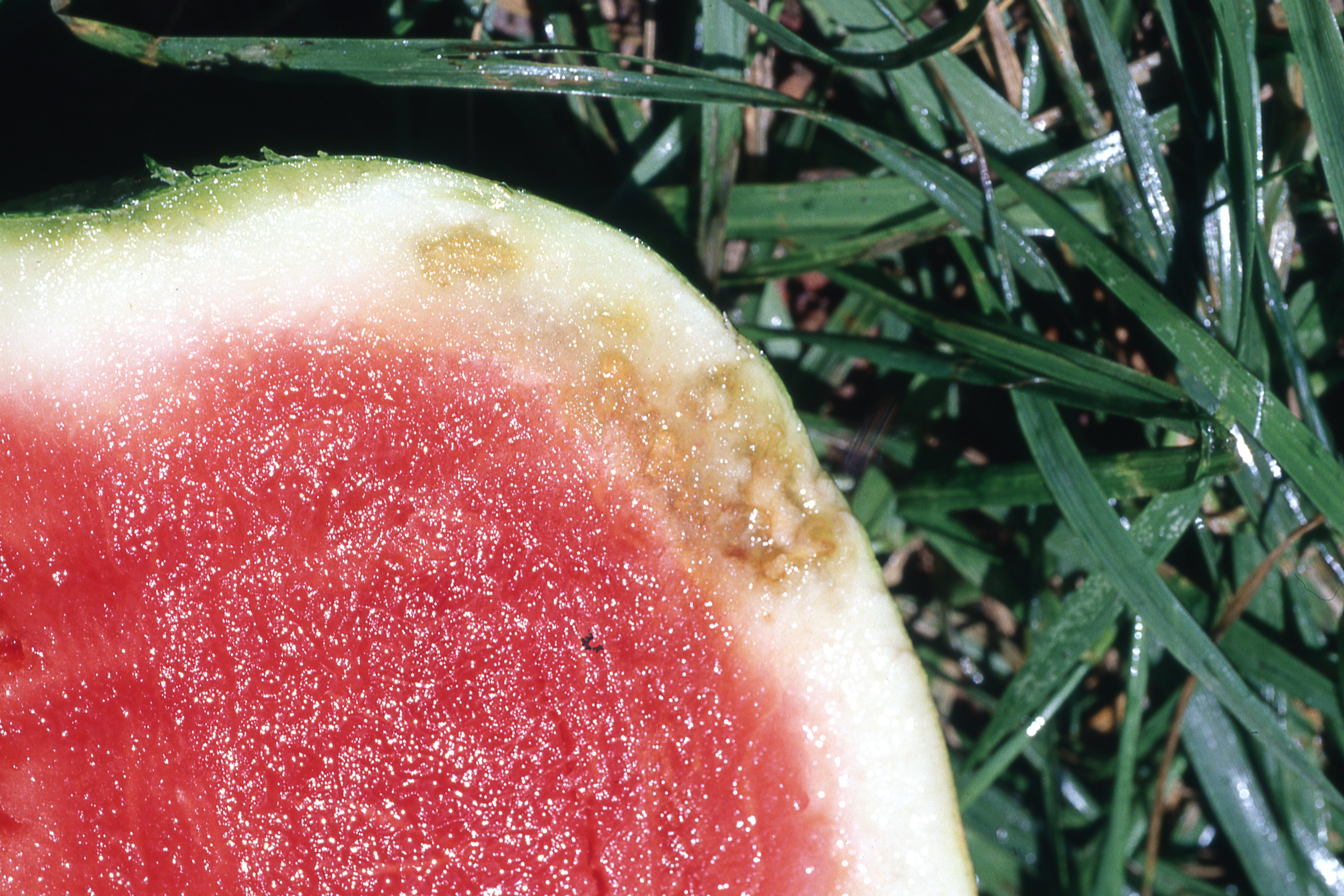 Figure 2. Rind necrosis of watermelon
 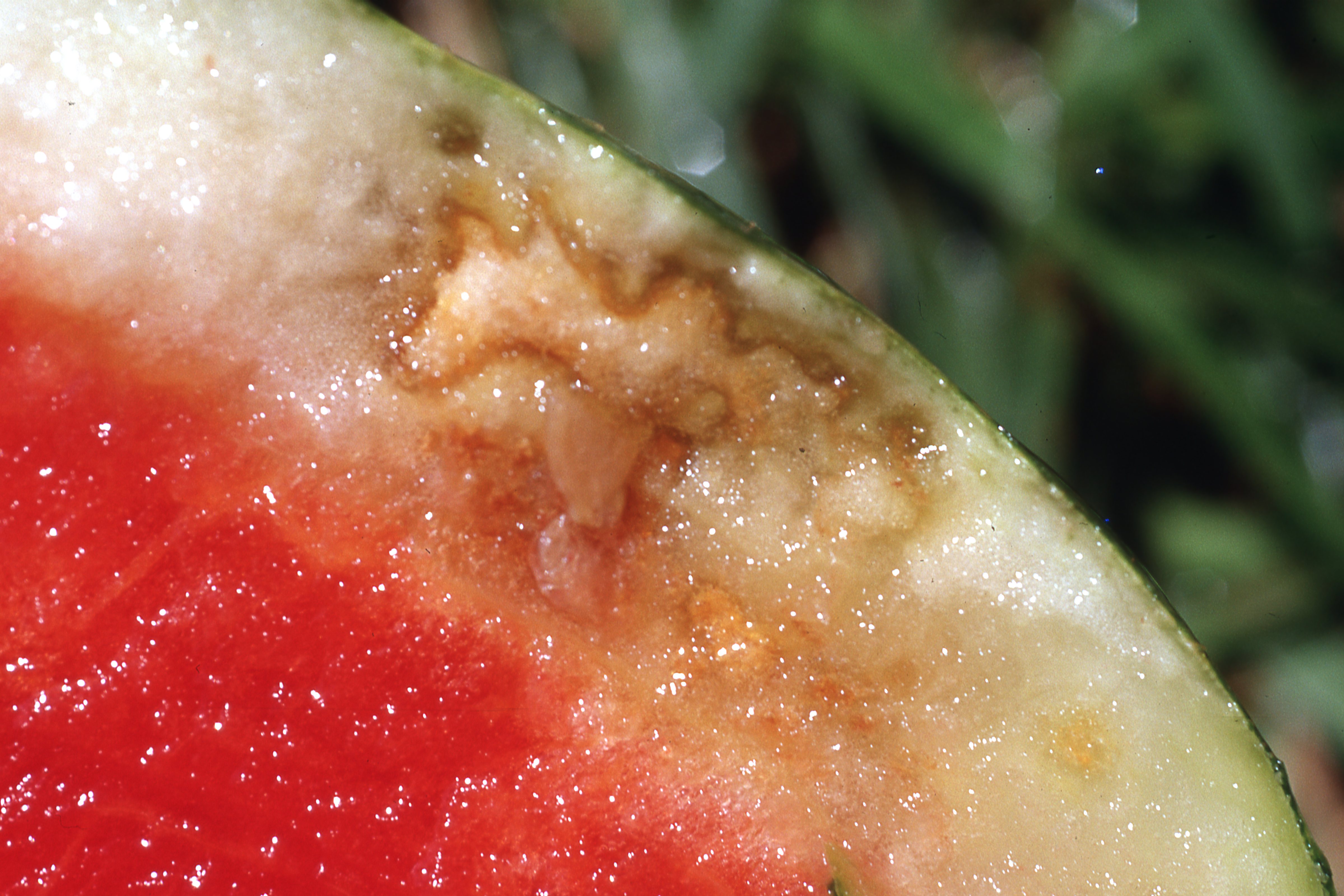 Figure 3. Rind necrosis of watermelon
 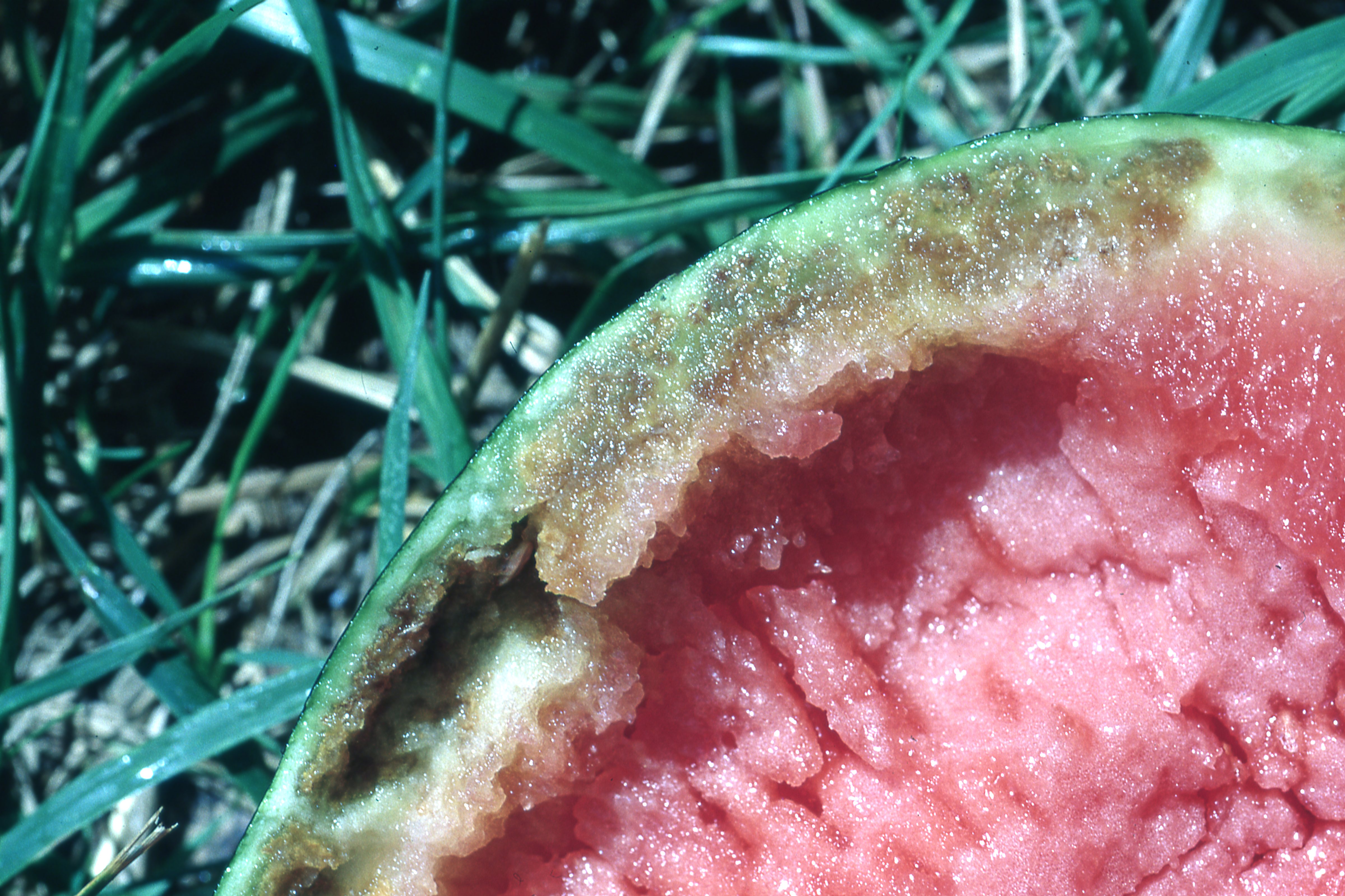 Figure 4. Severe rind necrosis of watermelon
 Root knot nematode
Root-knot nematode-Often the first evidence of this disease is the wilt and decline of the host plant. When the roots are sampled, the galls caused by the nematode are evident. Can be an important disease and is relatively widespread. Probably too often overlooked.
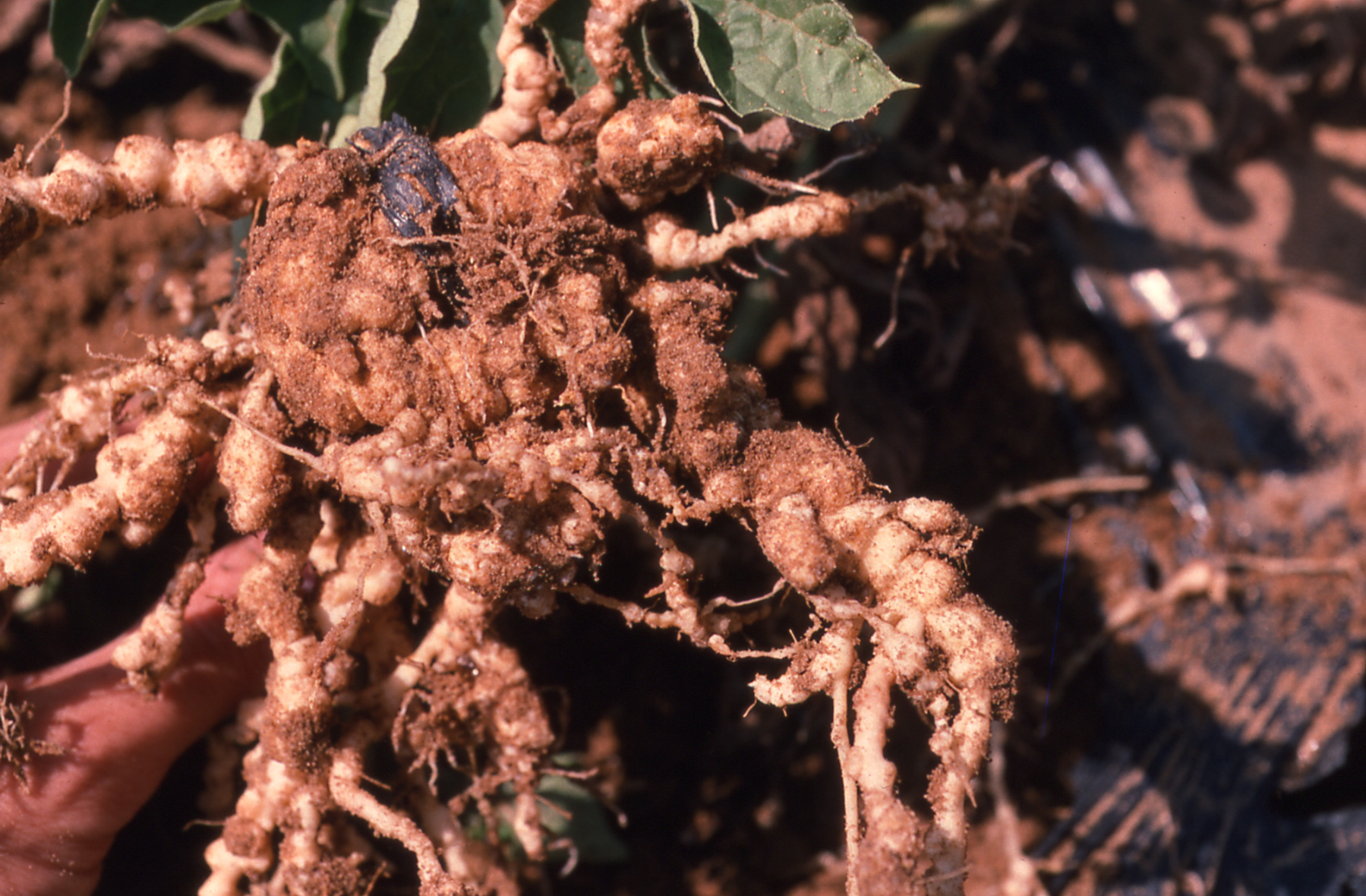 Figure 1. Roots of watermelon with galls due to root knot nematode.
 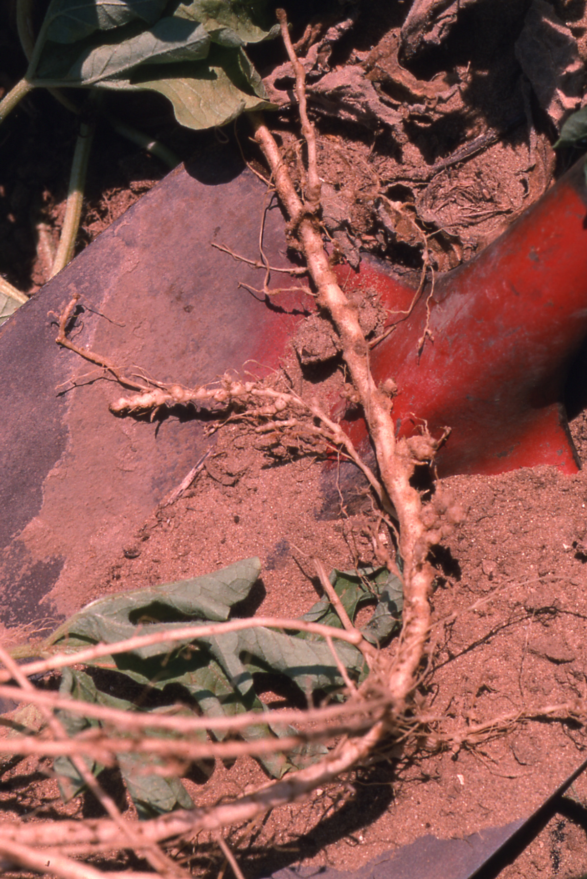 Figure 2. Galls of root knot nematode on watermelon roots.
 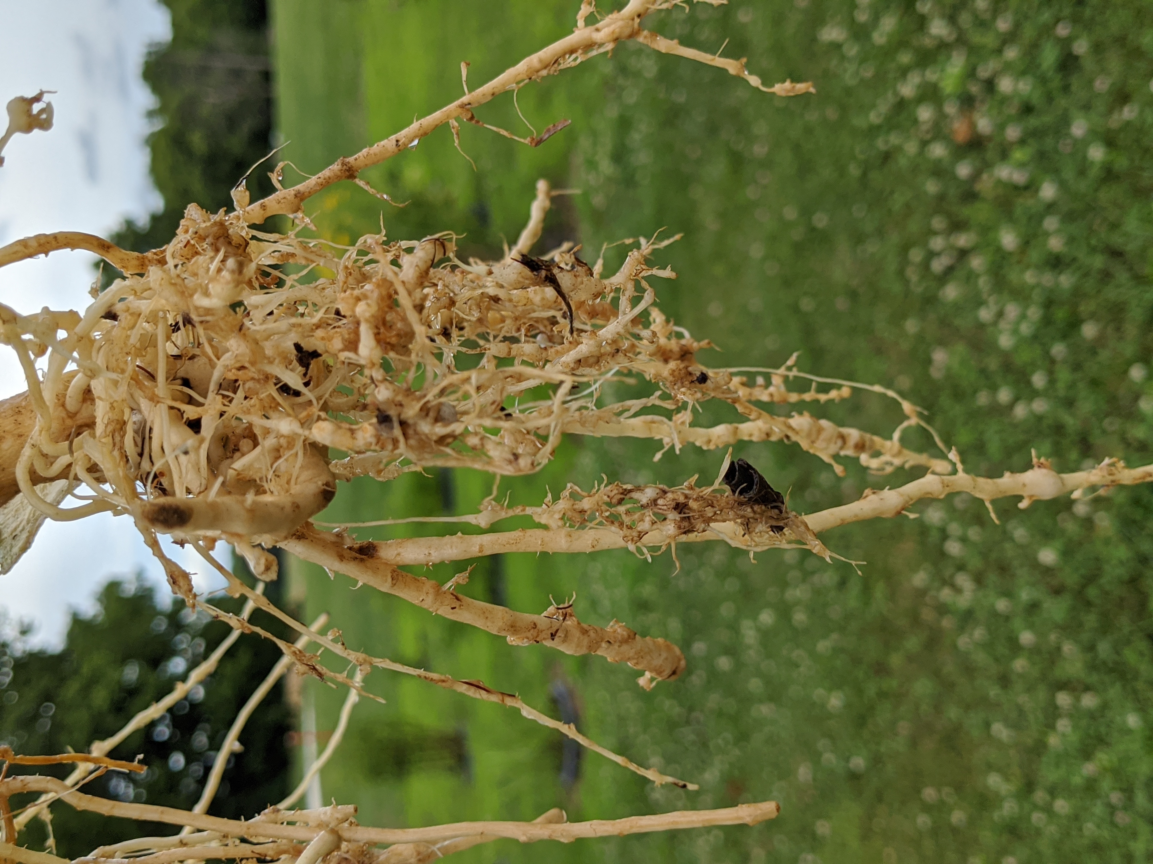 Figure 3. Root nematode galls on a watermelon root system that has been washed to reveal symptoms of root knot.
 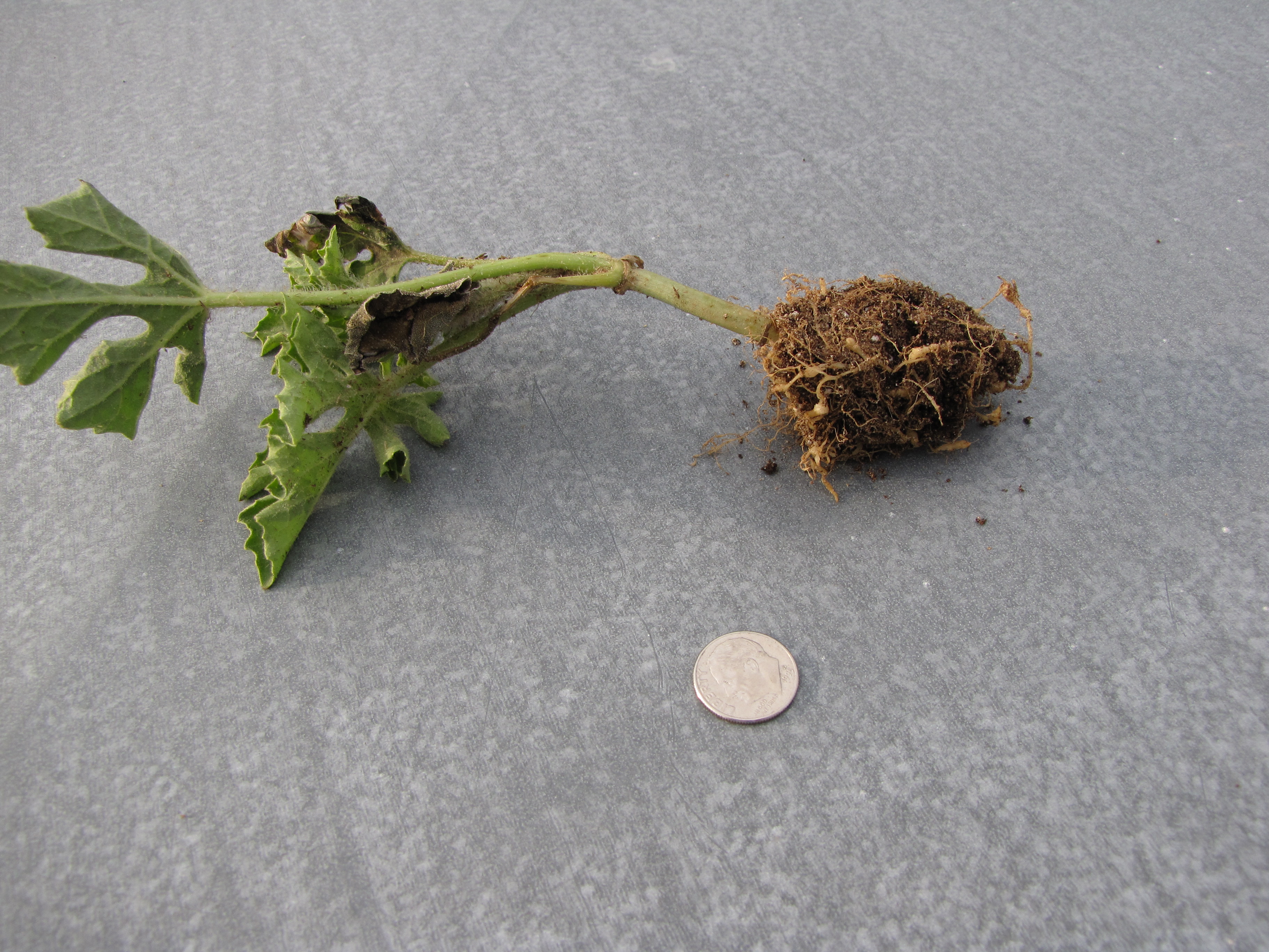 Figure 4. A watermelon seedling that has been recently transplanted in the field has root knot galls visible. Note that symptoms of decline of the plant foliage are visible.
 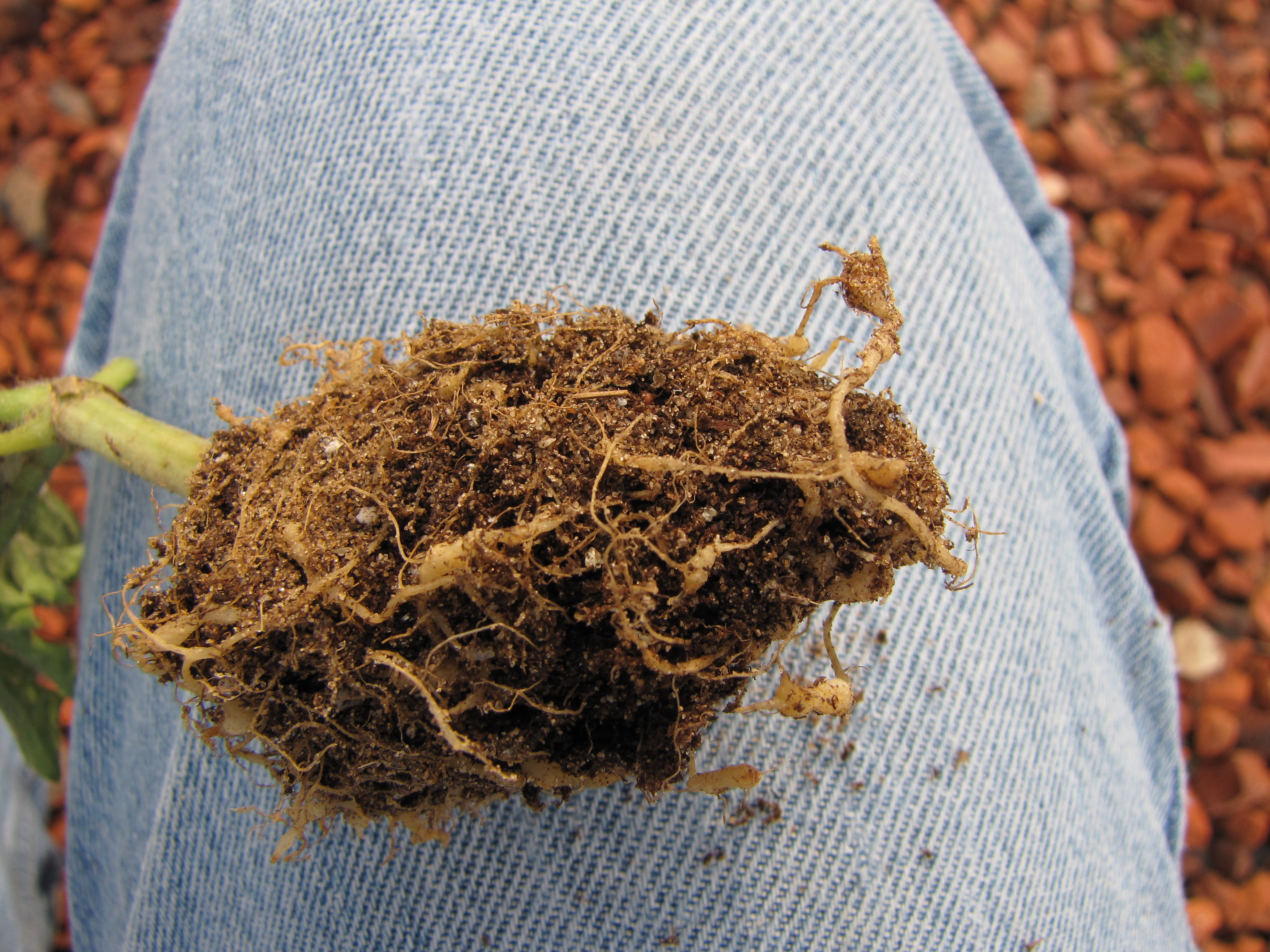 Figure 5. Close up of watermelon roots of transplant with galls.
 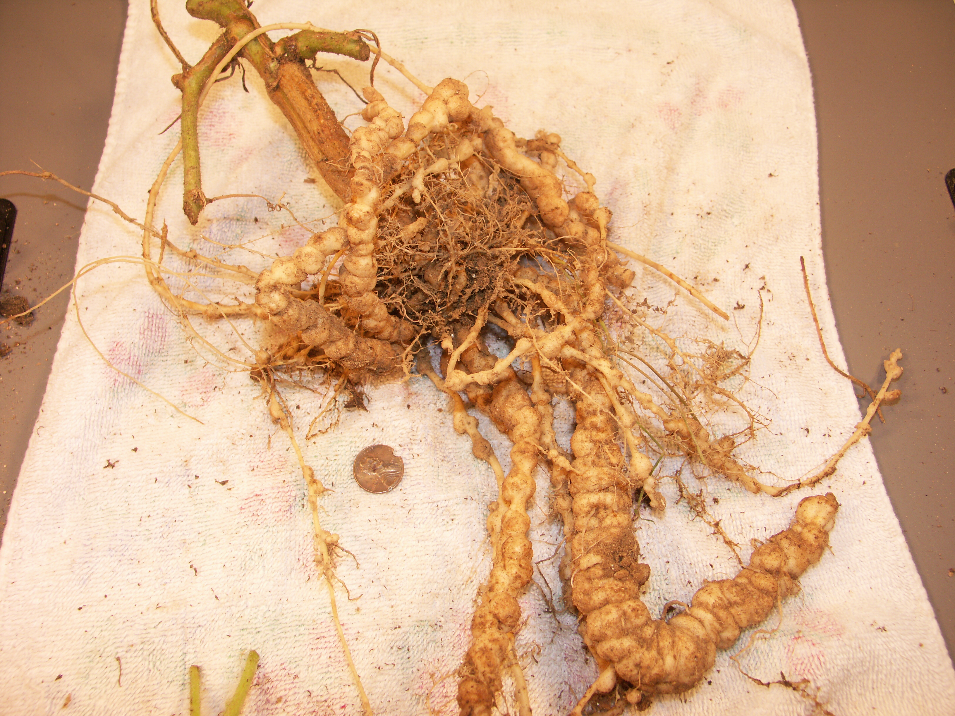 Figure 6. Severe root knot nematode galls on watermelon roots.
 Target cluster
Target cluster-This disorder is not well understood. It is apparently not an infectious disorder. However, in two instances documented below, the symptoms seem to be associated with a potyvirus infection. Target cluster is not a common or economically important disorder.
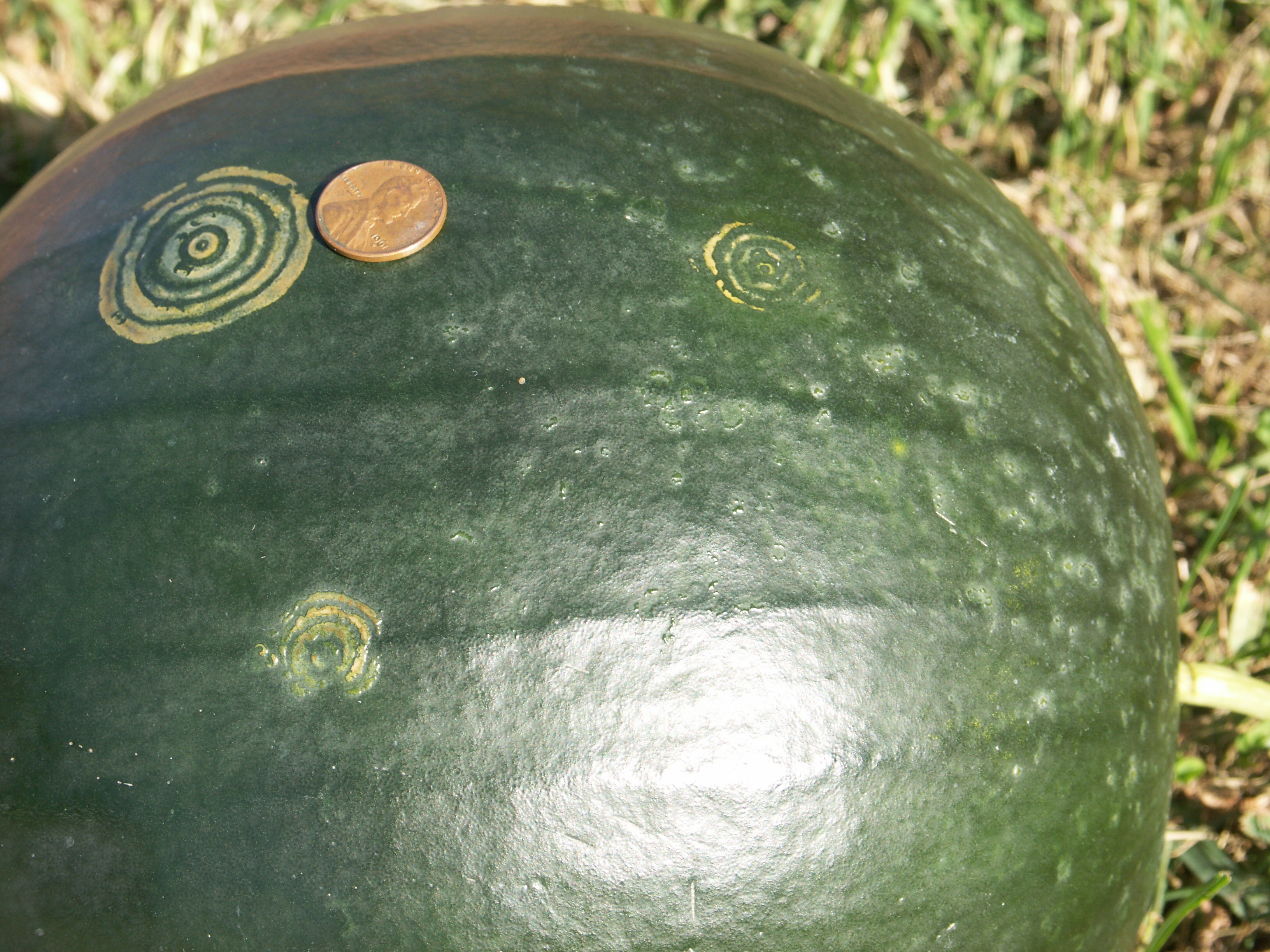 Figure 1. Target cluster of watermelon. Positive for potyvirus.
 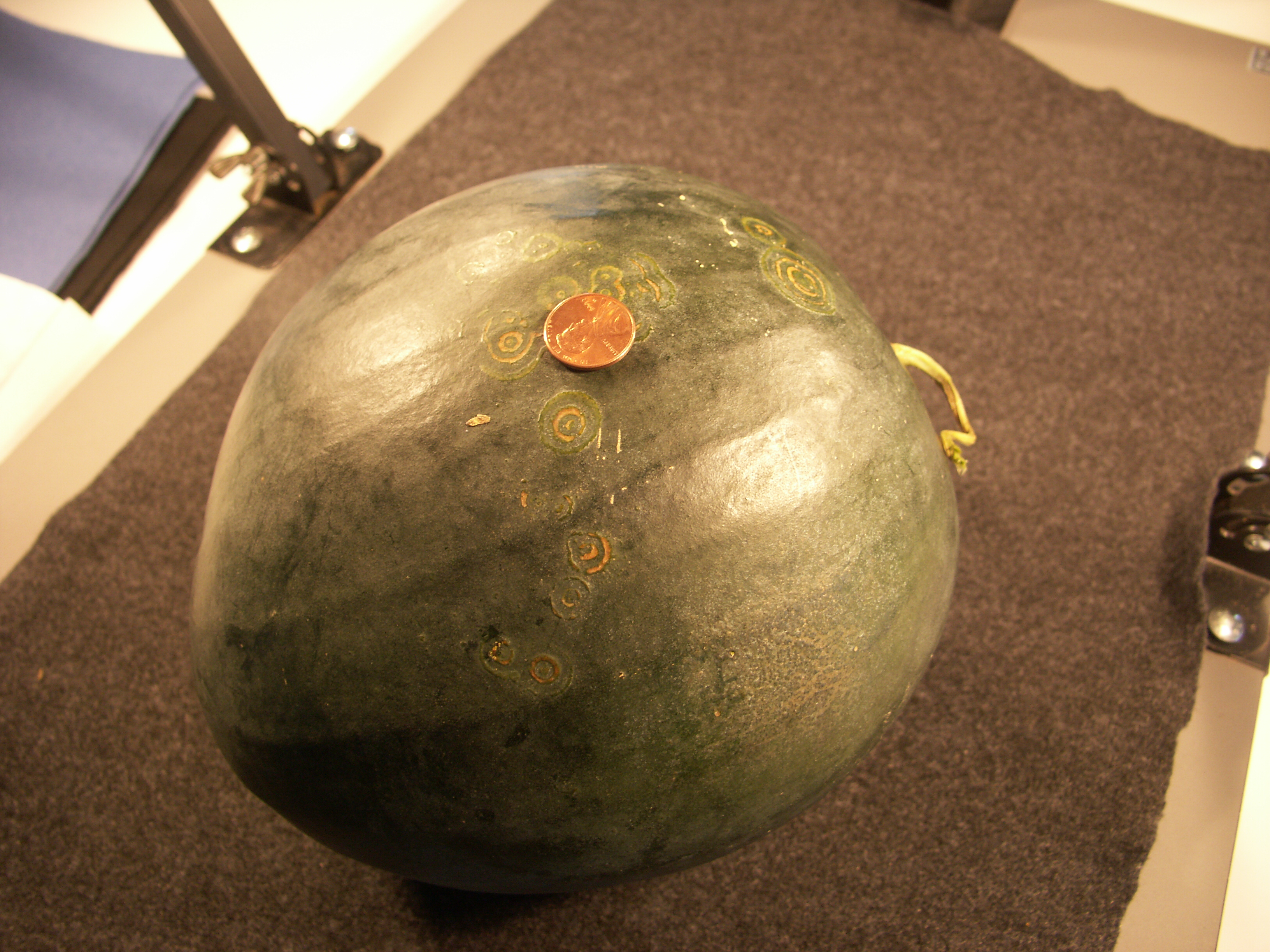 Figure 2. Target cluster of watermelon. Negative for potyvirus.
 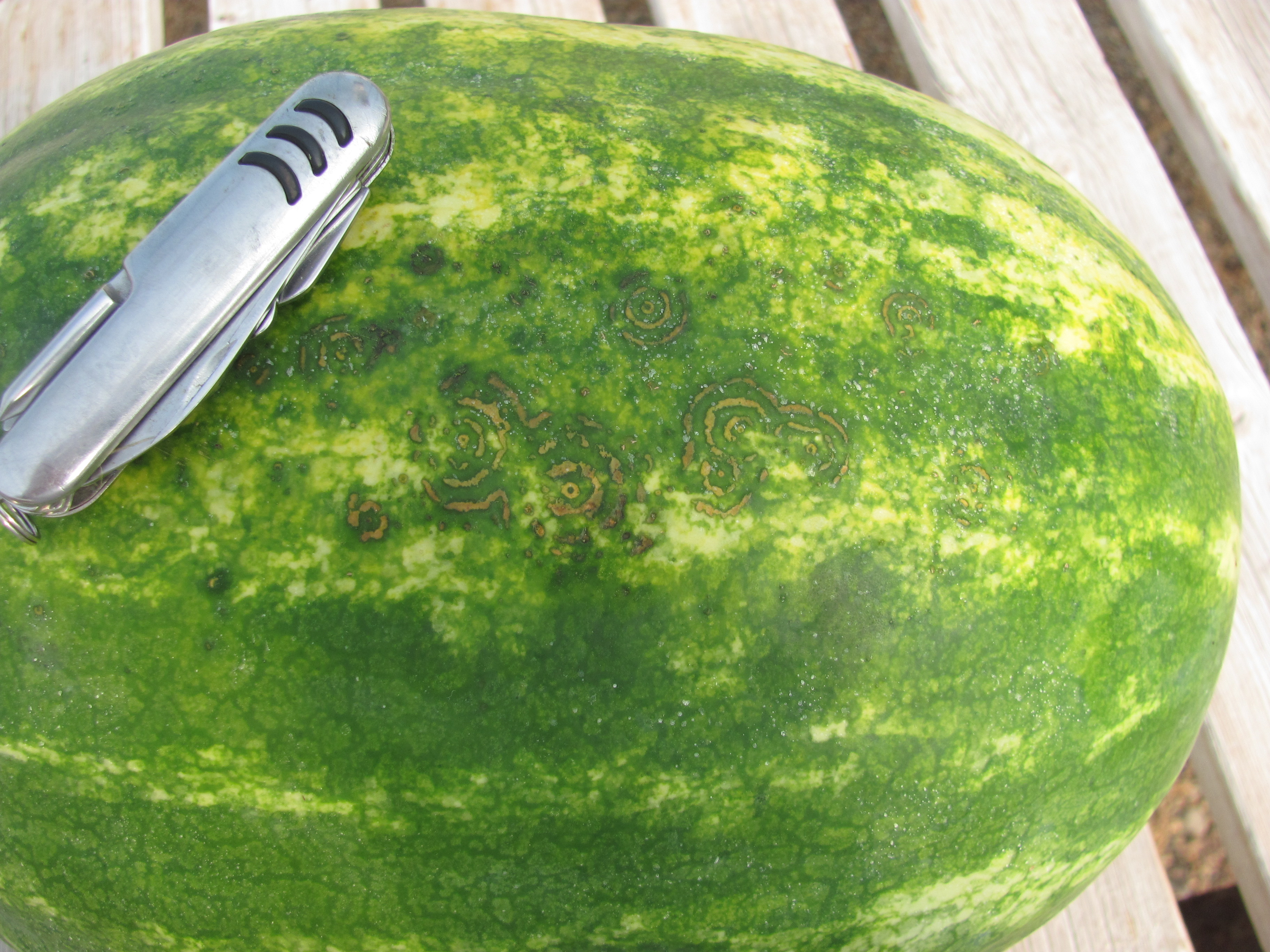 Figure 3. Target cluster of watermelon. Positive for papaya ringspot virus.
 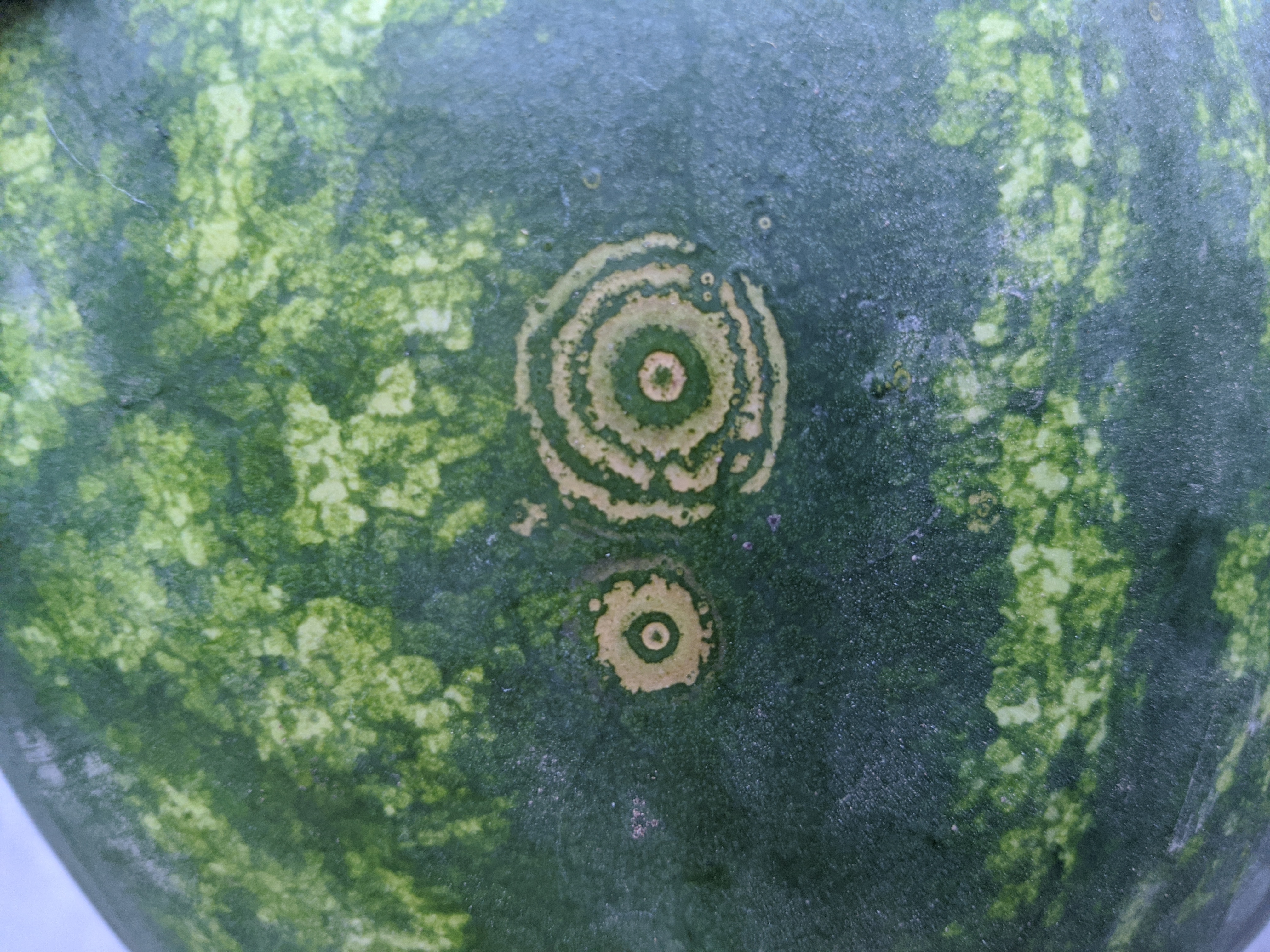 Figure 4. Target cluster of watermelon. Not analyzed for virus.
 Virus
Virus-The most common virus diseases of watermelon in Indiana are potyviruses. Watermelon do not have viral symptoms as much as pumpkins since the latter are grown later in the season. Symptoms on foliage may include mosaic and shoestring leaves. Symptoms on fruit are not common. Viruses do not usually cause economic damage to watermelon.
 Figure 1. Papaya ringspot virus of watermelon.
 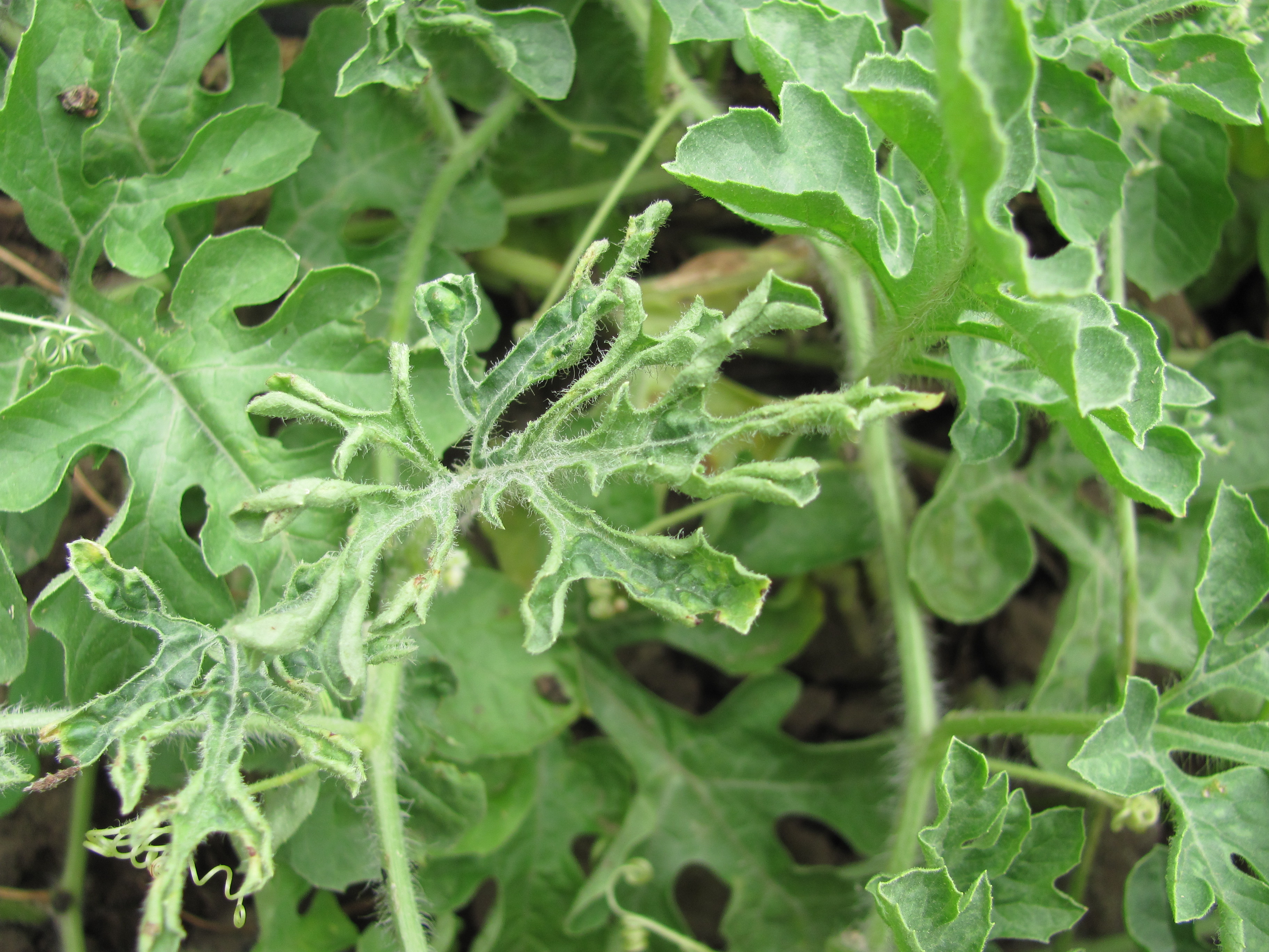 Figure 2. Papaya ringspot virus of watermelon.
 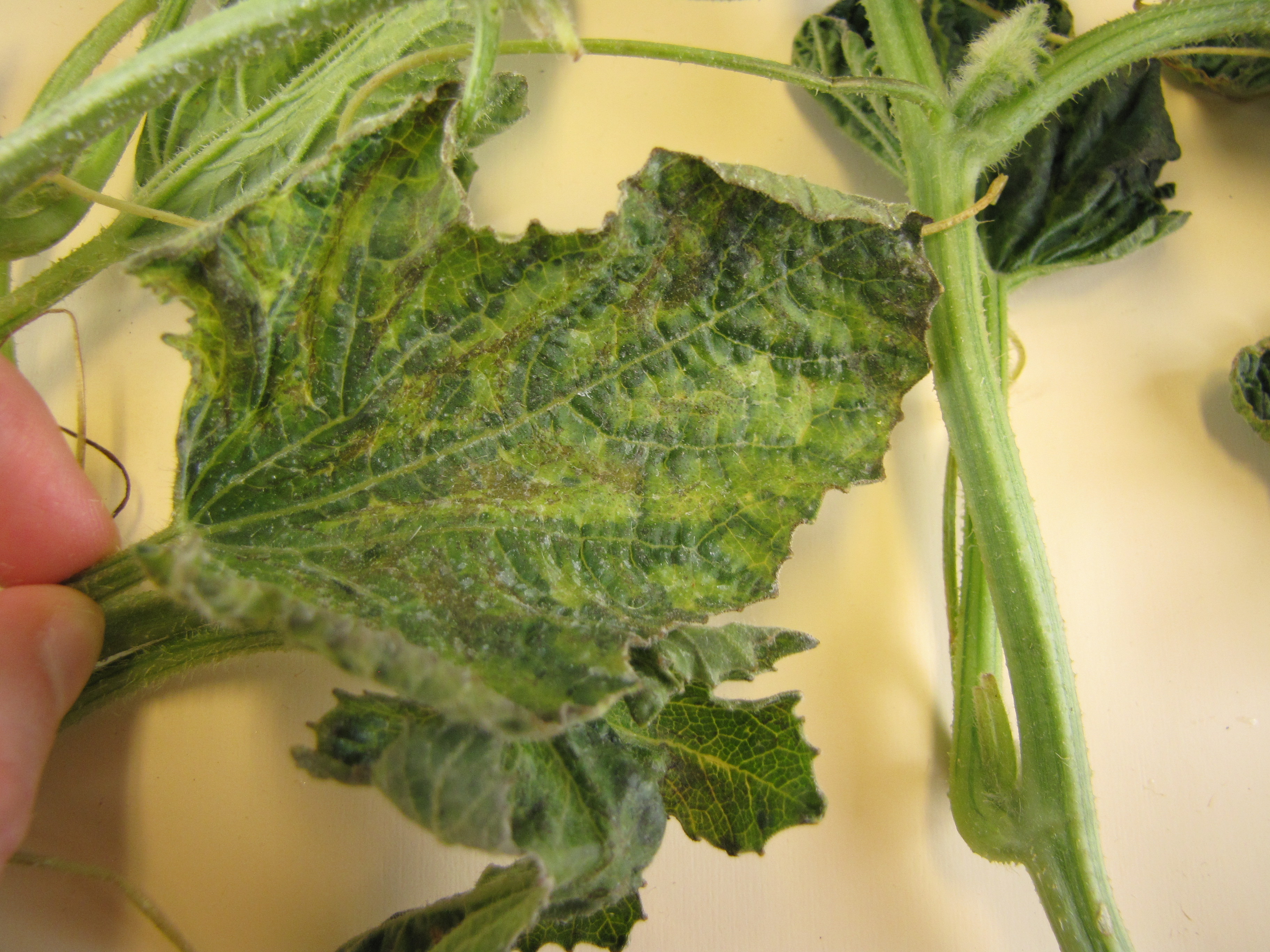 Figure 3. Watermelon leaf positive for potyvirus.
 White mold
White mold-While this disease has a huge host range, I have not known it to be an important problem on watermelon in Indiana. Below it is shown only in a transplant tray. However, I have observed vine death in the field due to white mold. Diagnostic is the irregular, dark sclerotia as in other crops.
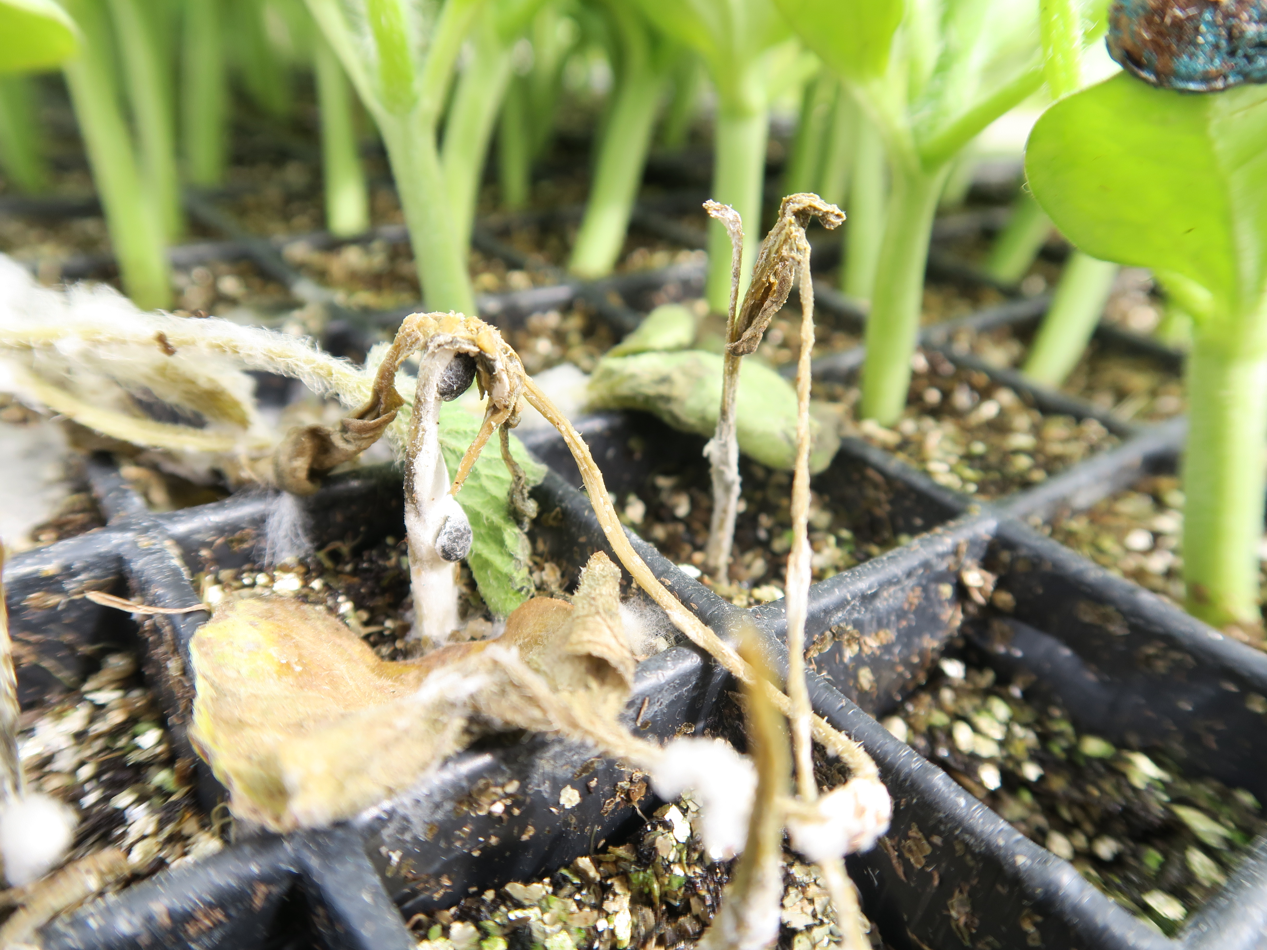 Figure 1. White mold of watermelon
 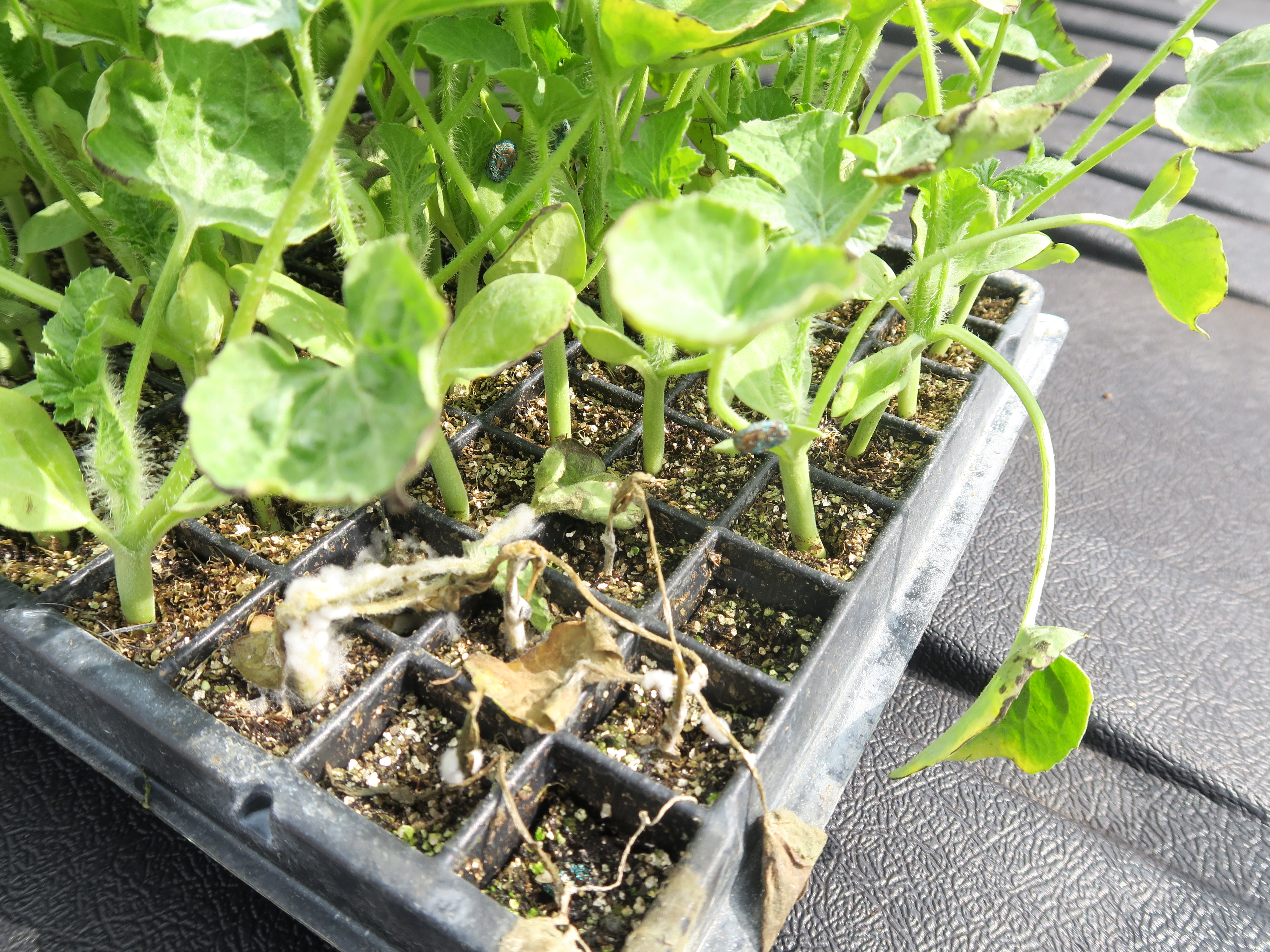 Figure 2. White mold of watermelon
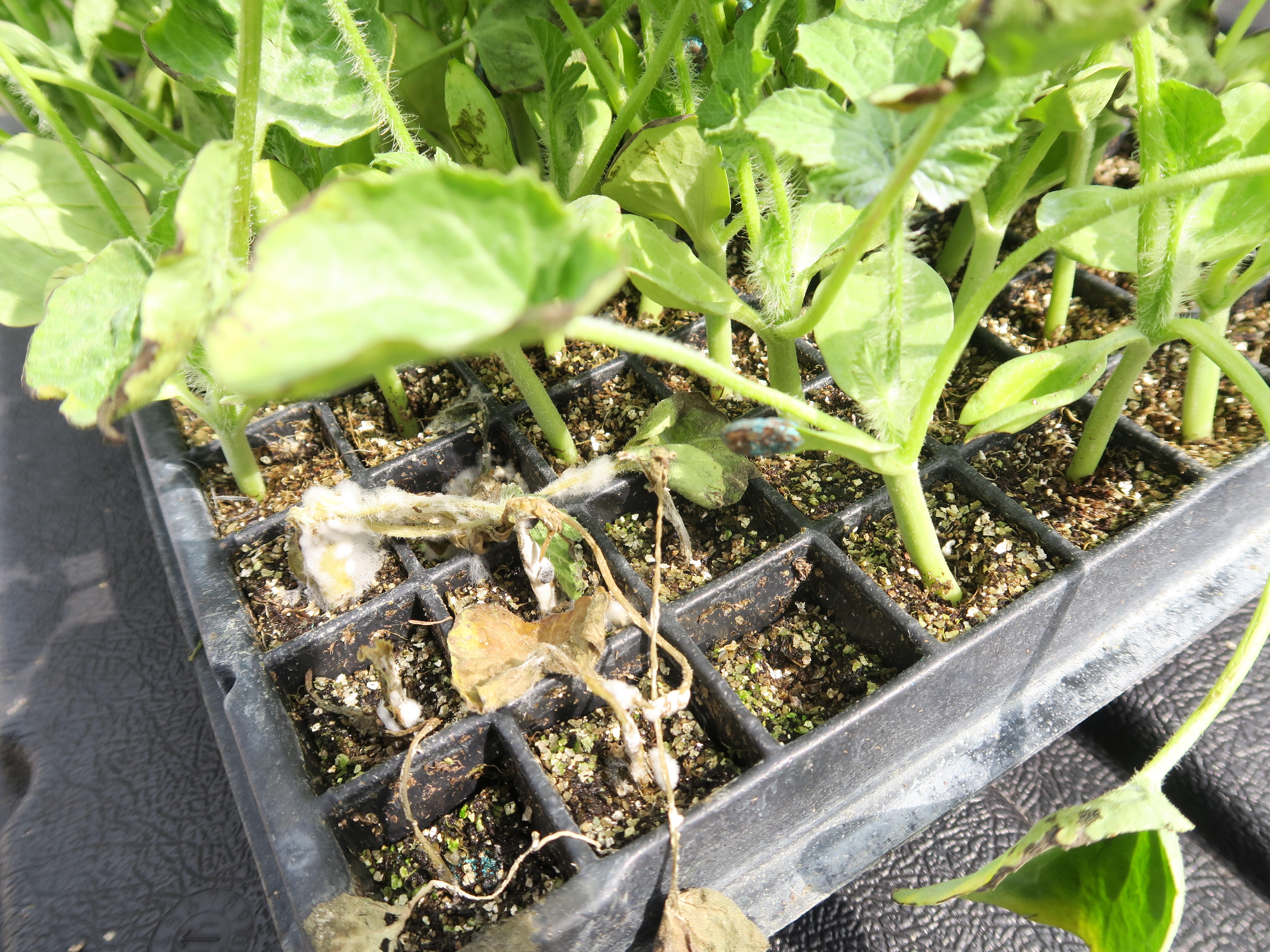 Figure 3. White mold of watermelon.