

pumpkin diseases
Pumpkins may be produced in relatively large acreage's for wholesale. Most pumpkin production is direct seeded. Increasingly, pumpkin production is no till or reduced till acreage.
Anthracnose
Anthracnose of pumpkin and squash-The lesions on fruit can cause the fruit to be unmarketable. However, the disease is not common. I have never observed lesions on leaves.
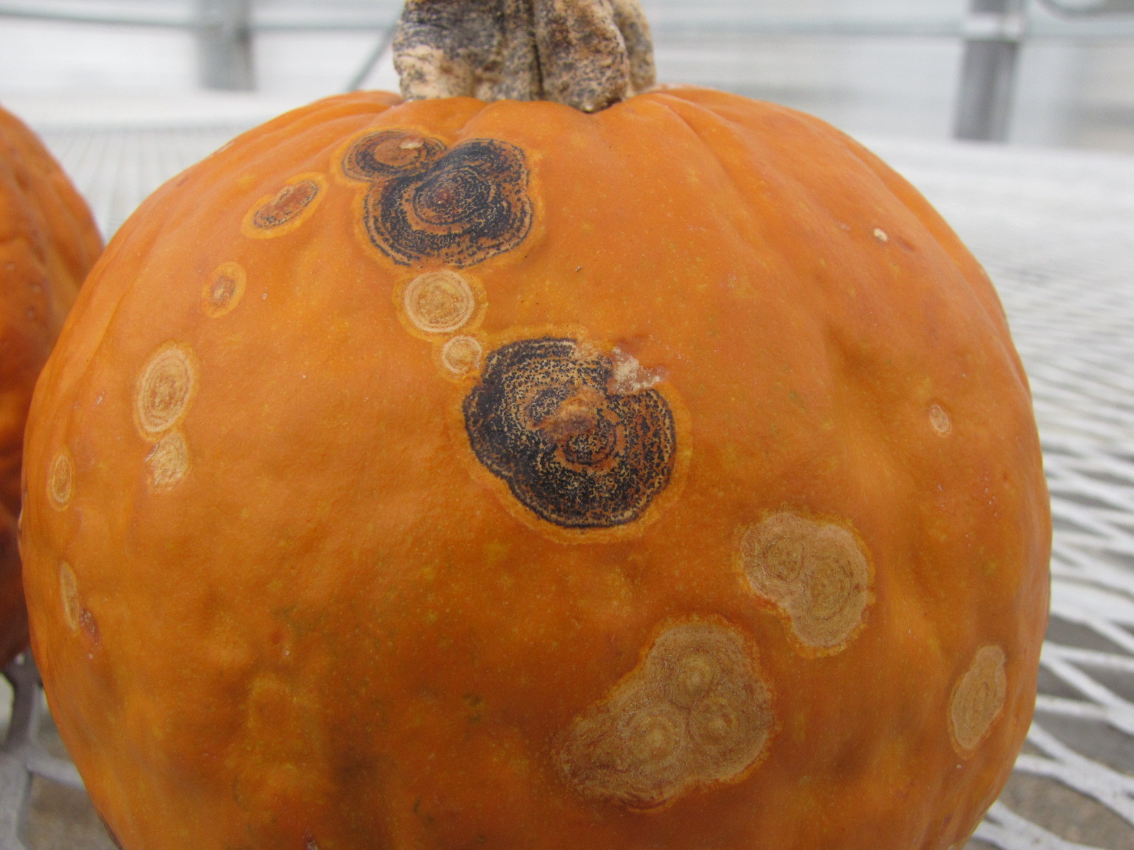 Figure 1. Lesions of anthracnose on pumpkin fruit often consist of round, dark rings on the fruit surface.
 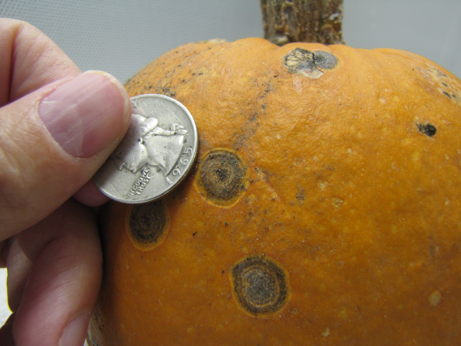 Figure 2. Anthracnose lesion on pumpkin in comparison to a quarter.
 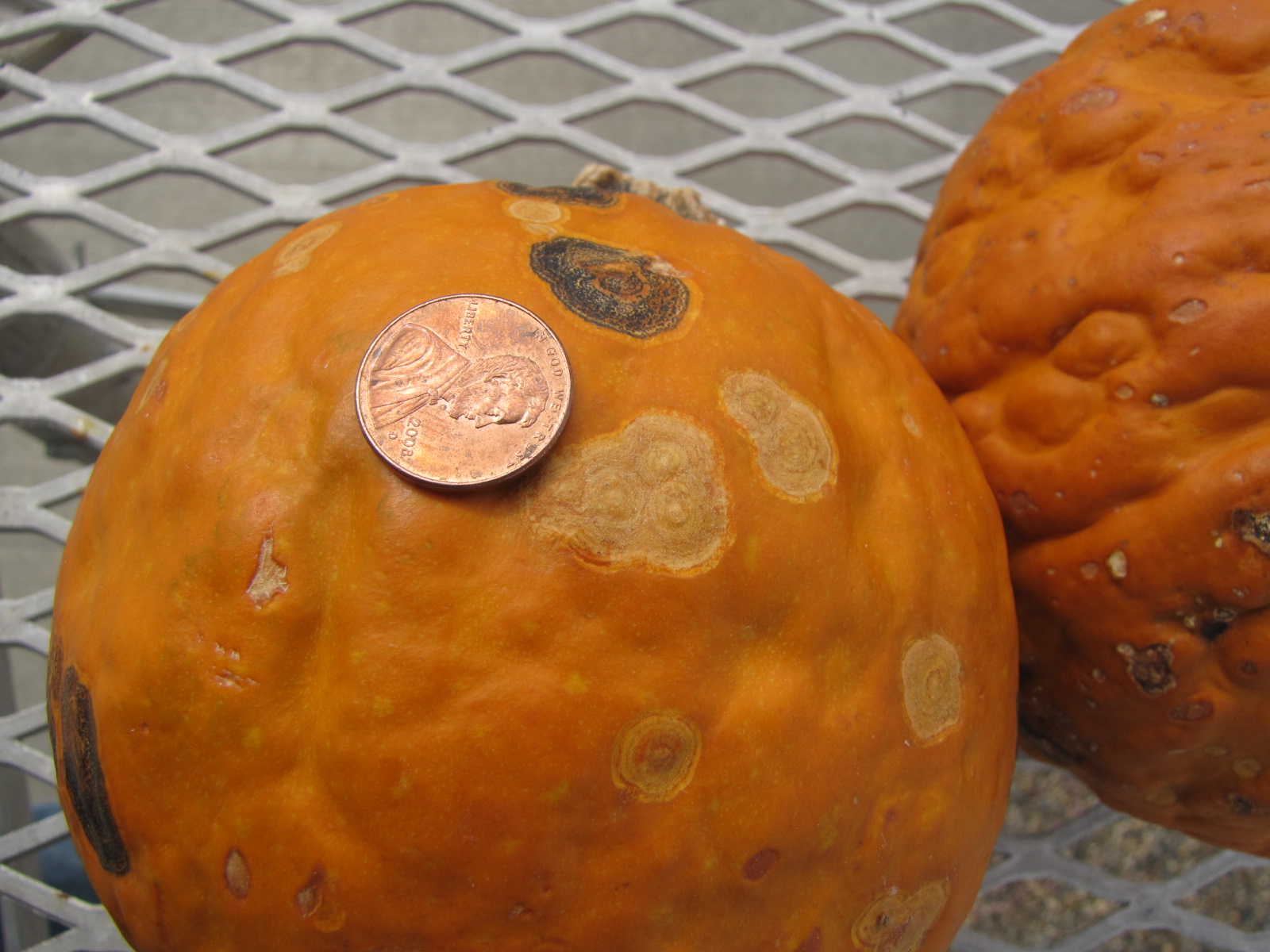 Figure 3. Anthracnose lesions of differing ages on pumpkin.
 Bacterial leaf spot
Bacterial leaf spot of pumpkin-Also known as Xanthomonas leaf spot. This continues to be one of the most important diseases of pumpkin. The lesions on leaves tend to be light brown and angular. However, lesions on leaves are not economically important. Leaf lesions do provide reservoirs of bacteria that may splash to fruit where they are responsible for raised, necrotic lesions often with water-soaked margins. Fruit lesions may affect marketability. In addition, lesions may be secondarily infected by fungi, creating enlarged lesions which may result in the rotting of the entire fruit.
 Figure 1. Lesions of bacterial leaf spot on a pumpkin leaf are often a light brown and maybe somewhat angular in shape.
 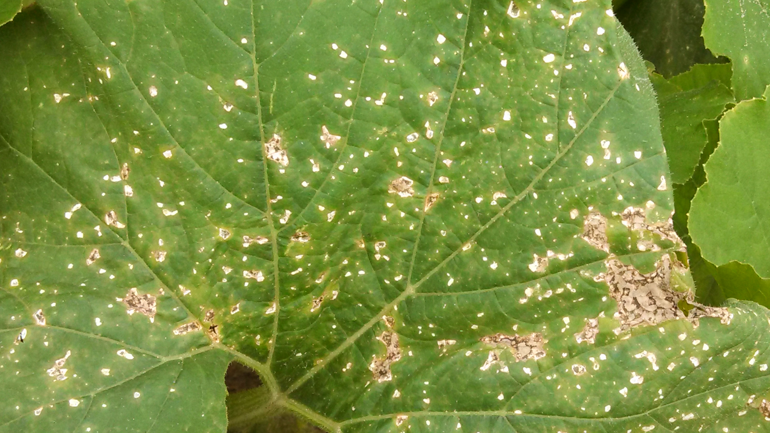 Figure 2. Close up of lesions on pumpkin of bacterial leaf spot.
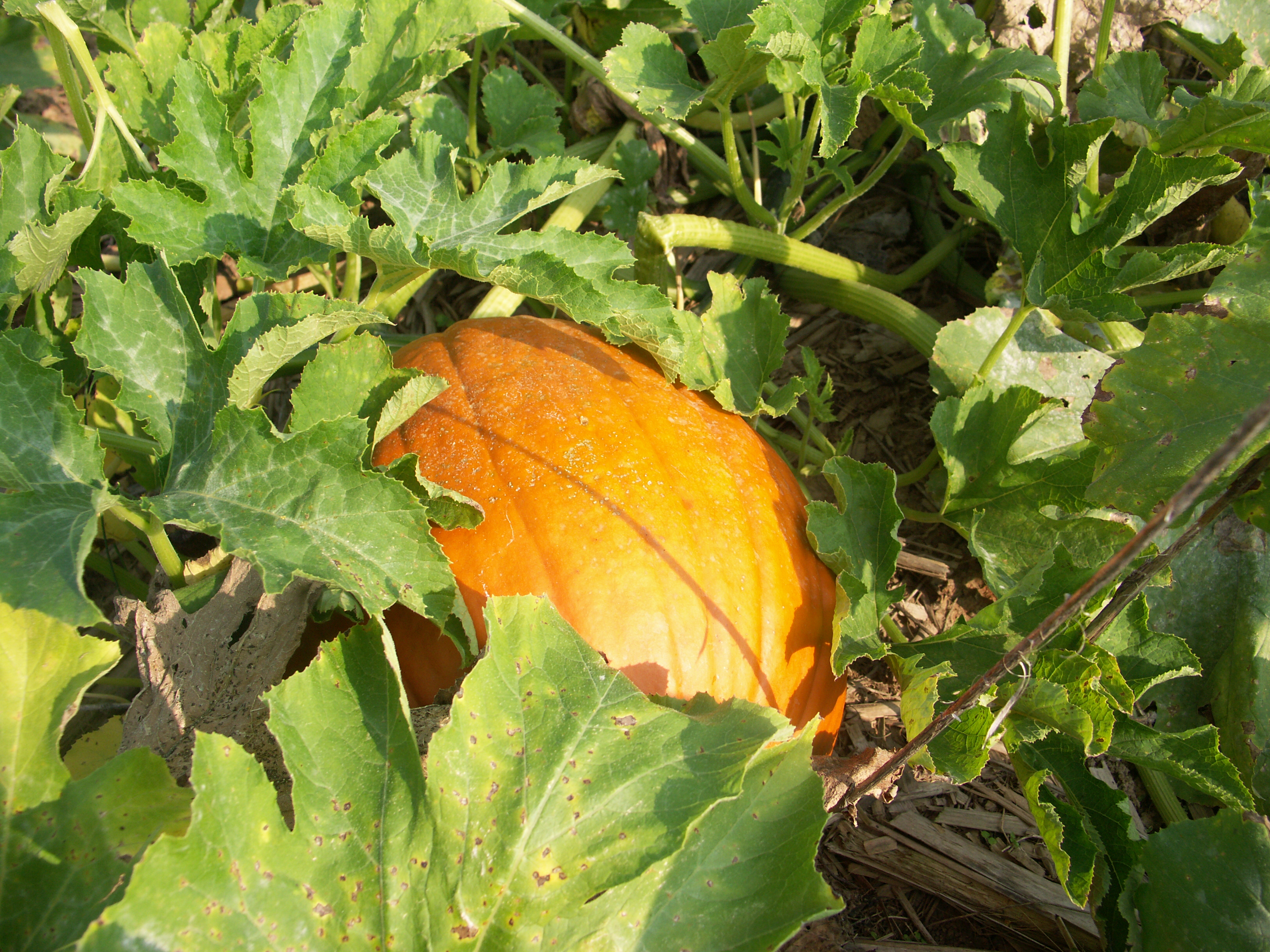 Figure 3. The bacterial spot lesions on the leaf in the foreground are a darker necrotic shade than the lesions in Figure 1 and 2.
 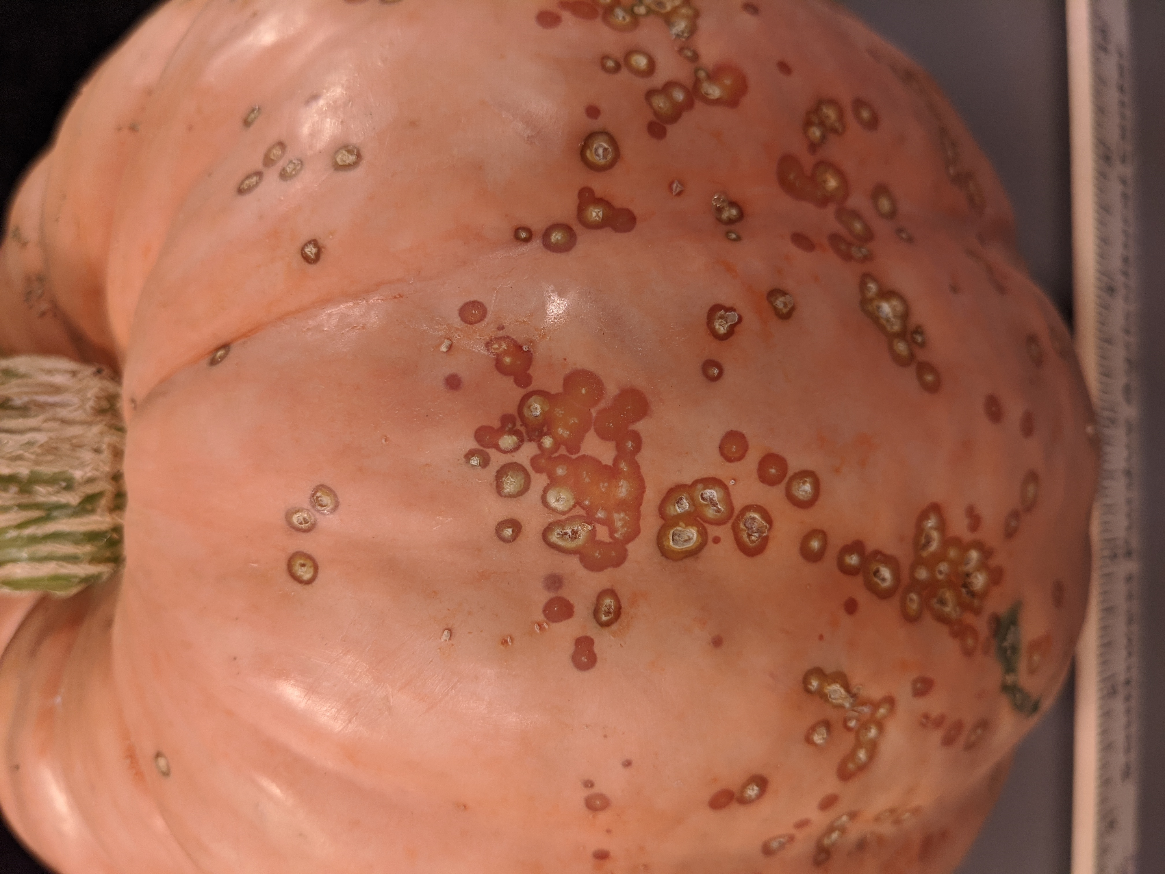 Figure 4. A specialty pumpkin with lesions of bacterial leaf spot of pumpkin. Note that lesions may have a water-soaked appearance. Older lesions may have a light necrotic center.
 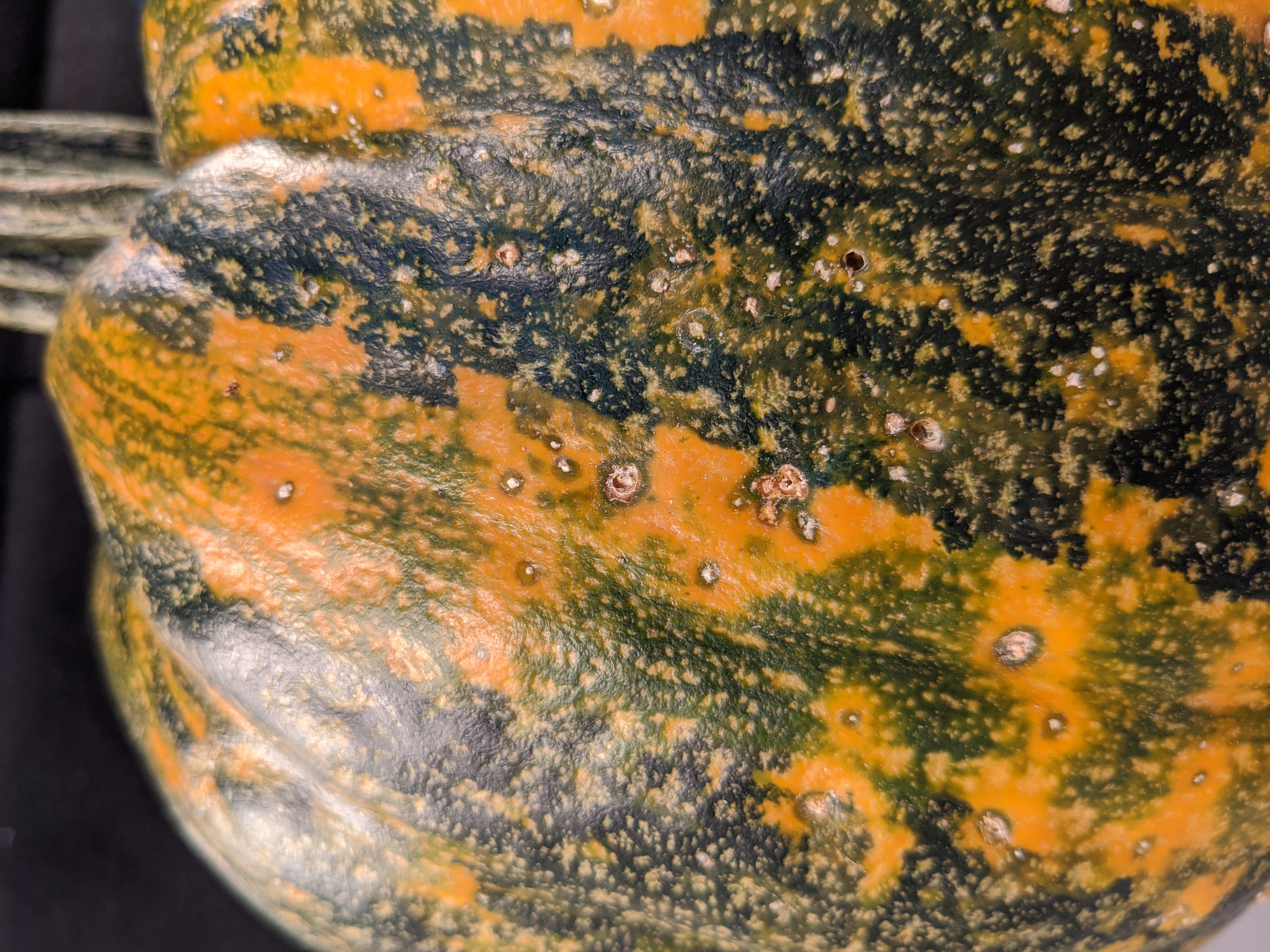 Figure 5. Lesions of bacterial spot on this pumpkin appear necrotic and may have small depressions in the center.
 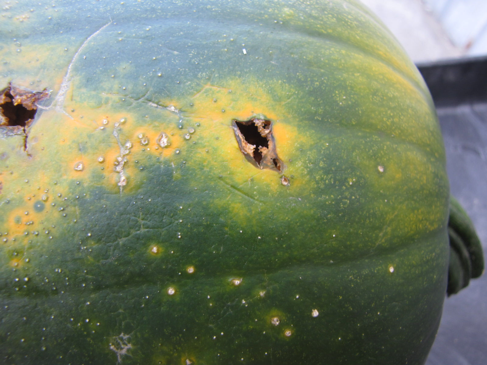 Figure 6. This immature pumpkin has lesions of bacterial spot of pumpkin. The lesions have the appearance of light necrotic scabs. The larger lesions are probably where one of the bacterial spot lesions became infected with a fungus that started in one of the bacterial spot lesions.
 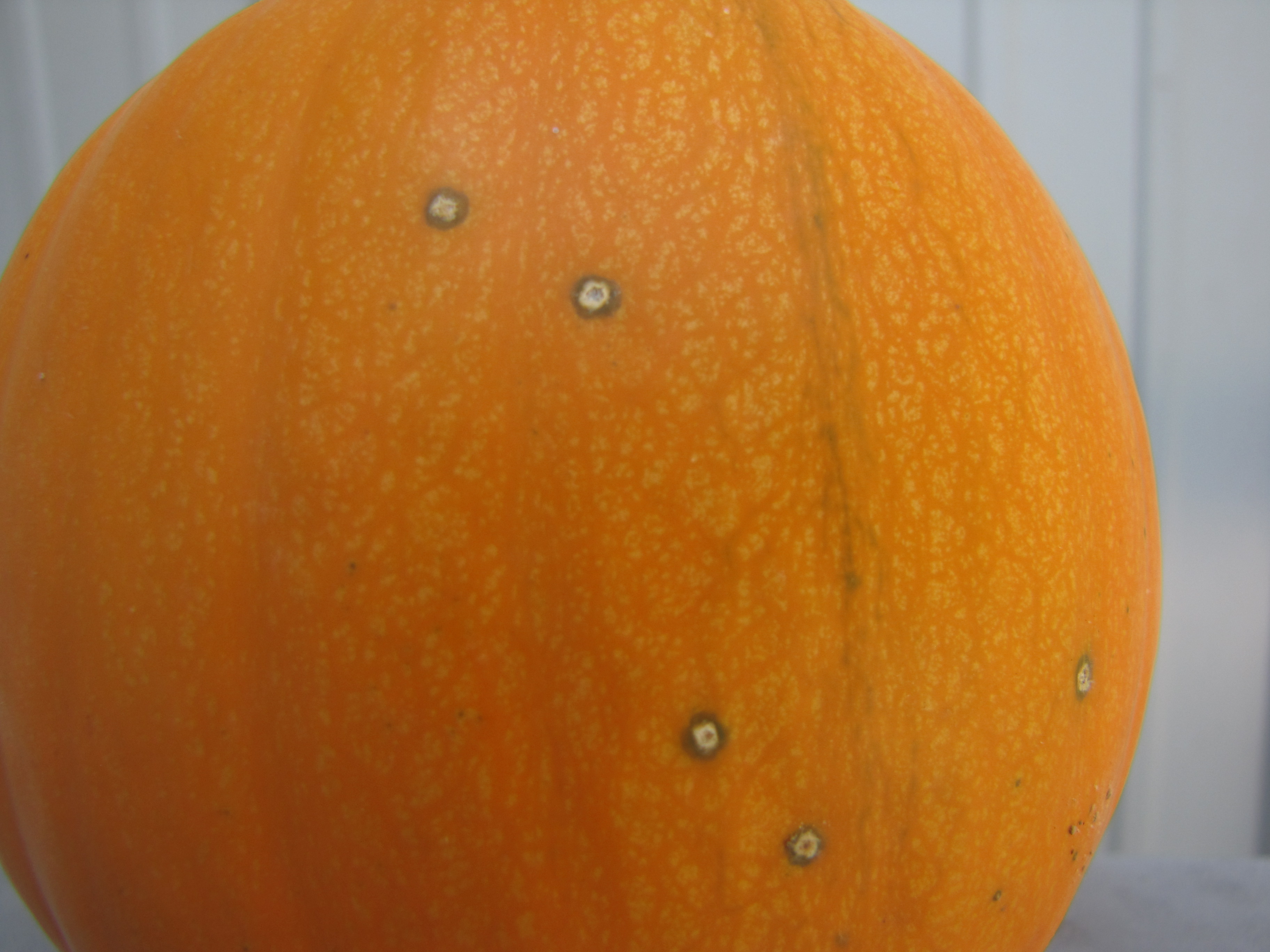 Figure 7. Bacterial spot lesions on a pie pumpkin.
 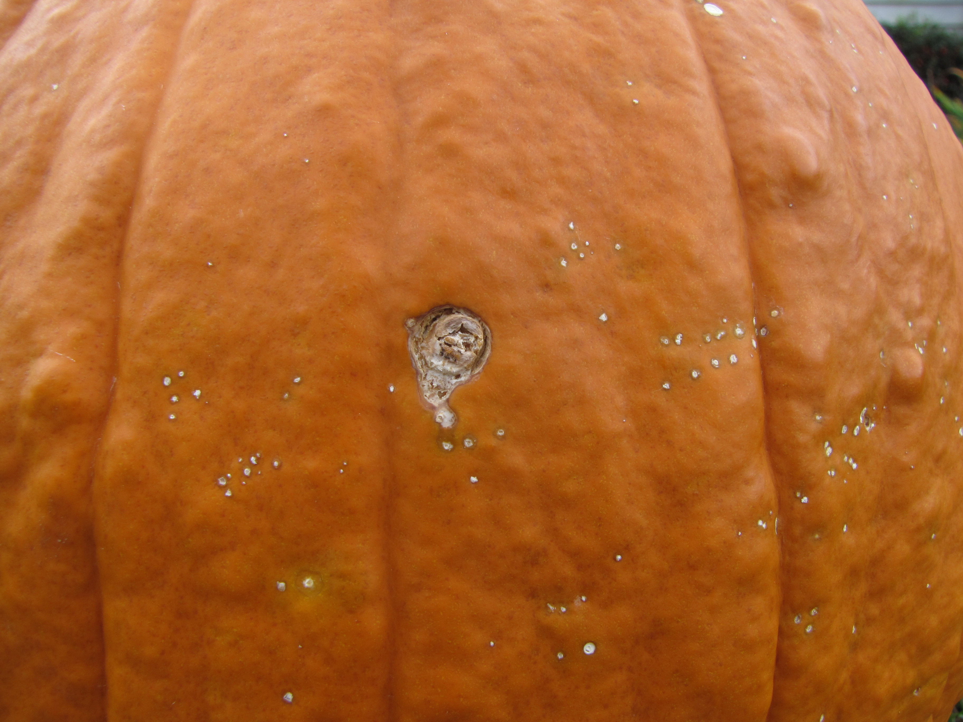 Figure 8. Several necrotic lesions caused by bacterial spot of pumpkin can be observed here. One lesion is much larger and has probably been infected by a secondary fungus.
 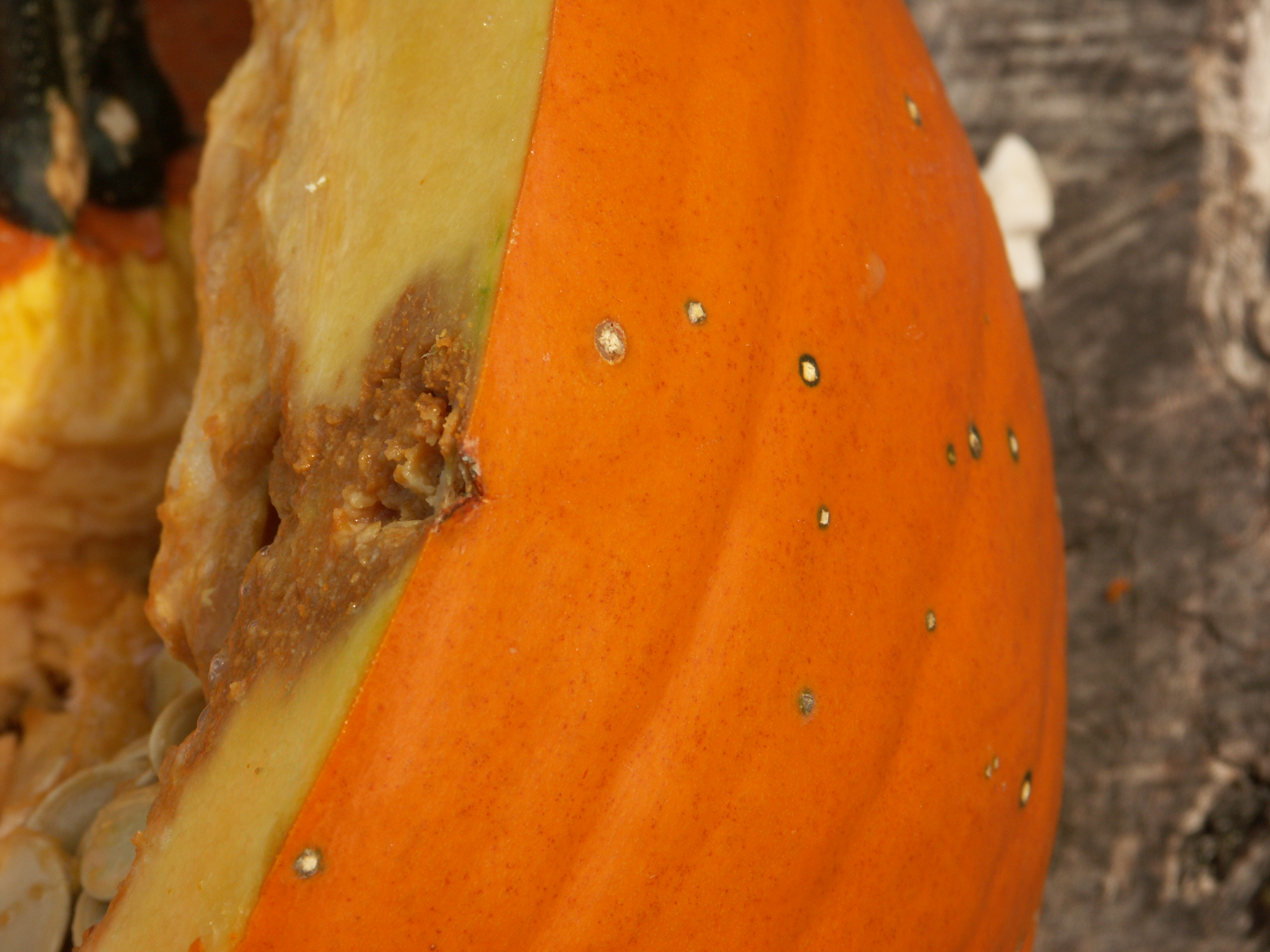 Figure 9. Typical lesions of bacterial spot can be observed on this pumpkin along with one that has been infected by secondary fungi causing it to rot through the pumpkin rind.
 Cercospora leaf spot
Cercospora leaf spot-Not a common disease nor an economically important one. However, it may be important to differentiate this disease from others such as bacterial leaf spot. Lesions on leaves are often a light brown and may be irregular in shape with no chlorotic halo. Fruit do not seem to be affected.
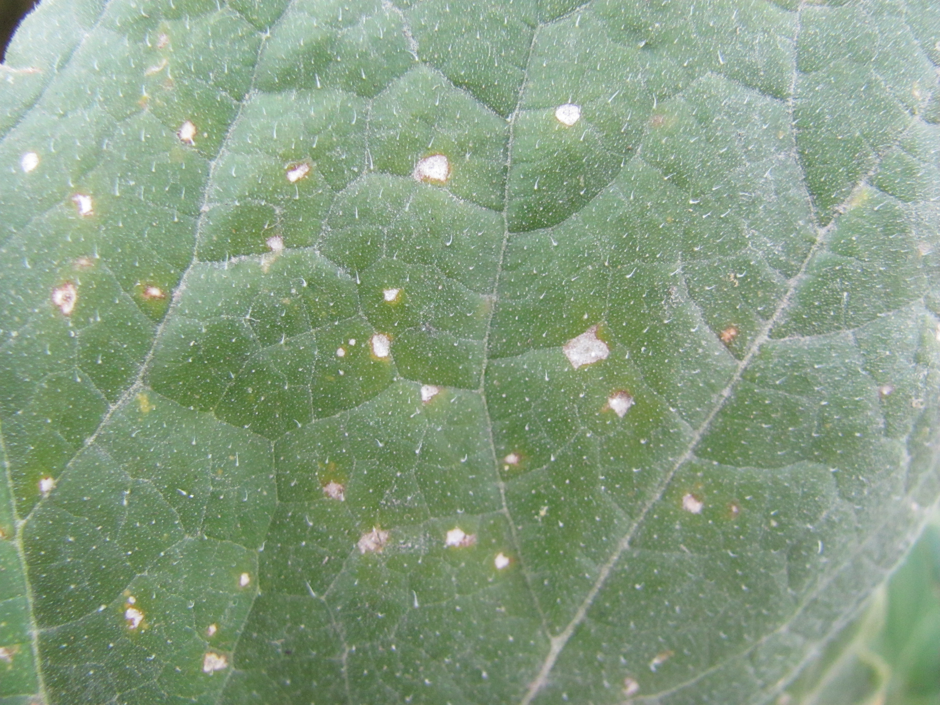 Figure 1. Cercospora leaf spot of pumpkin.
 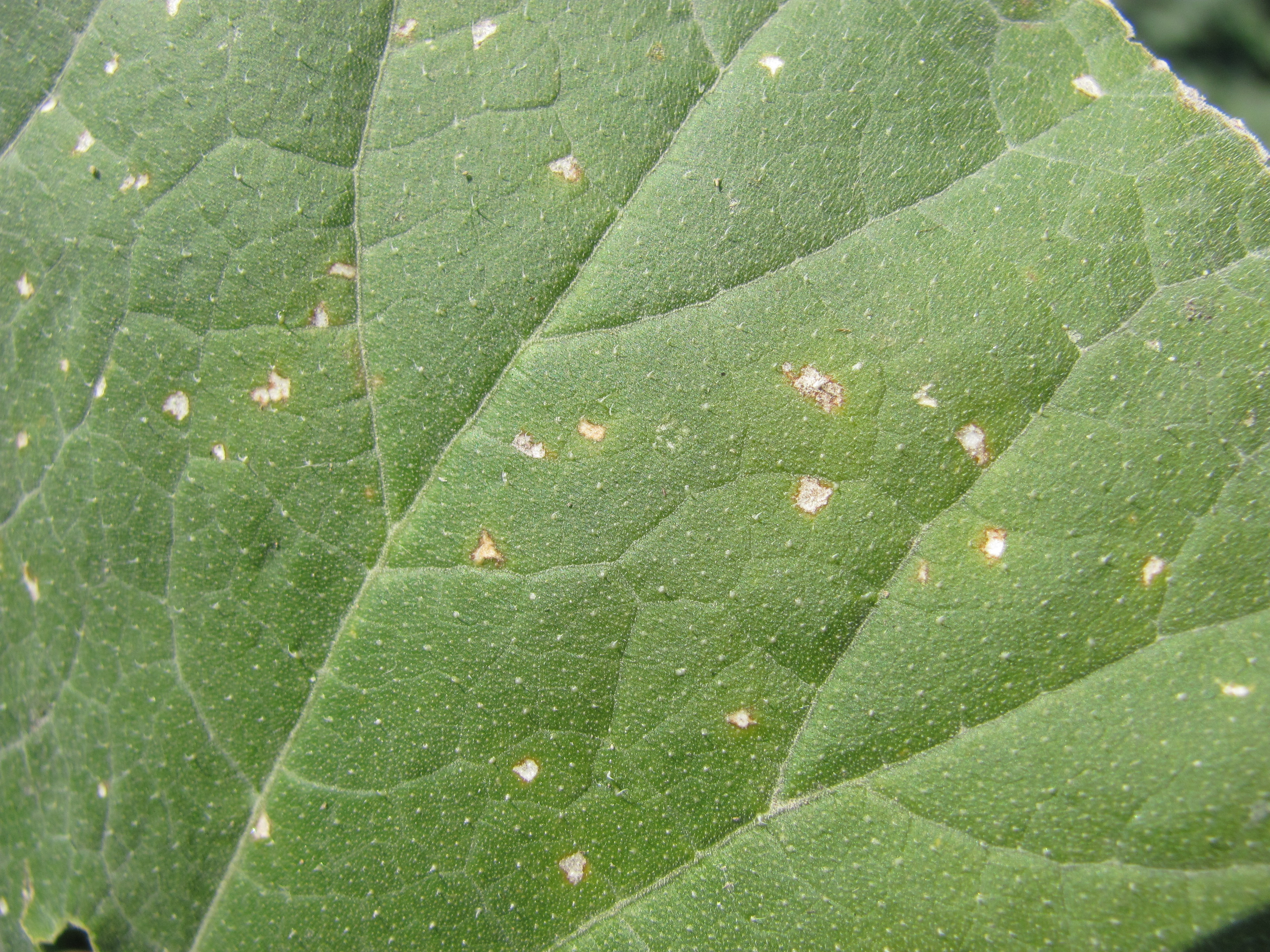 Figure 2. Cercospora leaf spot of pumpkin.
 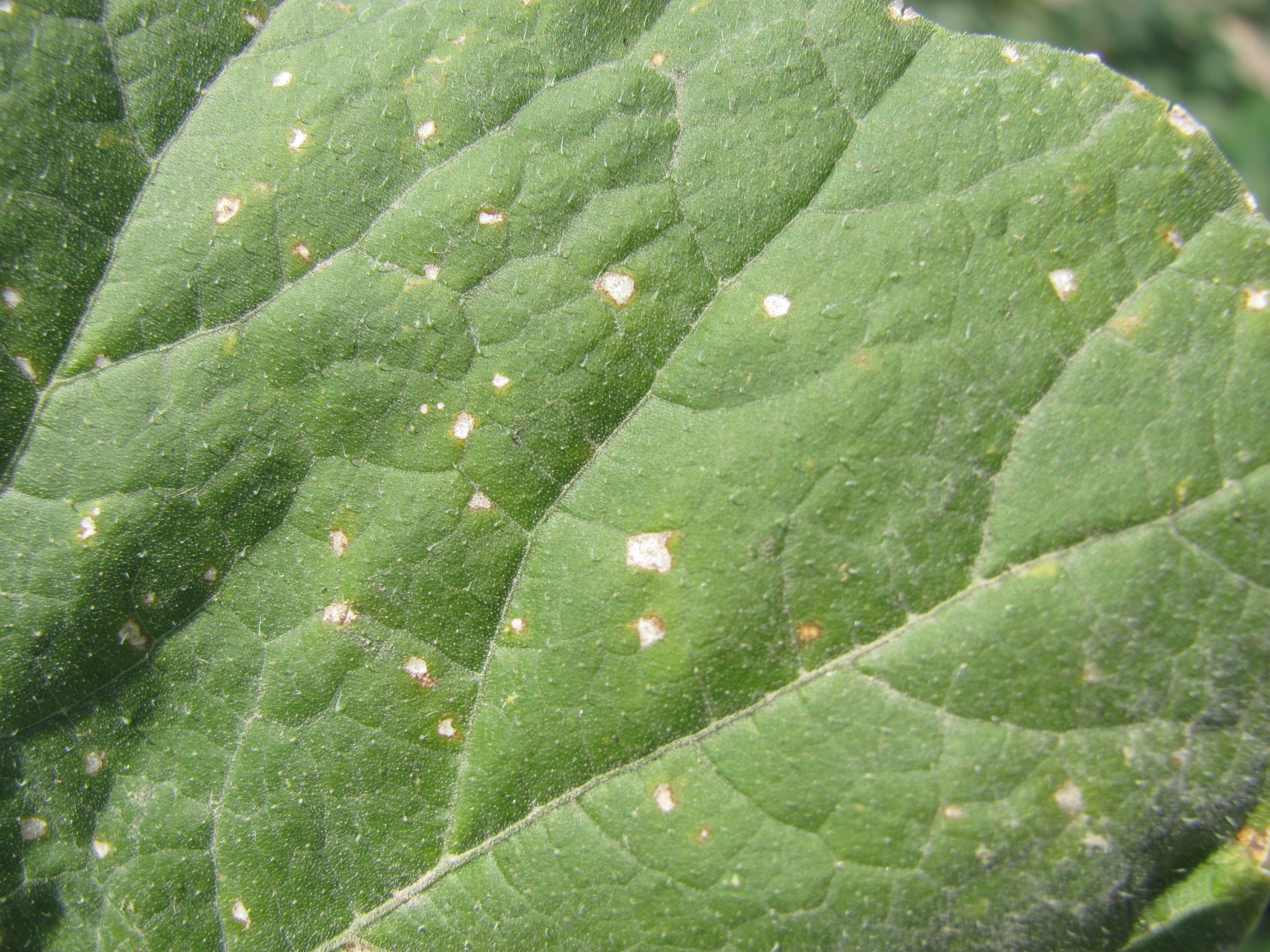 Figure 3. Cercospora leaf spot of pumpkin.
 Downy mildew
Downy mildew of pumpkin-Since the downy mildew causal organism doesn’t overwinter in Indiana, the disease doesn’t appear each year. However, the disease can be important if it appears in July through early September. On pumpkin, the lesions are often a mustard yellow and angular. Under moist conditions, sporulation can be observed on the underside of the leaf. When several lesions coalesce, the center of the enlarged lesion can turn necrotic. Fruit are not affected directly; therefore, late disease outbreaks may not require management.
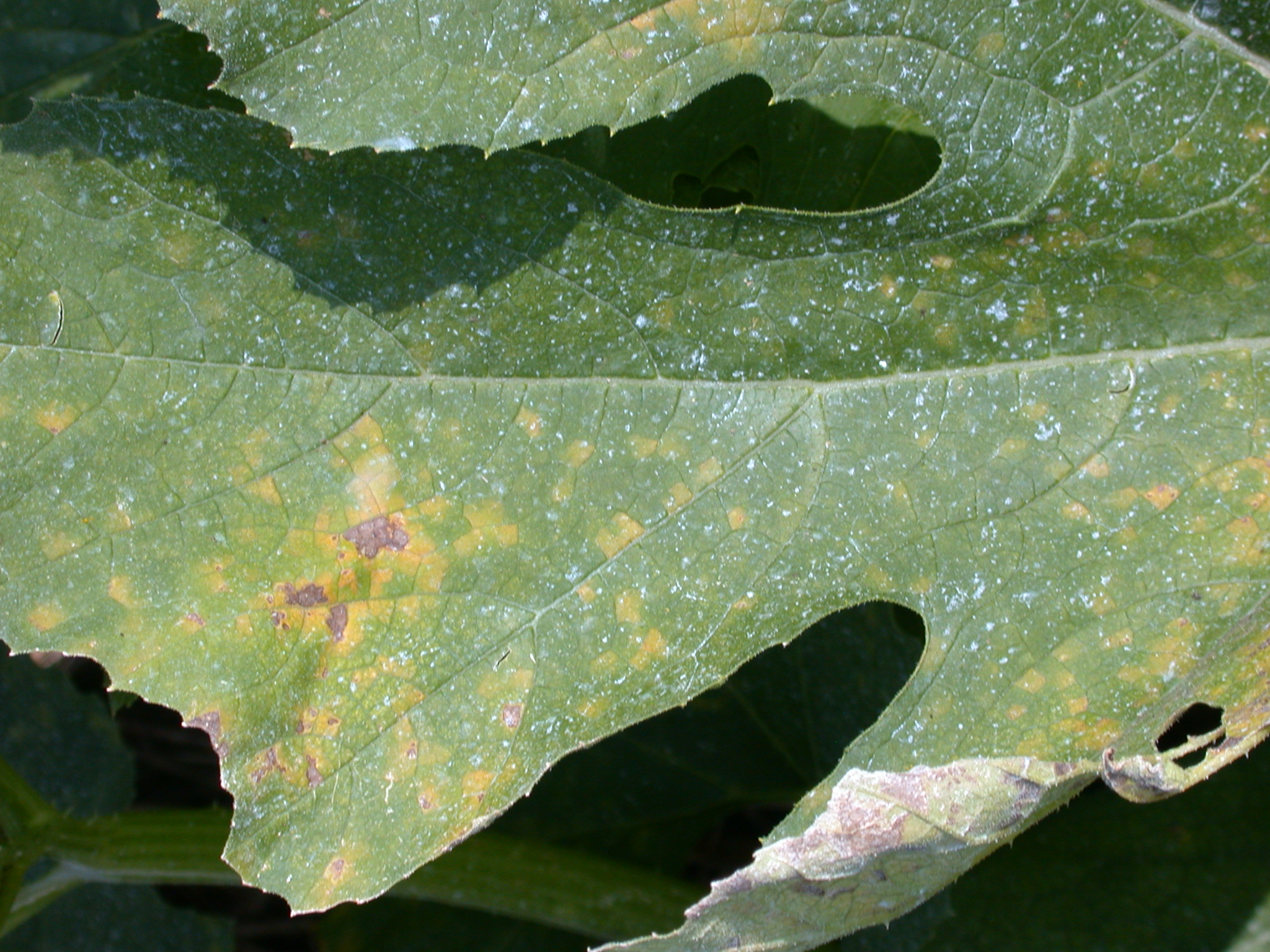 Figure 1. Downy mildew of pumpkin. Lesions tend to be angular and are initially chlorotic. Note older lesions have turned necrotic.
 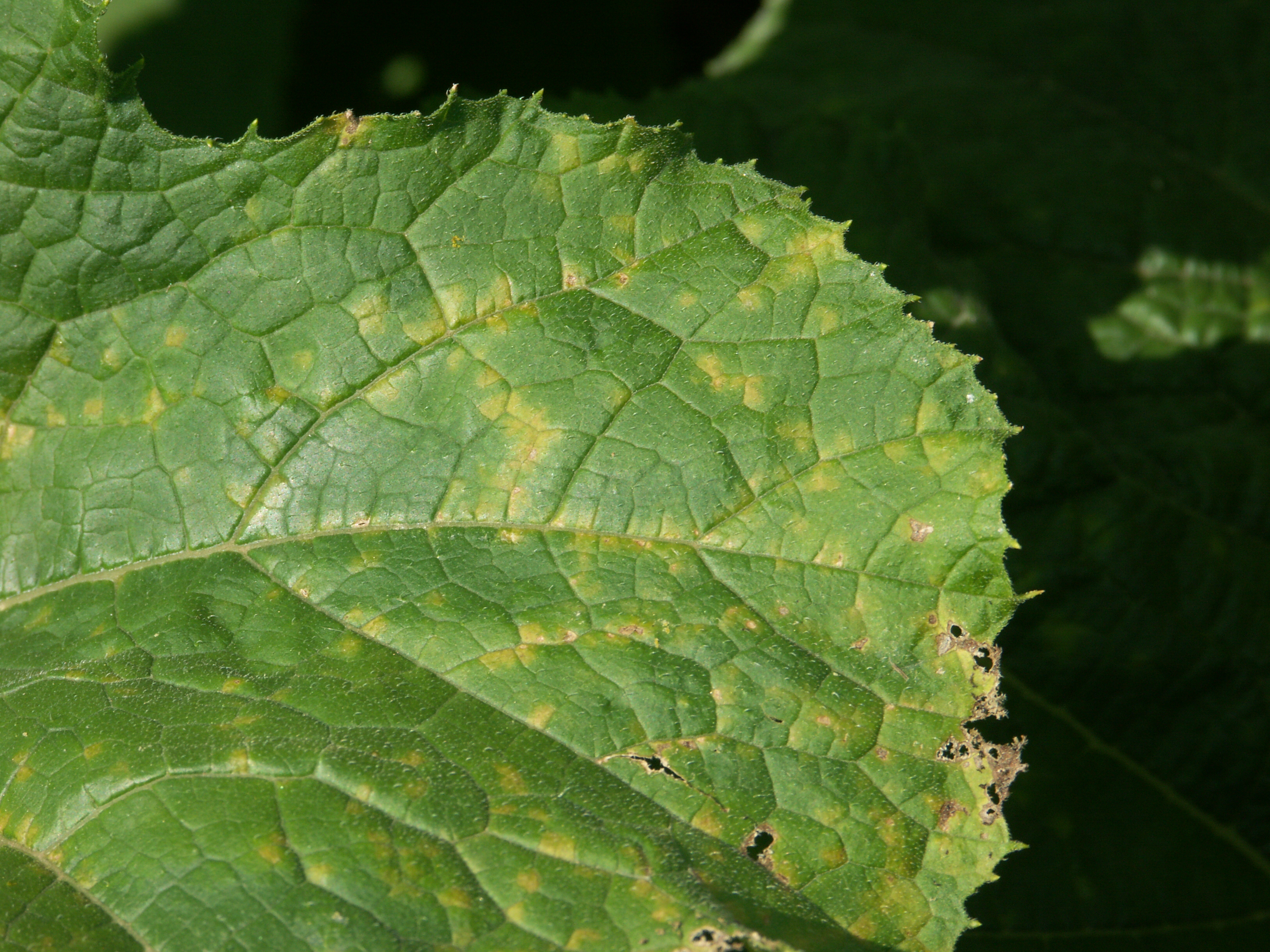 Figure 2. Downy mildew of pumpkin.
 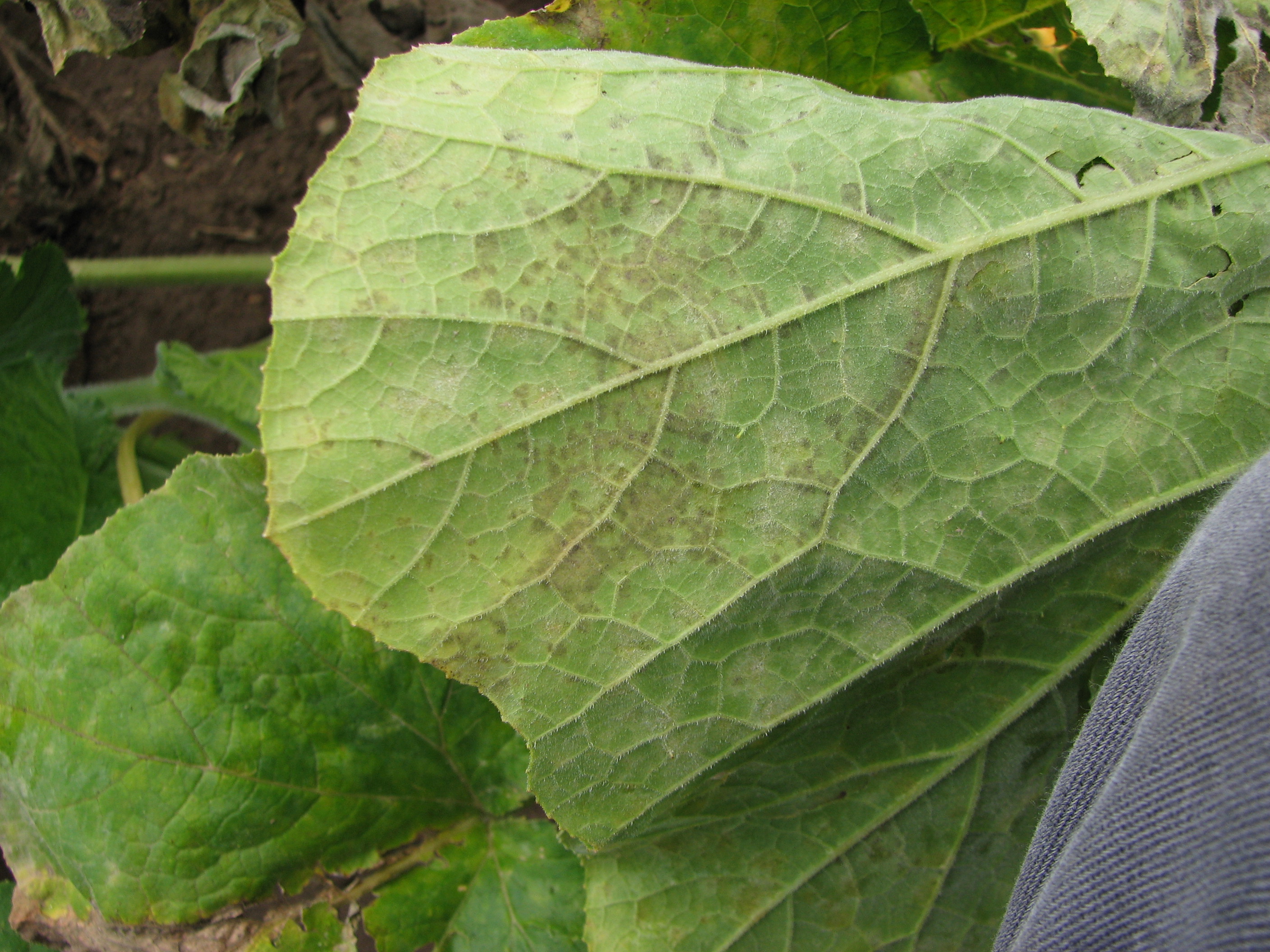 Figure 3. Under moist conditions, the causal fungus for downy mildew of pumpkin can be observed to sporulate on the underside of the leaf.
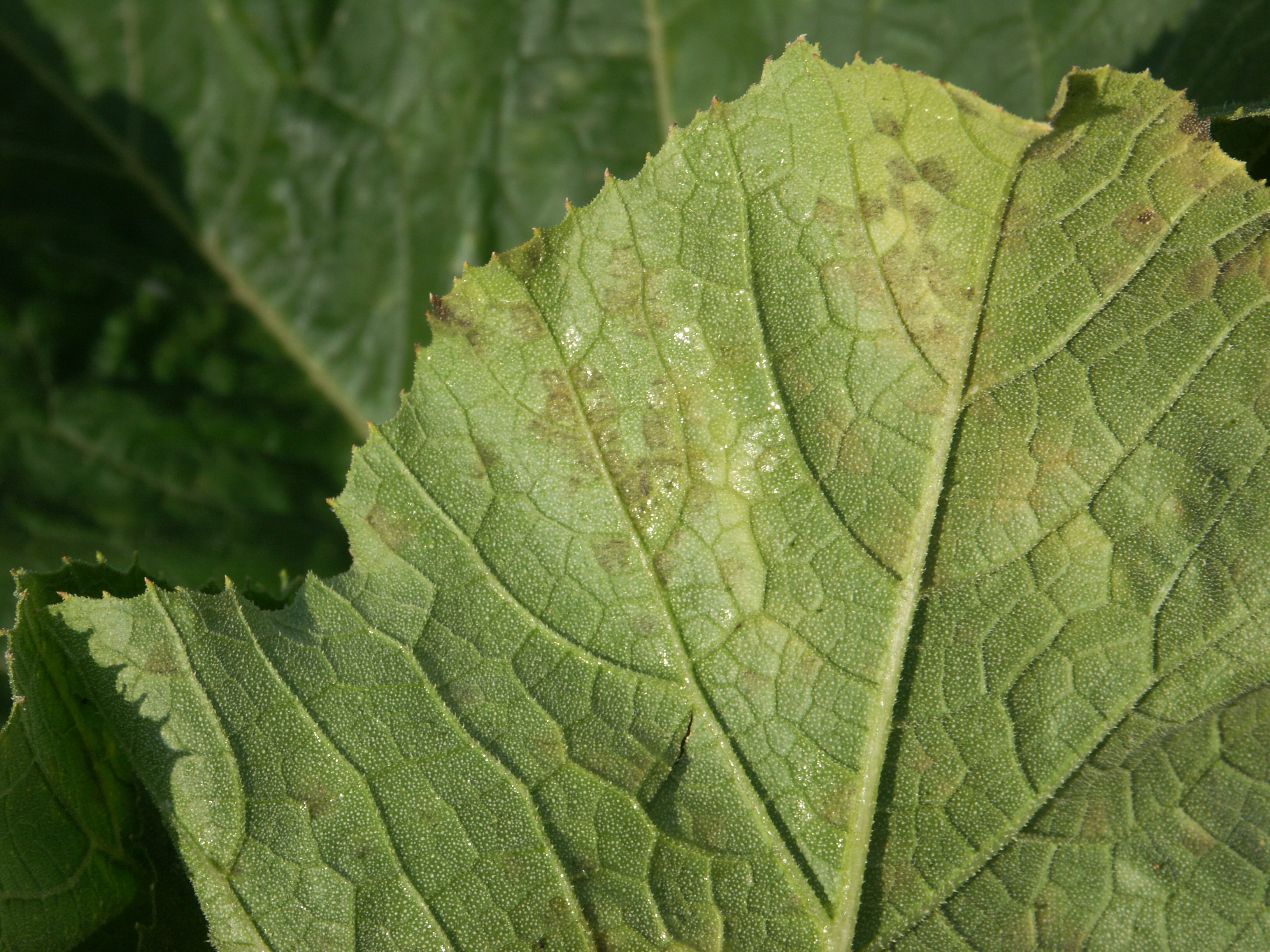 Figure 4. Sporulation on the underside of a pumpkin with downy mildew. Note pumpkin leaf is wet.
 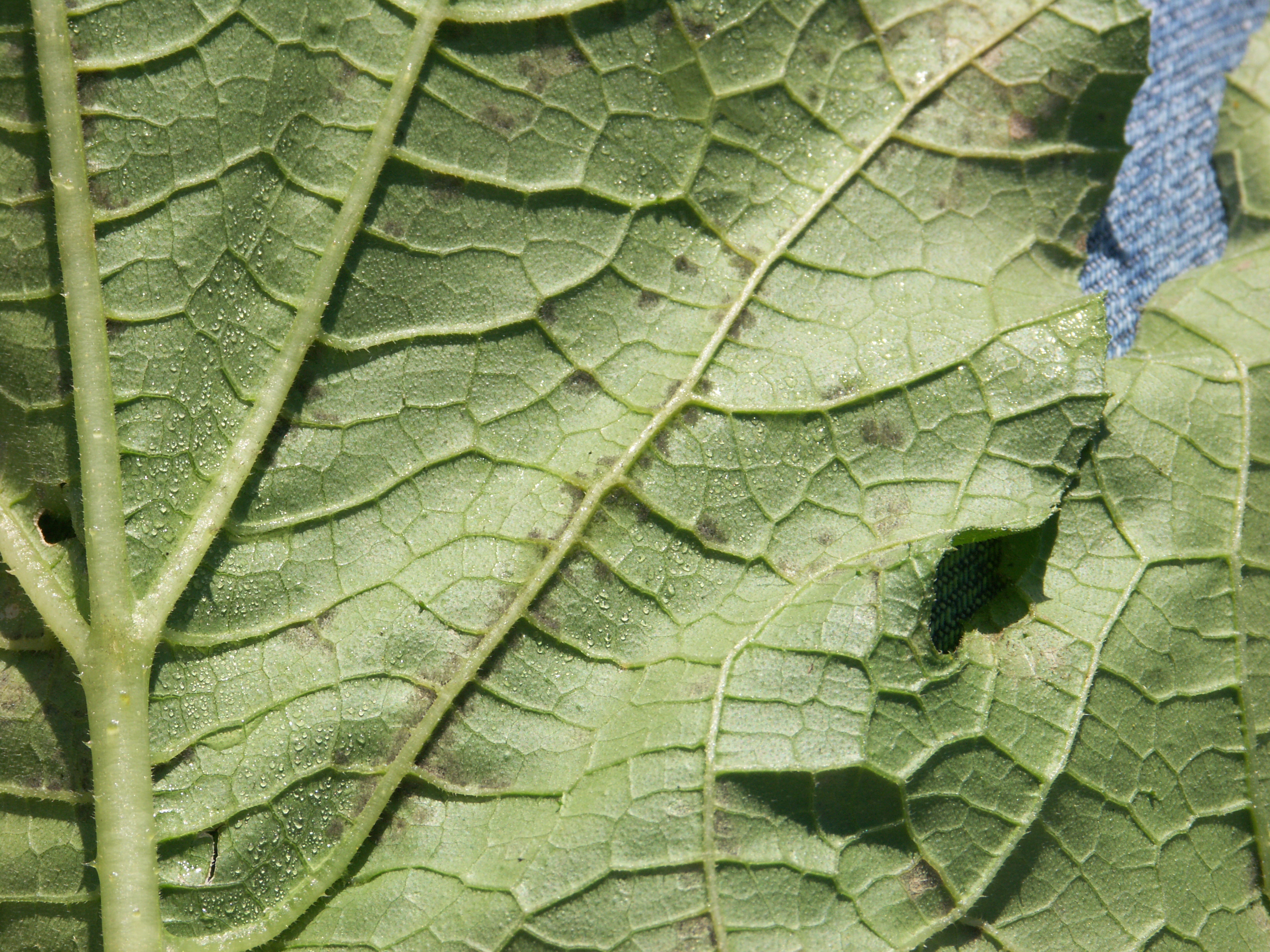 Figure 5. Downy mildew of pumpkin. Sporulation is visible on the underside of the leaf near the vein where moisture has accumulated.
 Fusarium fruit rot
Fusarium fruit rot-Fruit may initially seem to have a soft area. The lesion will become sunken. A white sporulation may be observed. Lesions of Fusarium fruit rot may be as a result of an initial bacterial leaf spot infection.
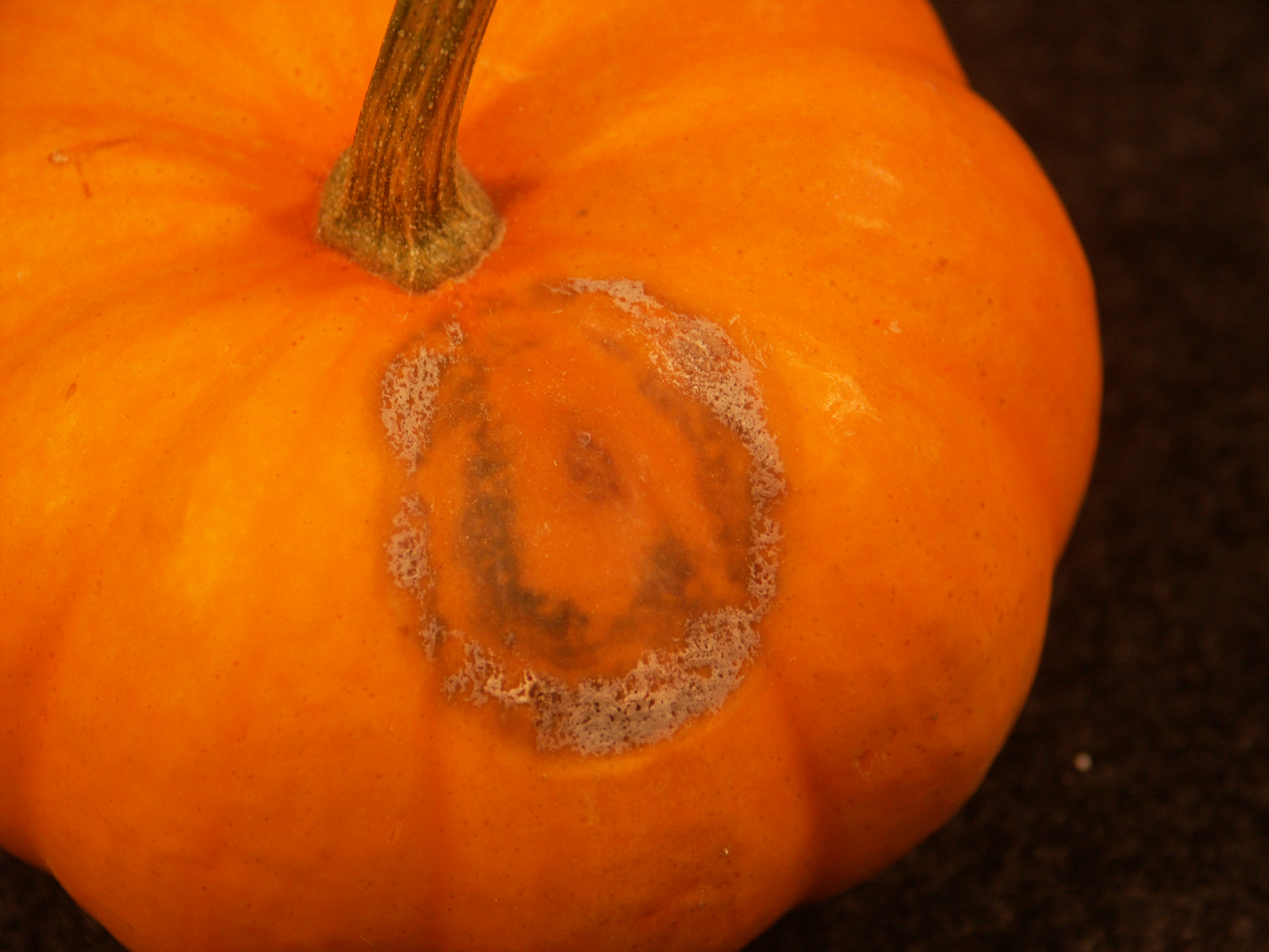 Figure 1. Fusarium fruit rot of pumpkin. Note gray/white sporulation.
 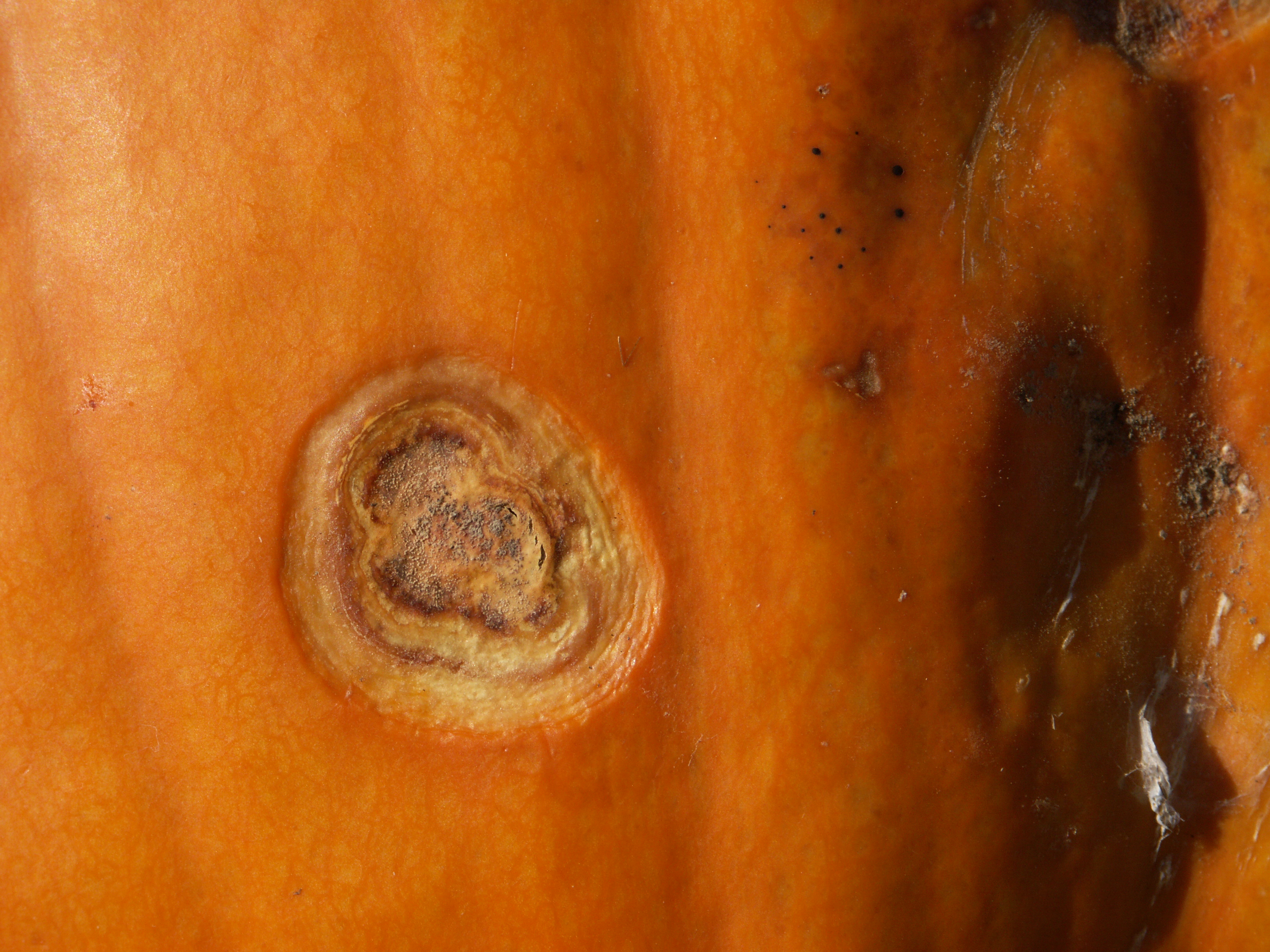 Figure 2. Fusarium fruit rot of pumpkin. Note sporulation in center of lesion.
 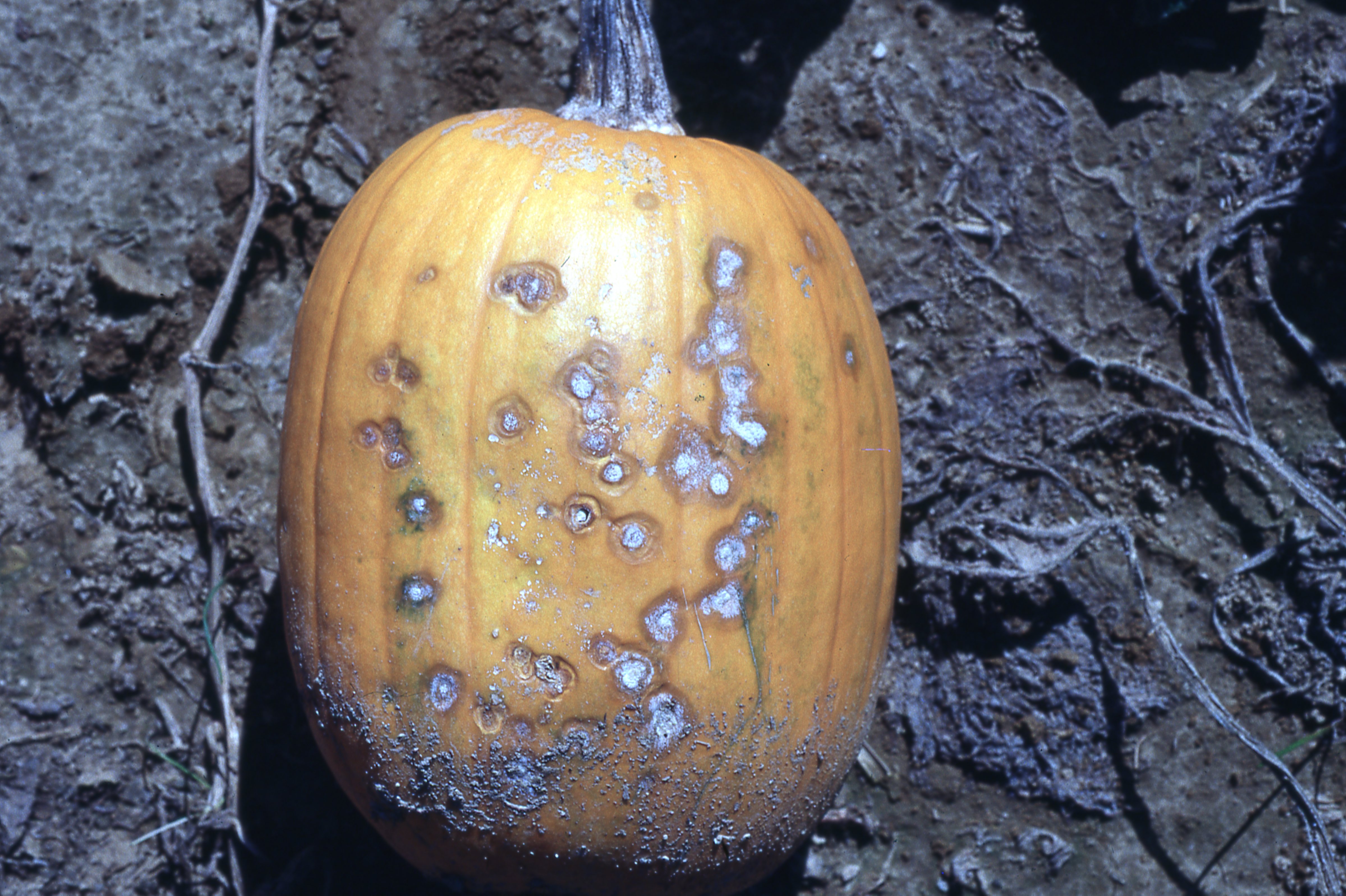 Figure 3. Fusarium fruit rot of pumpkin.
 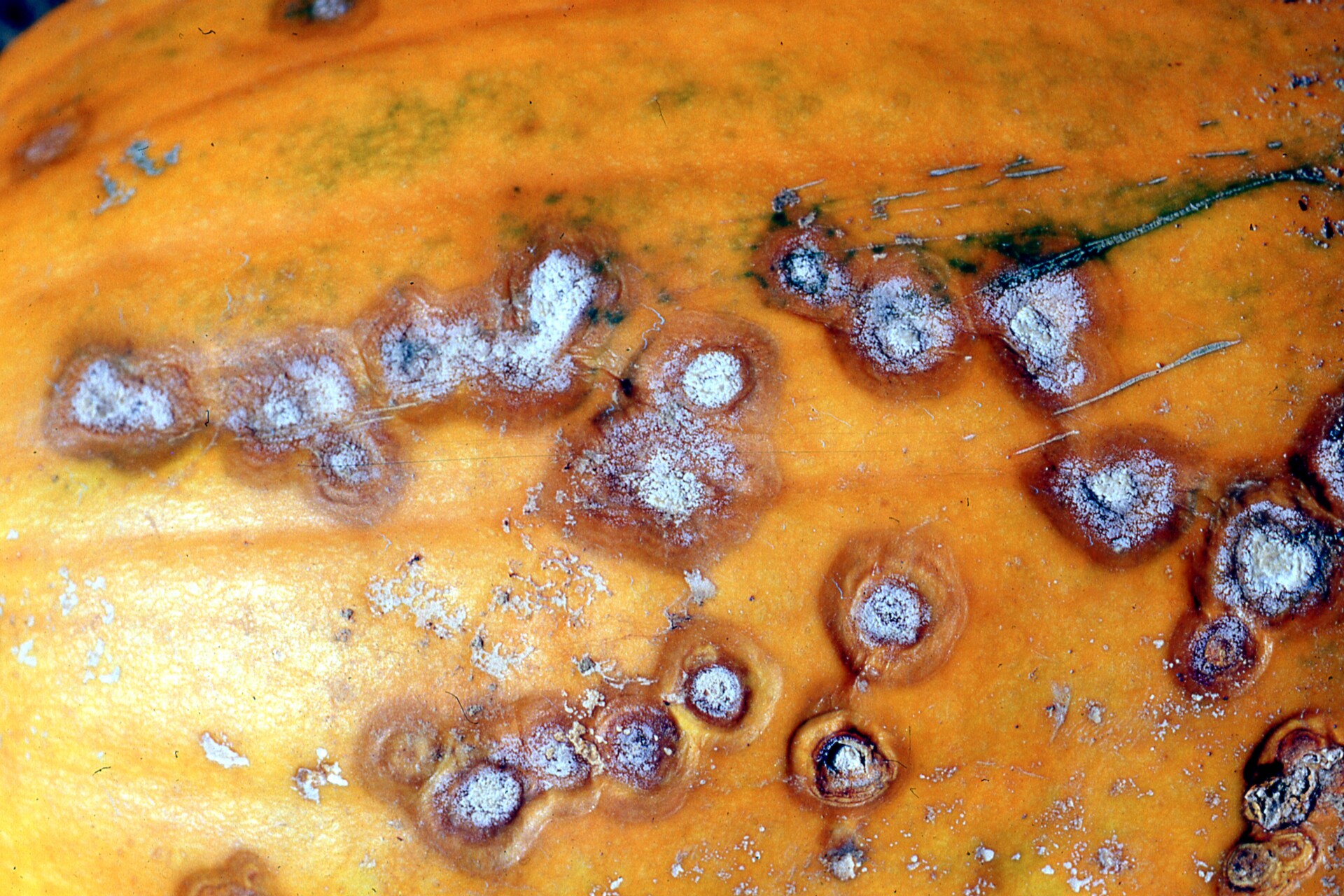 Figure 4. Fusarium fruit rot of pumpkin.
 Gummy stem blight / black rot of pumpkin
Gummy stem blight/black rot-Gummy stem blight is the name for symptom on leaves or stems. Black rot is the name for symptoms due to the same pathogen on fruit. Gummy stem blight symptoms are not economically important on pumpkin in Indiana. Black rot problems occasionally cause marketability issues. The latter symptoms can sometimes appear as a ring structure which may appear to be virus-like symptoms. However, dark fruiting bodies in the lesions are diagnostic.
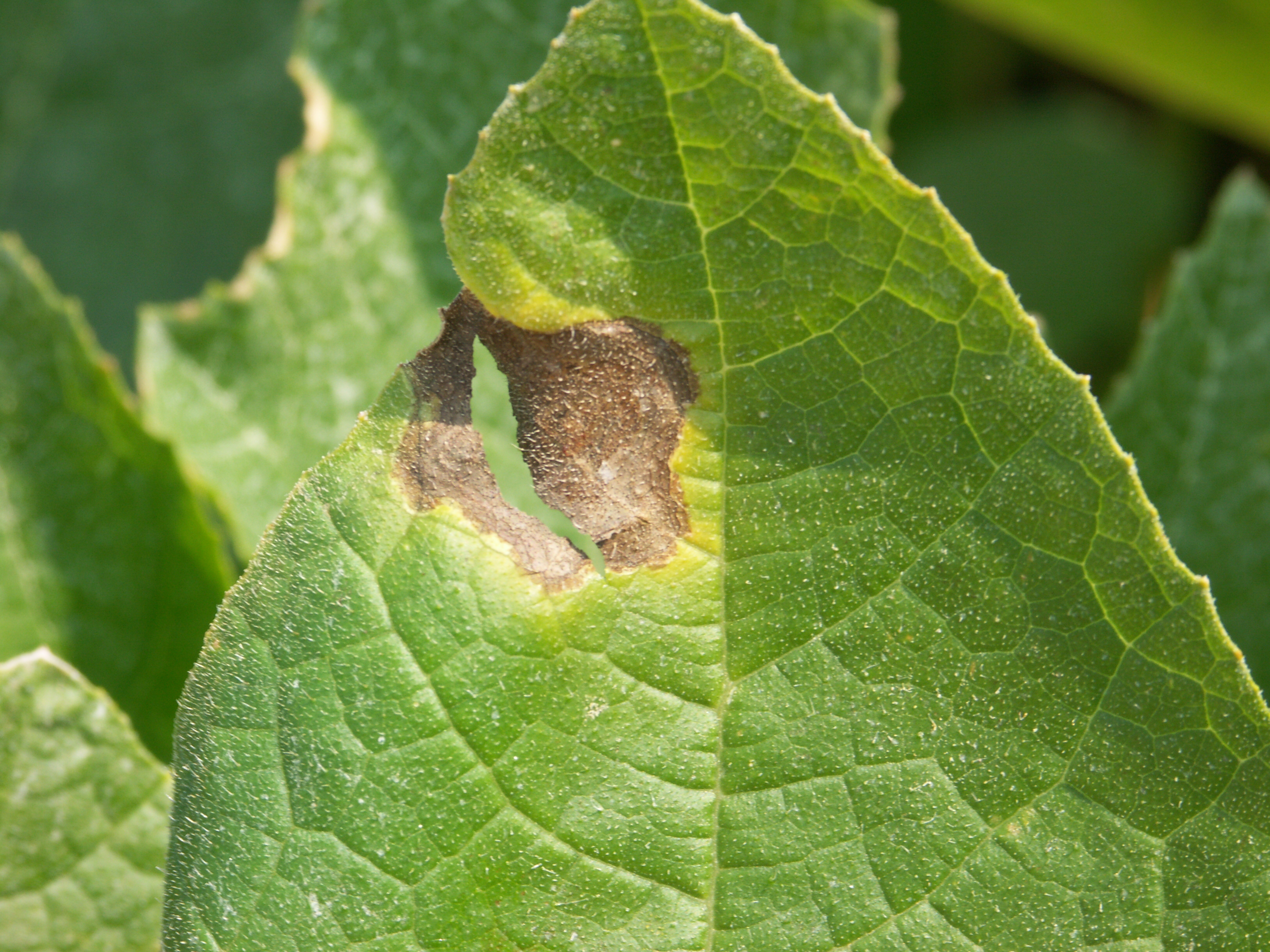 Figure 1. Gummy stem blight lesion on a pumpkin leaf.
 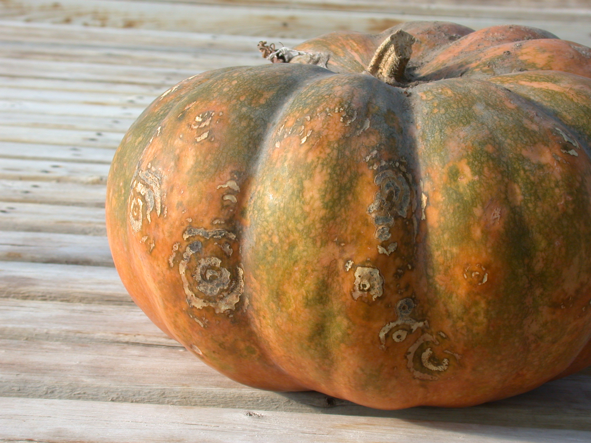 Figure 2. Black rot of pumpkin. Note target spot-like pattern of lesion.
 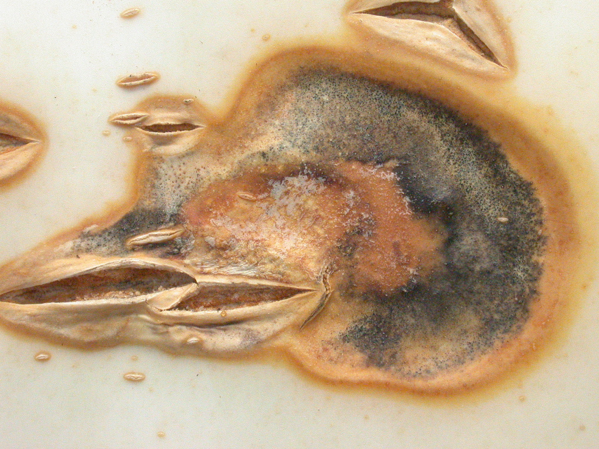 Figure 3. Close-up of black rot lesion on pumpkin.
 Phytophthora fruit rot
Phytophthora fruit rot of pumpkin-This important disease can cause fruit rot and a disease of the vine. Thus, the observer may first notice a wilt of vines. Lesions may occur on vines or on leaves. A damping-off may be observed. Fruit may become soft and develop white mold on the lesion, often toward the ground.
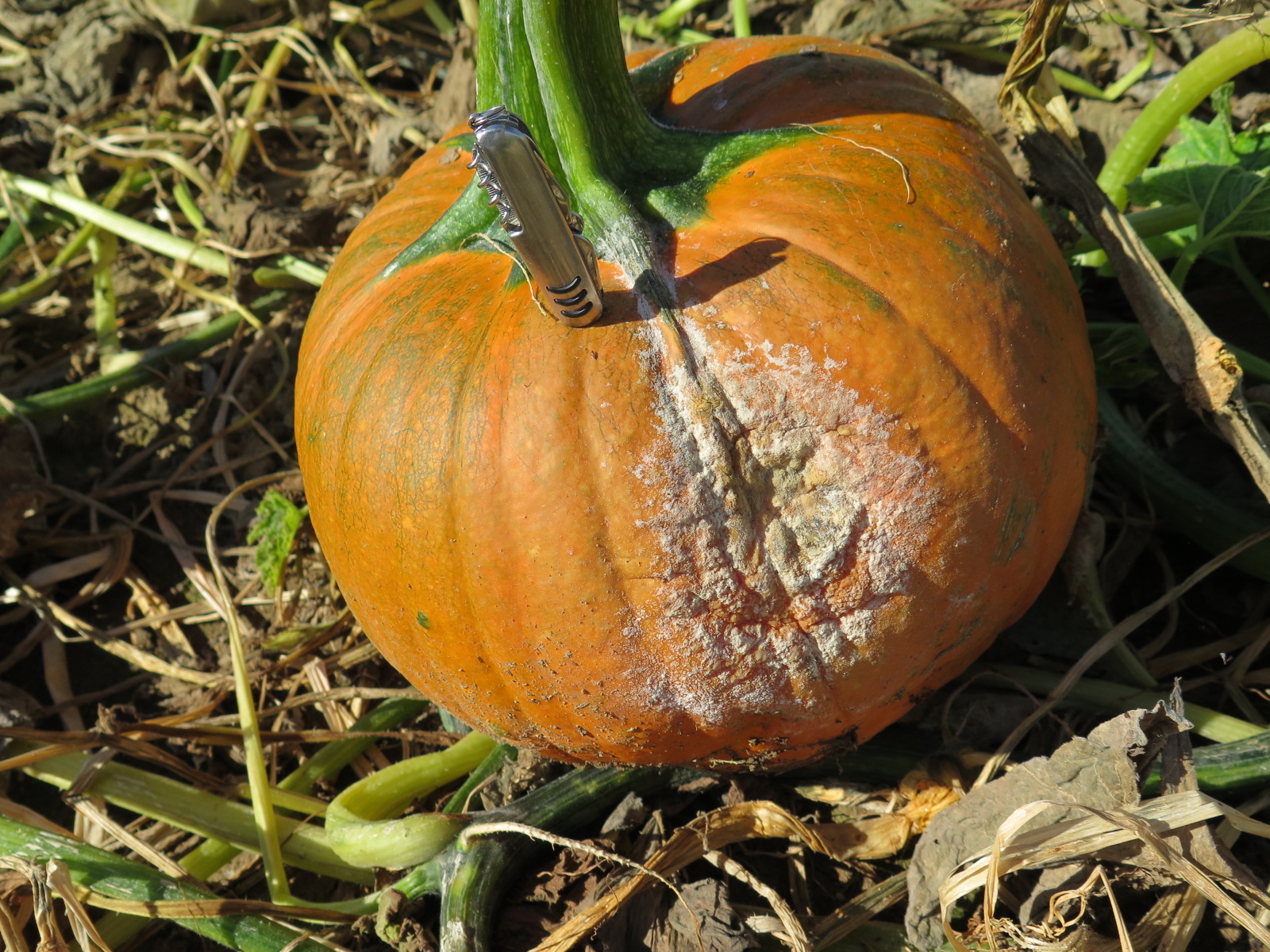 Figure 1. Phytophthora fruit rot of pumpkin.
 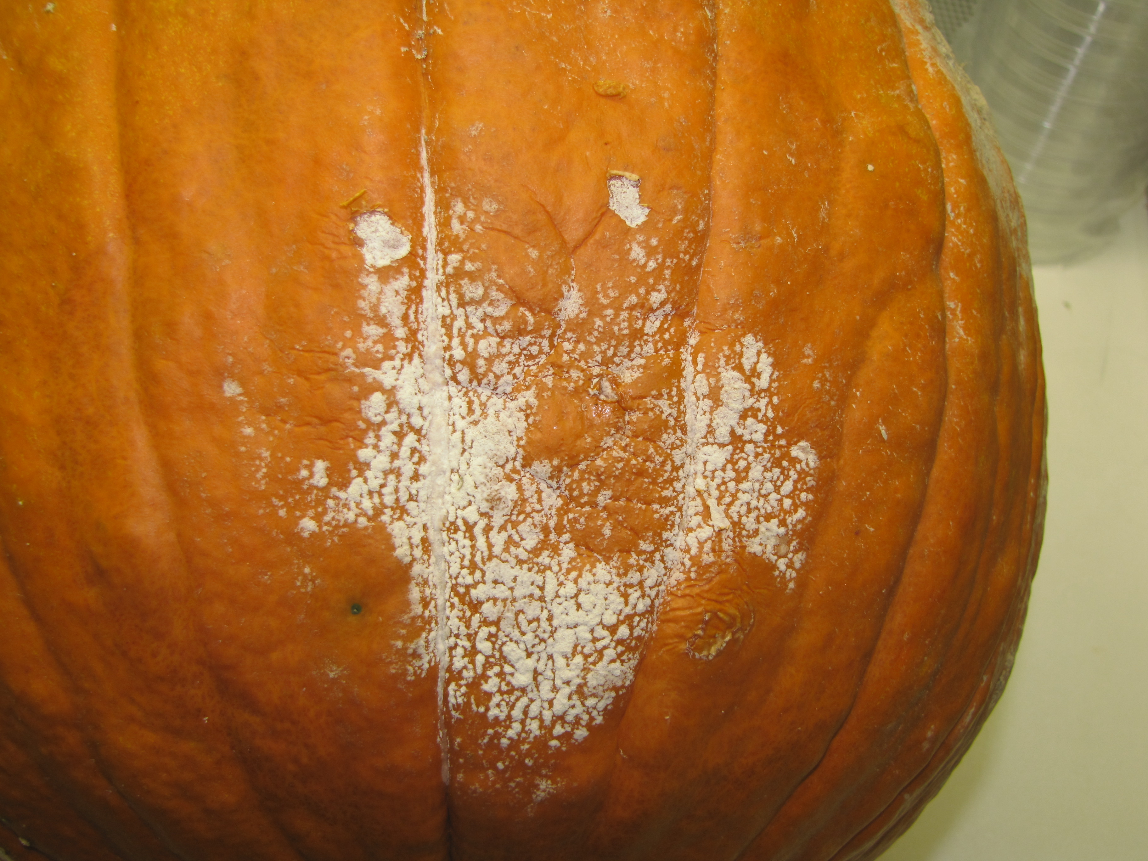 Figure 2. Phytophthora fruit rot of pumpkin.
 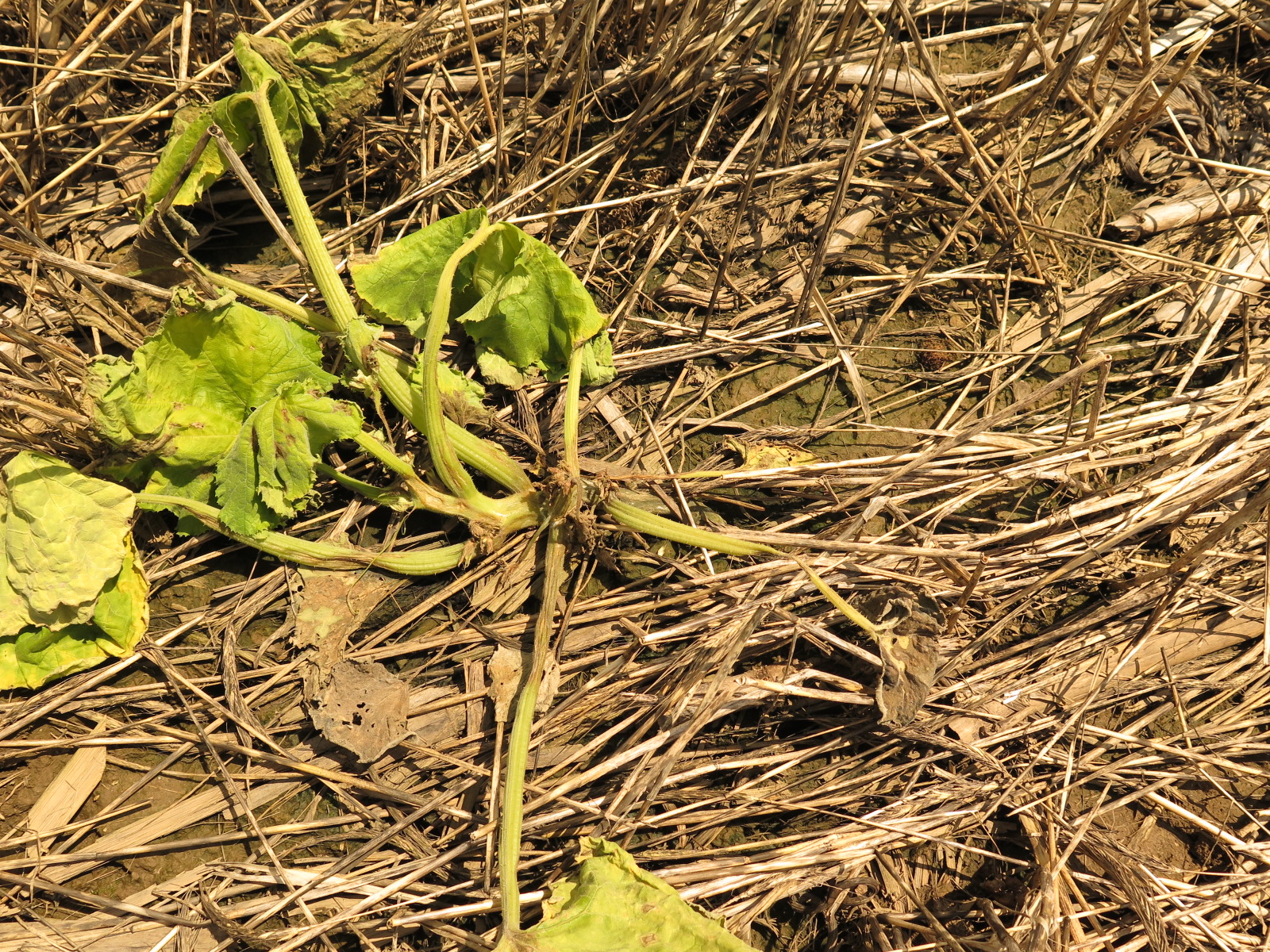 Figure 3. Phytophthora damping off of pumpkin.
 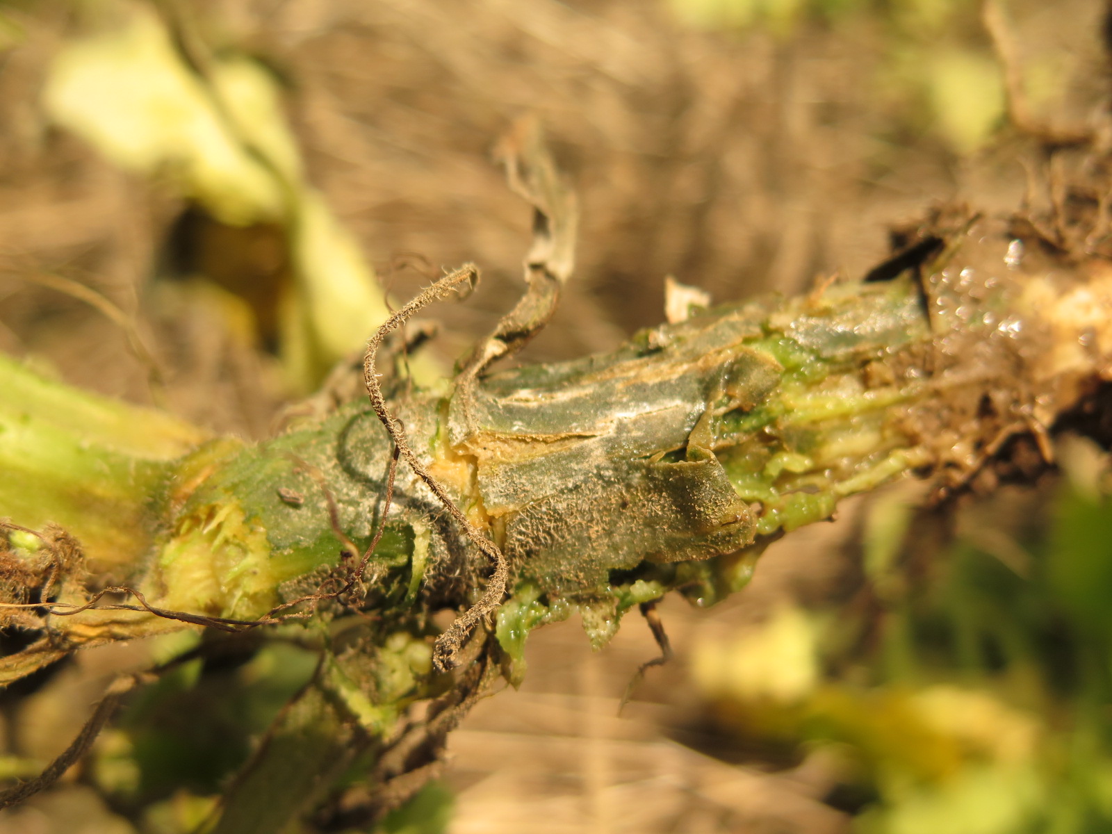 Figure 4. Phytophthora blight of pumpkin on crown.
 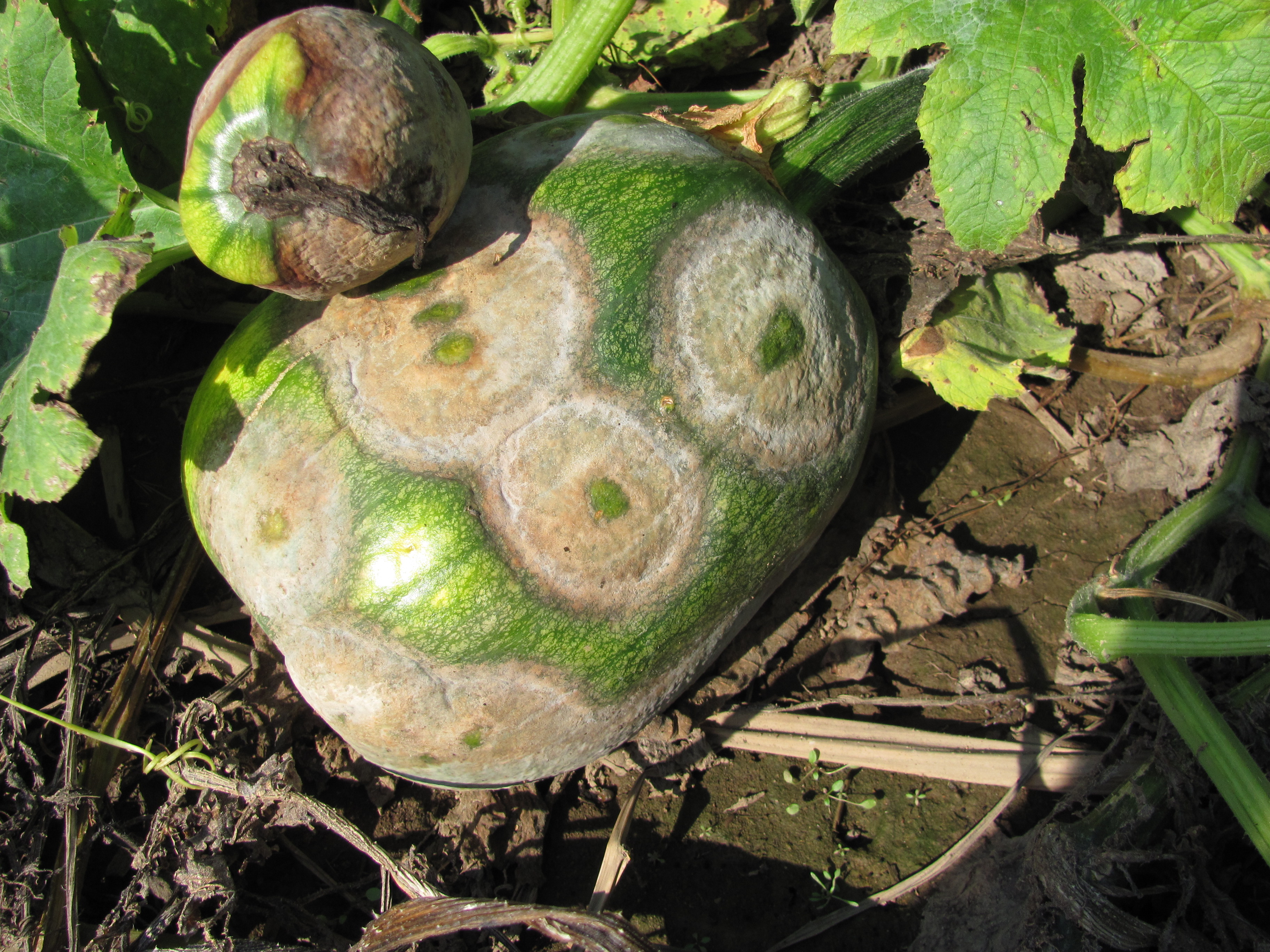 Figure 5. Phytophthora fruit rot.
  Figure 6. Phytophthora fruit rot.
  Figure 7. Phytophthora fruit rot. Growth and sporulation of causal fungus is often on the underside of fruit due to increased moisture.
 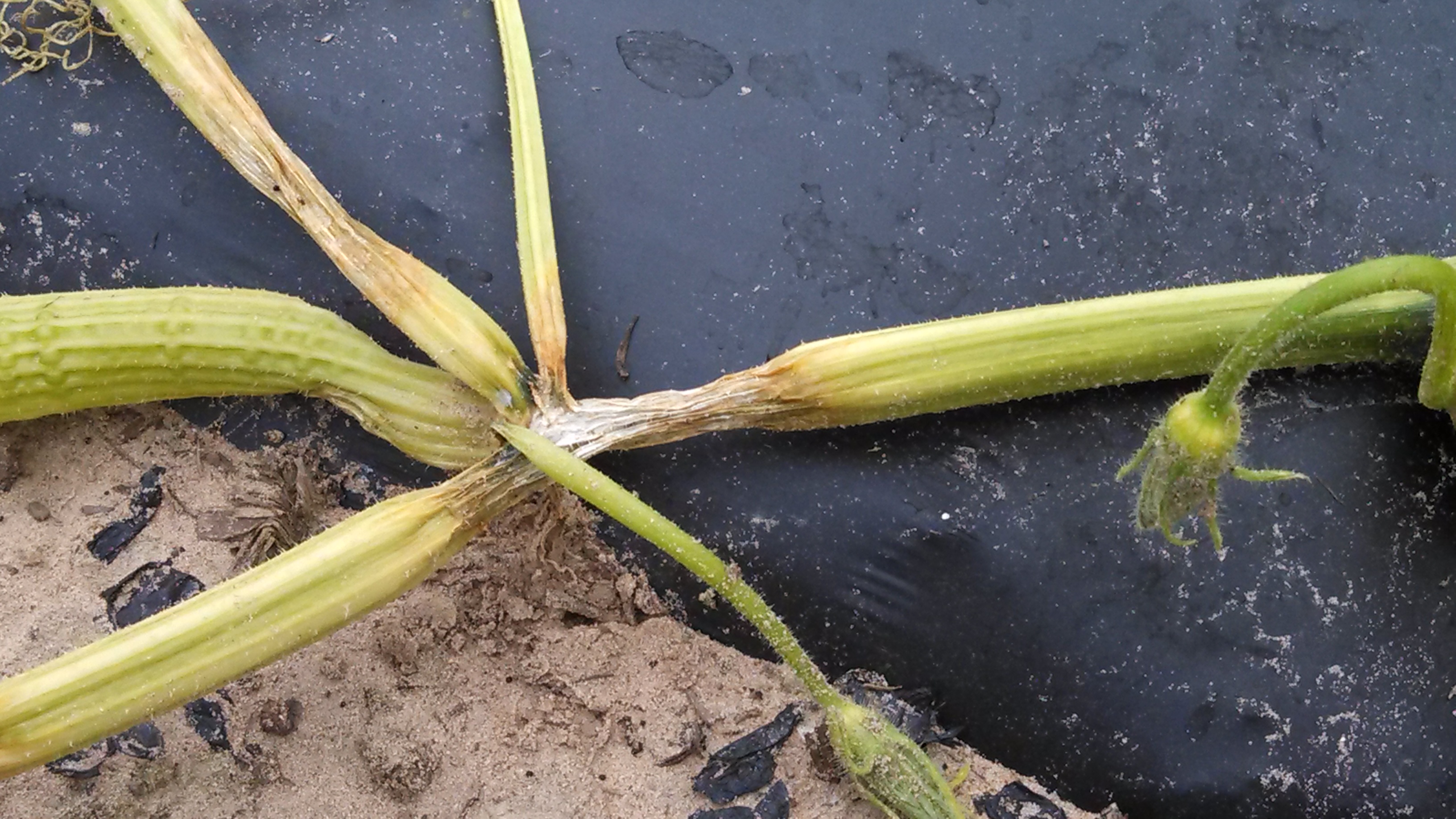 Figure 8. Phytophthora blight lesion on stem.
 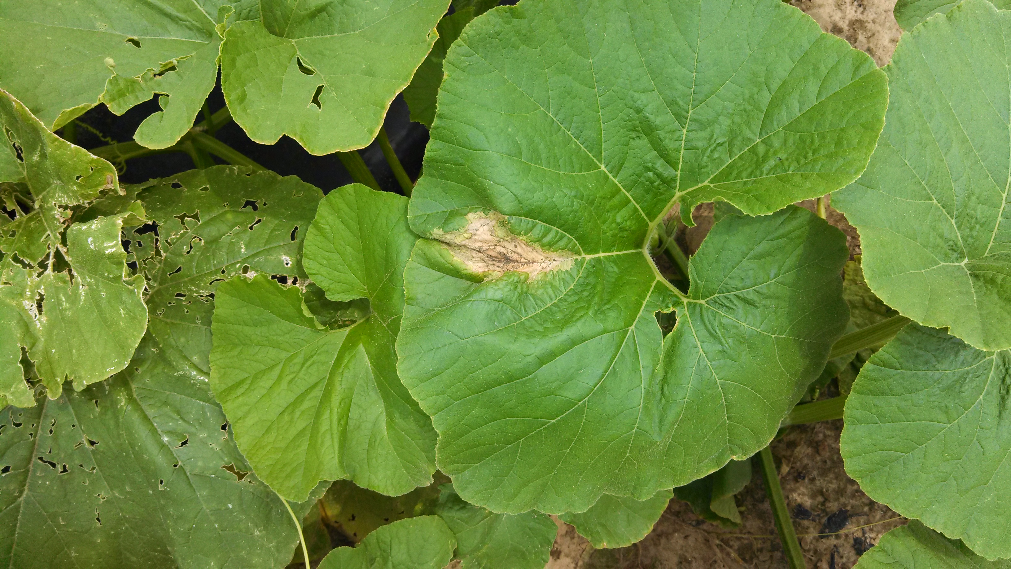 Figure 9. Phytophthora blight lesion on stem.
 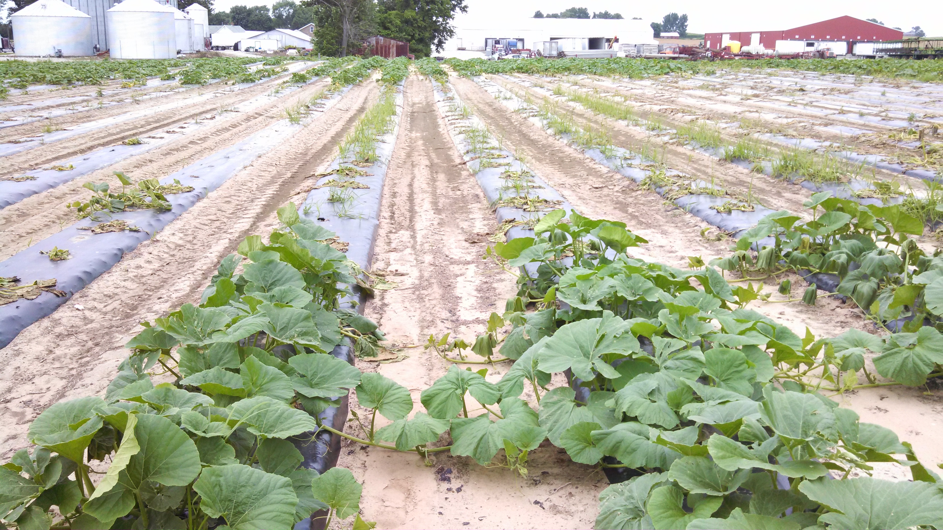 Figure 10. Phytophthora blight has affected the pumpkin plants in the lower area of this field. Note the wilted and dead plants in the low area shown here.
 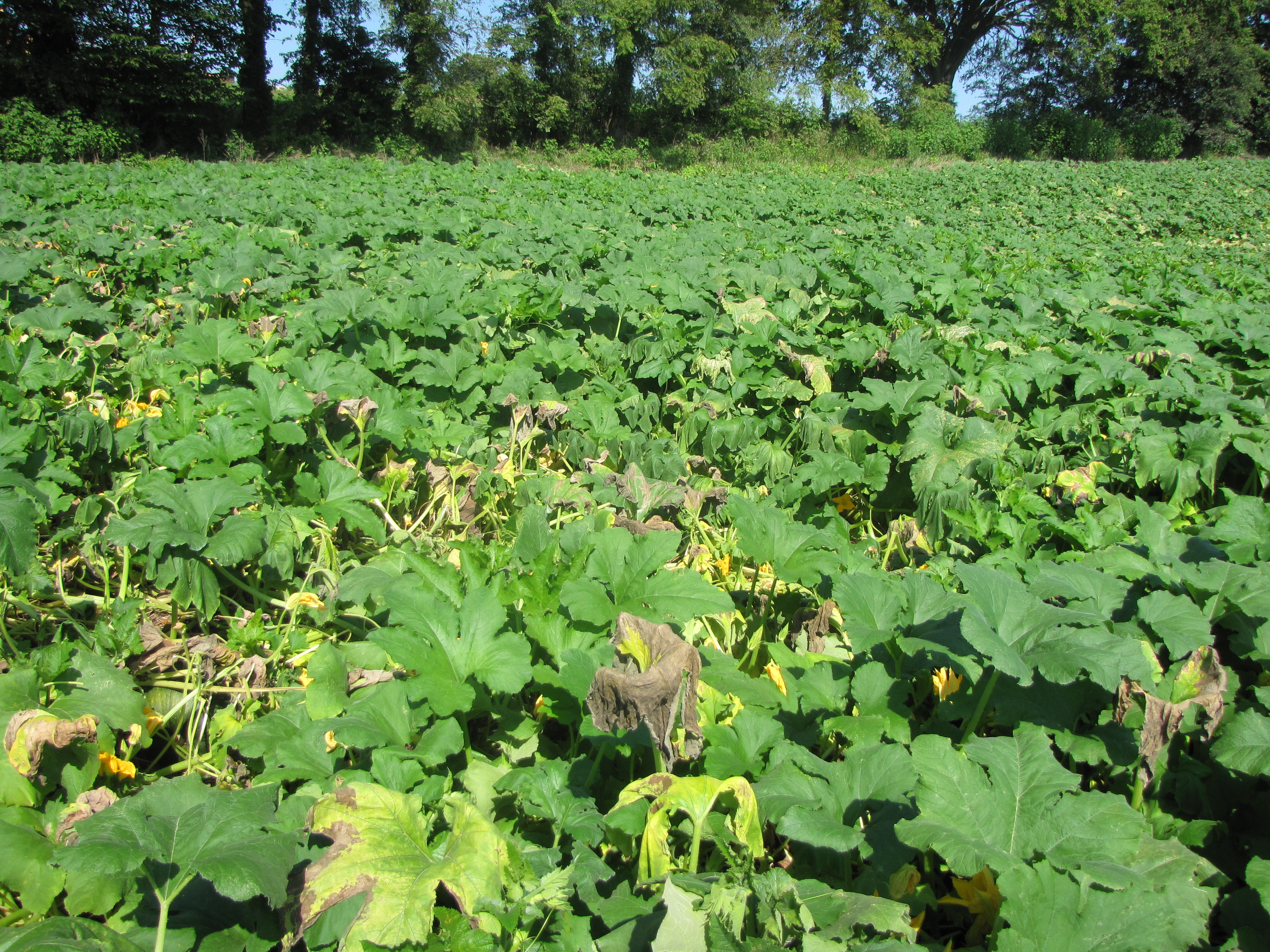 Figure 11. Often the first symptom of Phytophthora blight of pumpkins is the wilt and decline of plants caused by lesions on stems.
 Plectosporium blight
Plectosporium blight of pumpkin-Spindle shaped, white or off-white lesions may be observed on the stem including the area known as the handle of the pumpkin. In severe cases, the fruit may be covered with scabby lesions. The occurrence of this disease is sporadic: although the disease is not common, when it appears, it can prove economically important.
 Figure 1. Plectosporium blight of pumpkin. Lesions are most common on the handle or lower stem.
 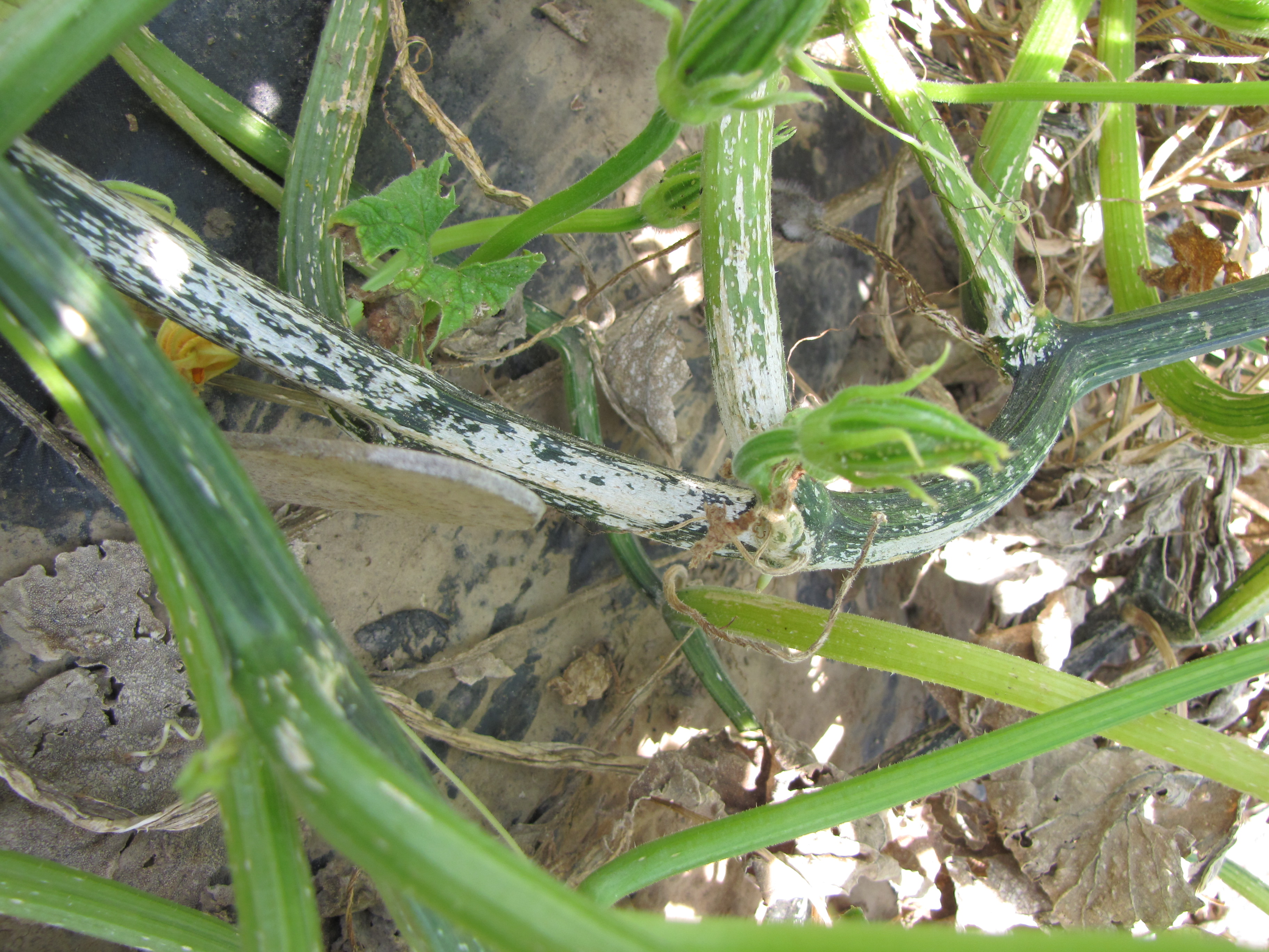 Figure 2. Plectosporium blight of pumpkin on stem.
 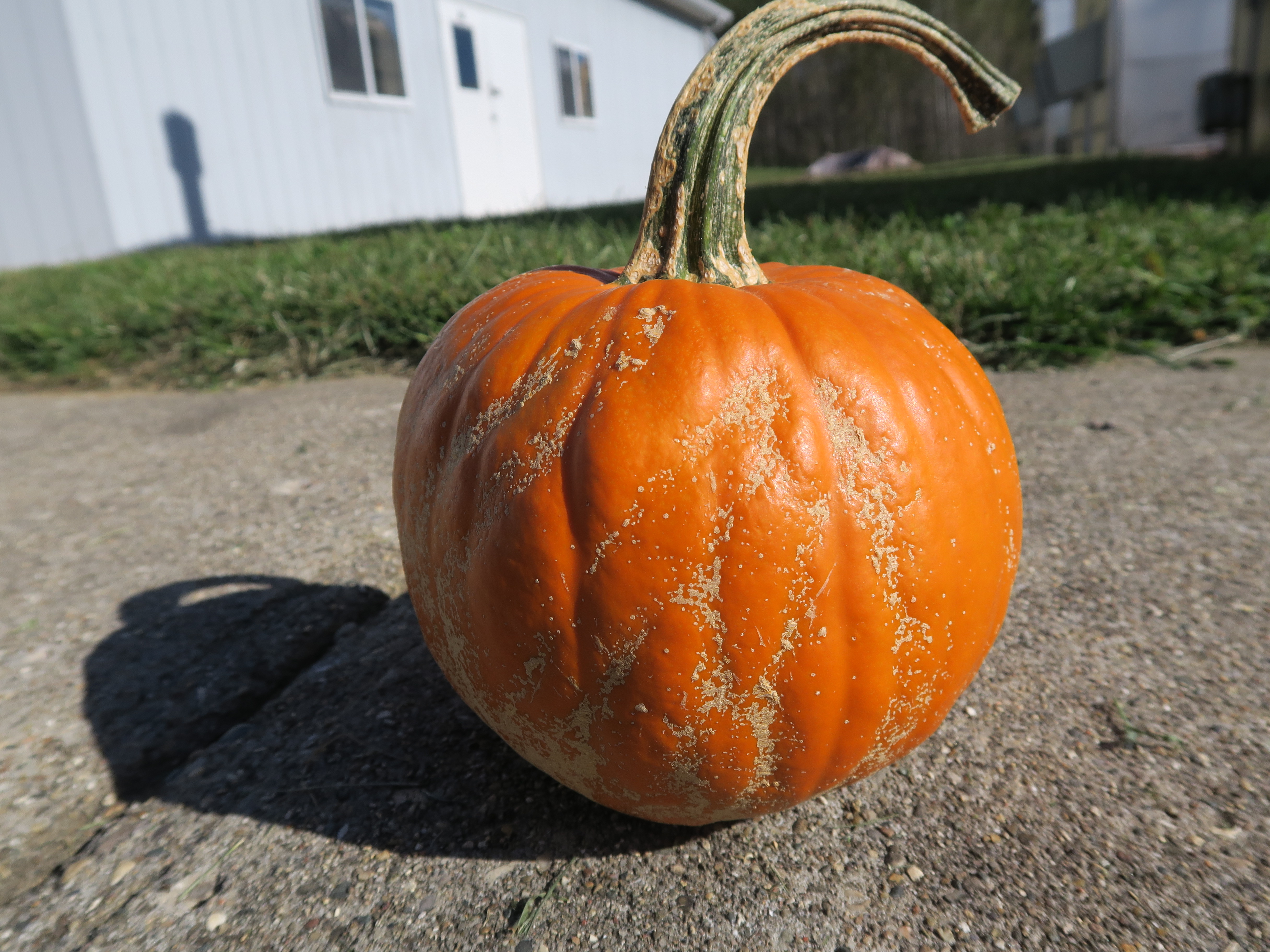 Figure 3. Plectosporium blight of pumpkin.
 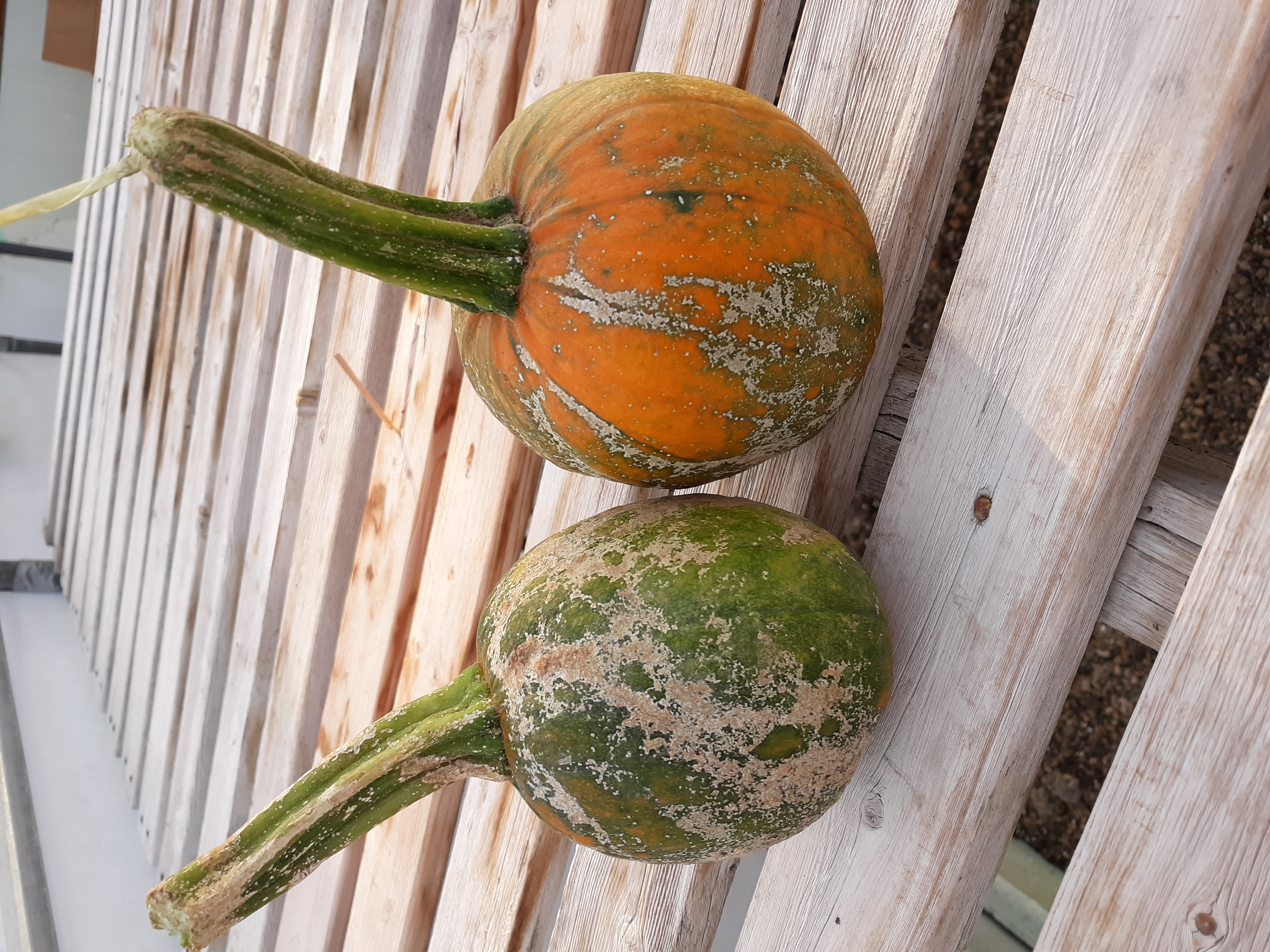 Figure 4. Plectosporium blight of pumpkin.
 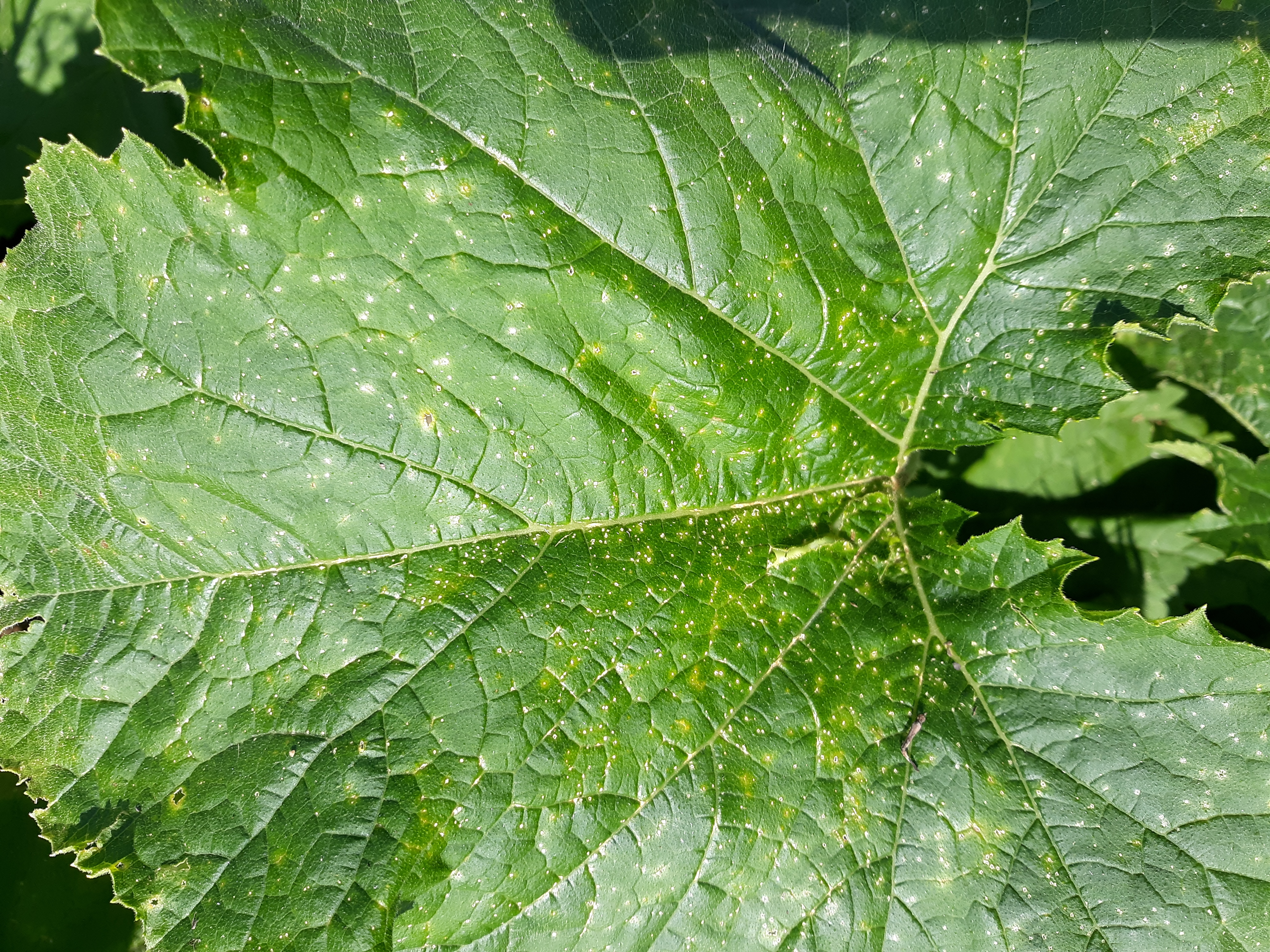 Figure 5. Plectosporium blight of pumpkin on leaf.
 Powdery mildew
Powdery mildew of pumpkin-Powdery mildew of pumpkins is easily recognized and may be observed to some extent in most pumpkin plantings. While powdery mildew is usually not severe, pumpkins that are not managed for this disease may have lower yields and/or lower quality fruit. Most growers use systemic fungicides and varieties of pumpkin with partial resistance to powdery mildew.
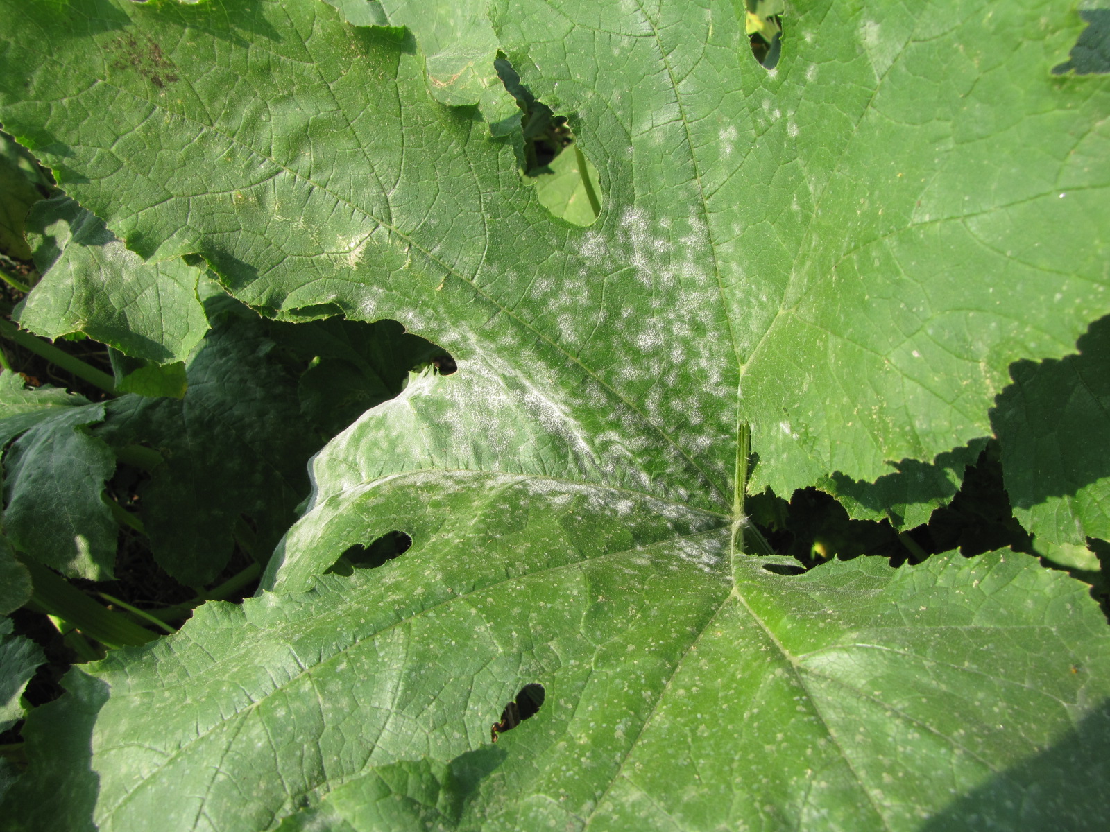 Figure 1. Powdery mildew of pumpkins can be easily recognized by the talc-like lesion on the upper and lower surface of leaves.
 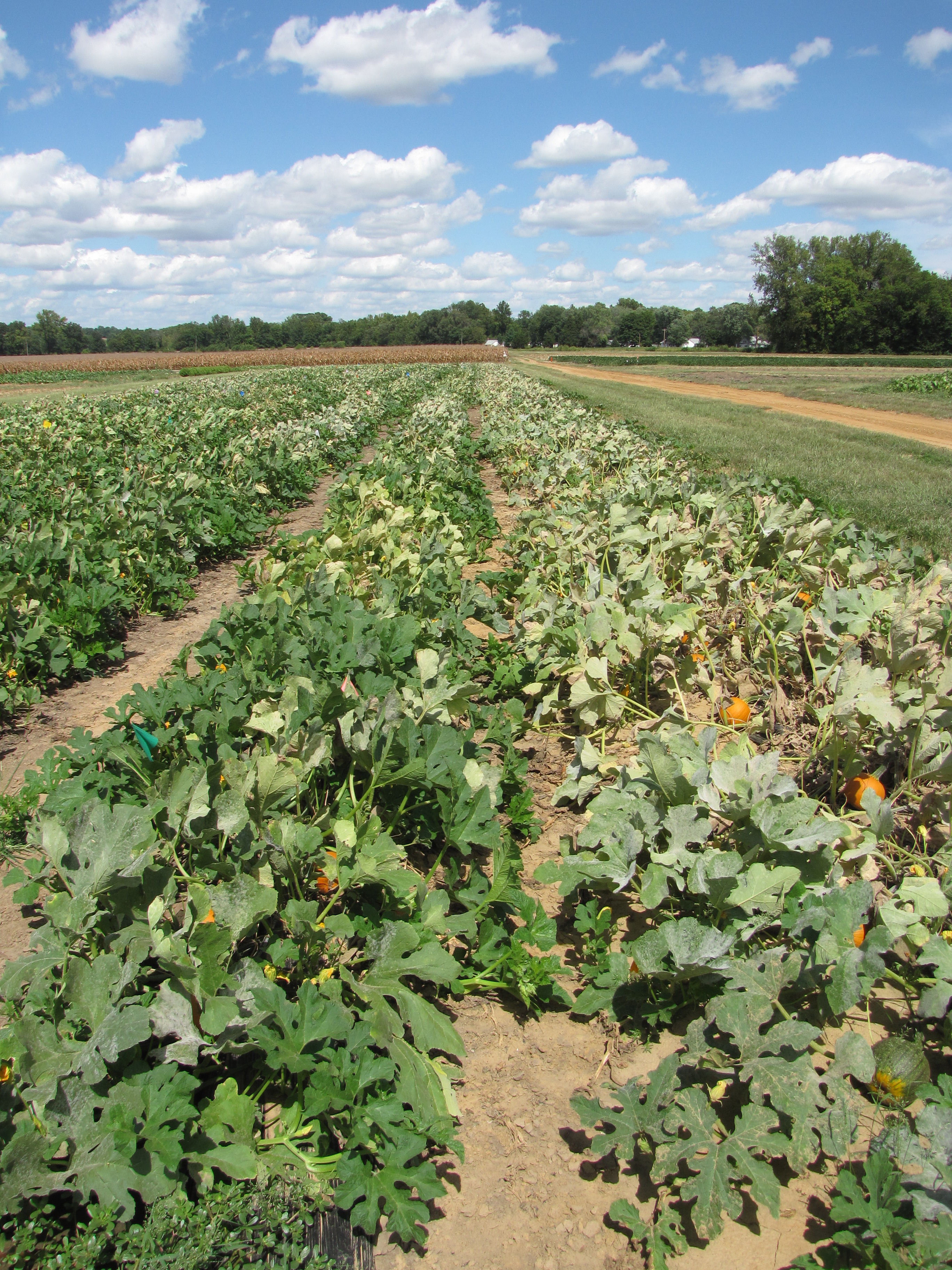 Figure 2. A fungicide trial for products for powdery mildew of pumpkin. The untreated row on the right has significant symptoms of powdery mildew. The adjacent row to the left has been treated with a systemic fungicide and has relatively mild symptoms.
 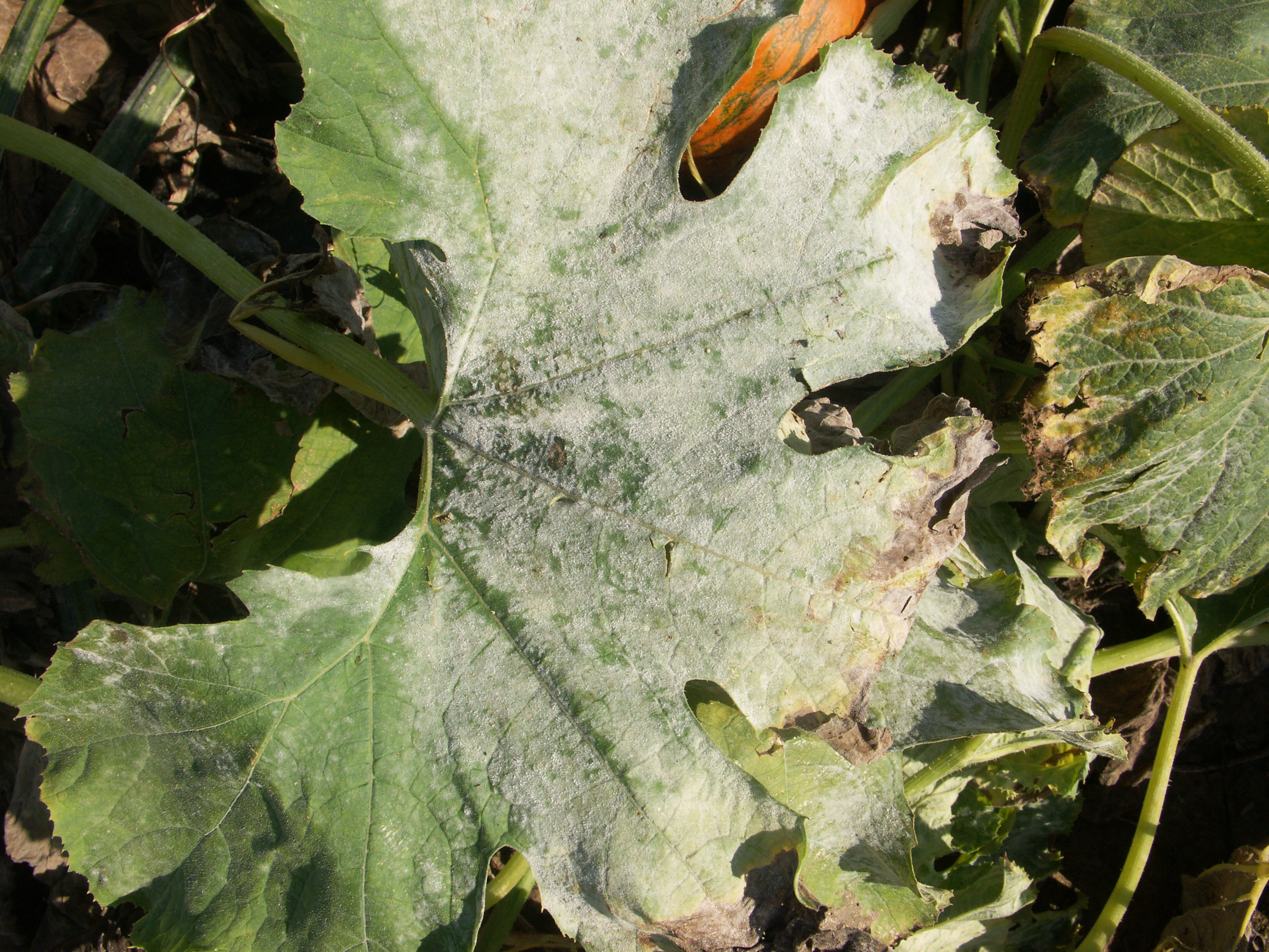 Figure 3. Severe symptoms of powdery mildew on a pumpkin leaf.
 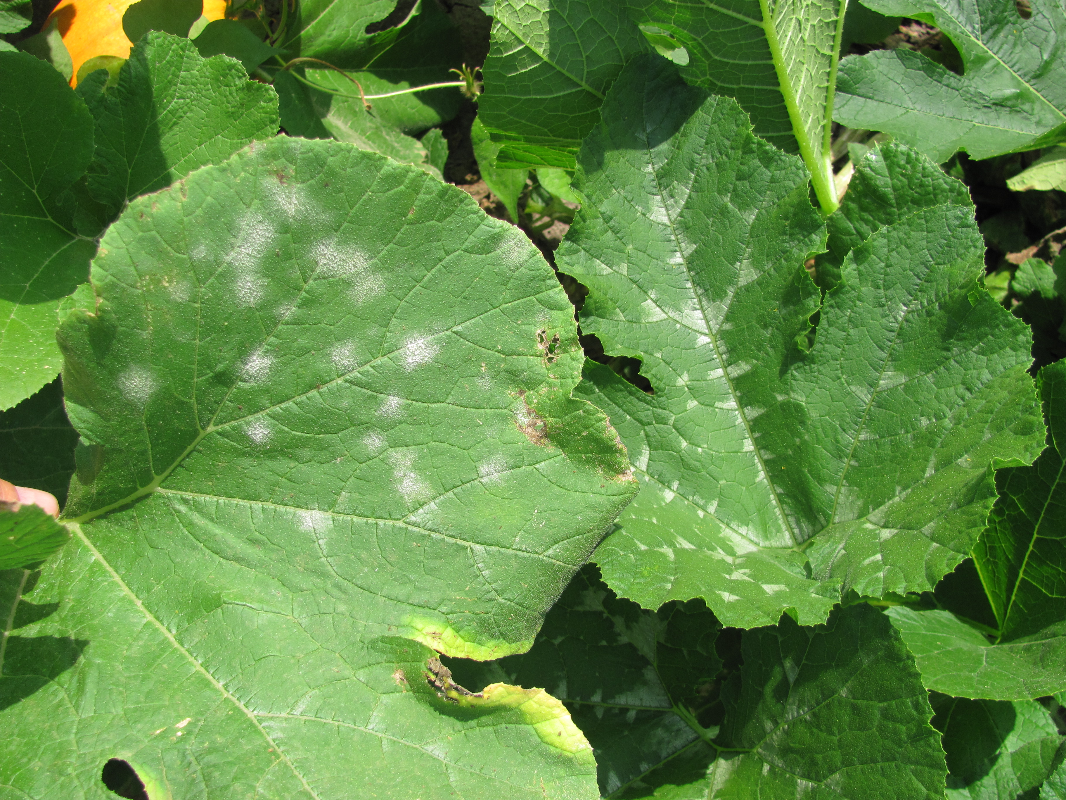 Figure 4. Powdery mildew lesions can be observed on the lower leaf in this photo. The upper leaf has light colored variegation that are sometimes mistaken for powdery mildew.
 Virus
Virus-The most common virus diseases of pumpkins in Indiana are aphid borne potyviruses. Symptoms on foliage may include mosaic and shoestring leaves. Pumpkin fruit may be undersized, misshapen, and/or display uneven ripening patterns. The importance of Potyviruses in Indiana depends on when symptoms appear in relation to fruit set. Early infections are more likely to lead to yield or quality loss.
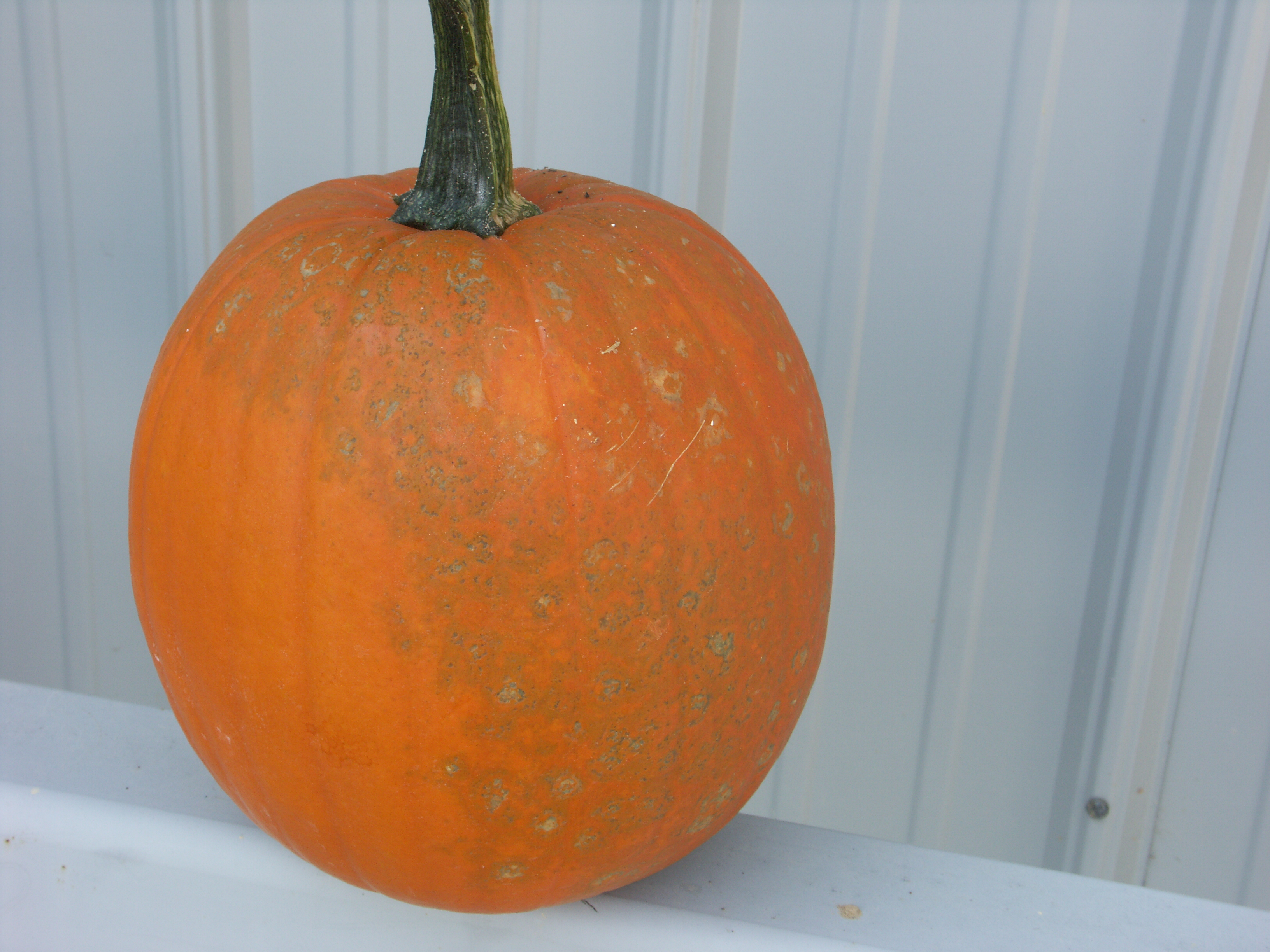 Figure 1. This pumpkin tested positive for Watermelon mosaic virus 2 and zucchini mosaic virus, both poty viruses. Note the sunken, gray, mostly circular lesions.
 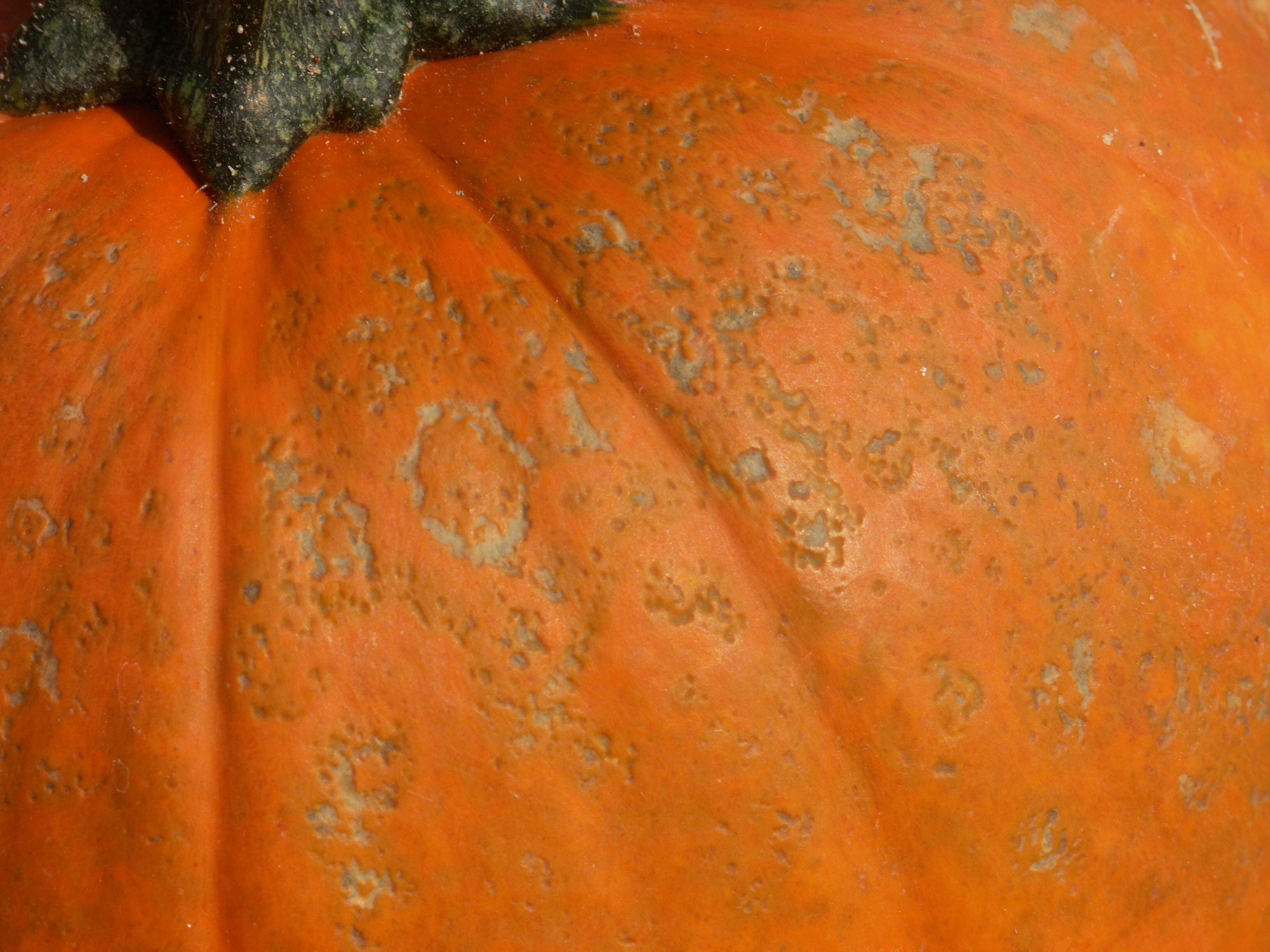 Figure 2. Close up of pumpkin in figure 1.
 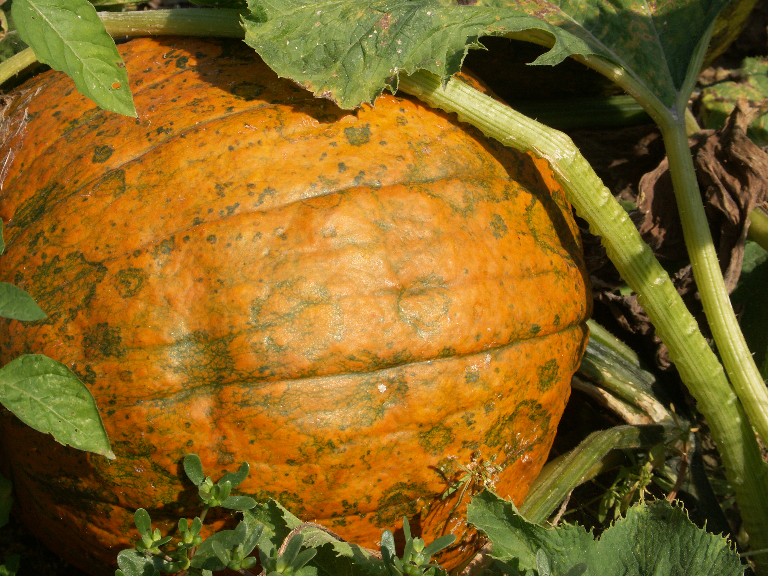 Figure 3. Poty virus on pumpkin.
 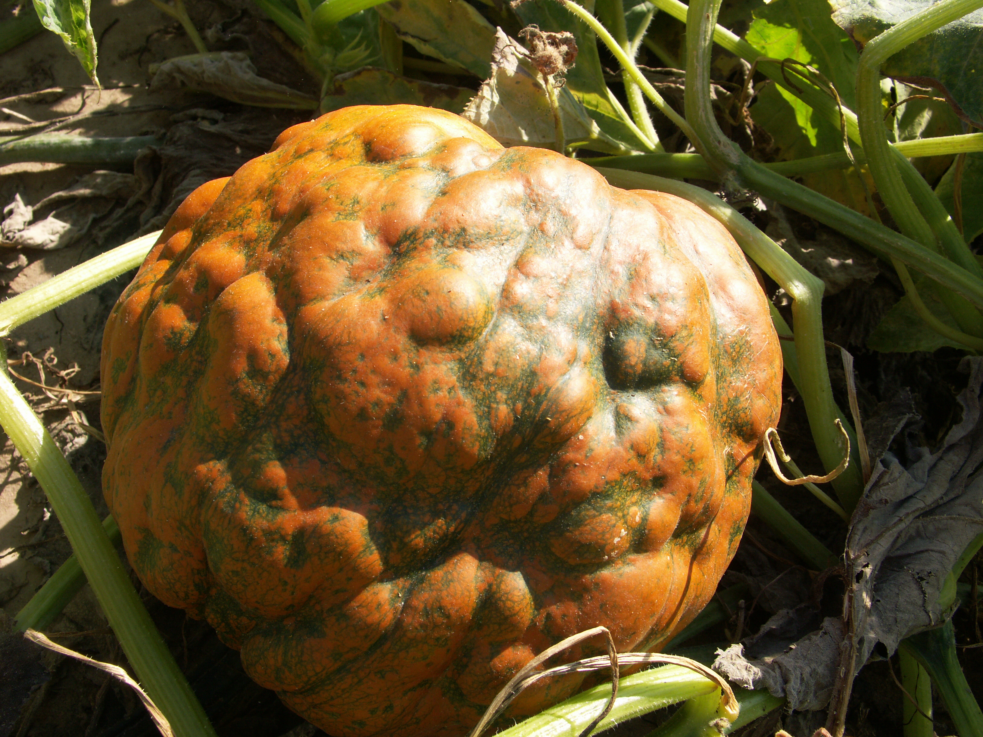 Figure 4. Poty virus on pumpkin.
  Figure 5. Pumpkins affected by papaya ringspot virus, a potyvirus.
 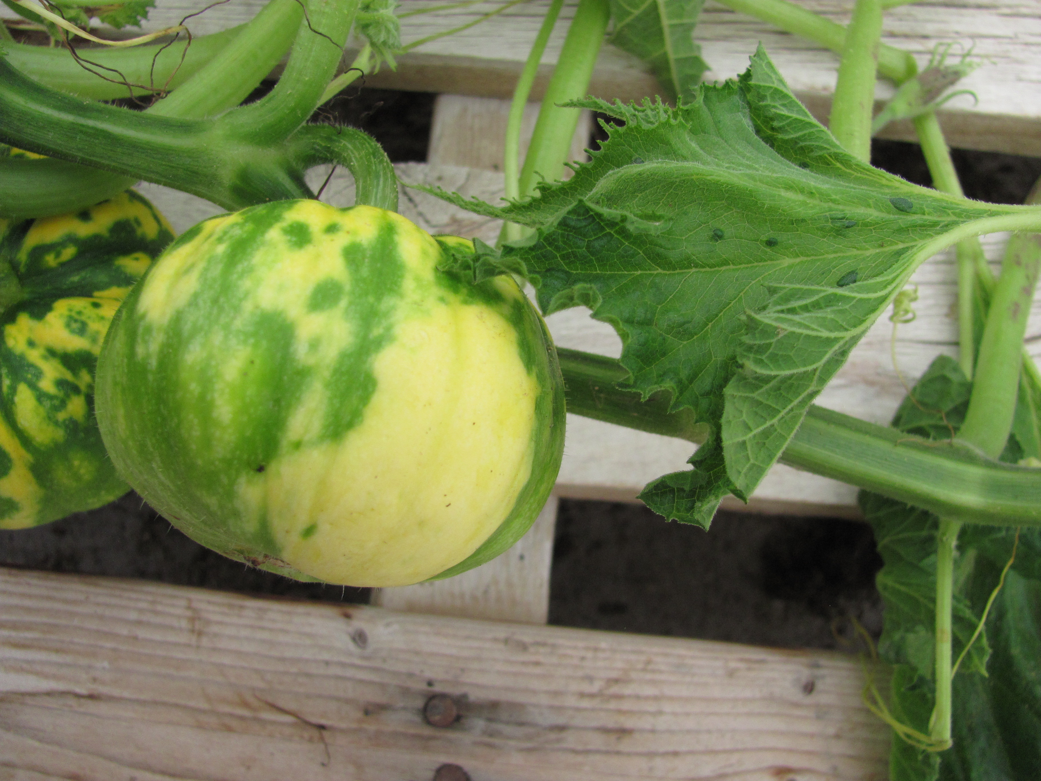 Figure 6. Pumpkins affected by papaya ringspot virus, a potyvirus.
 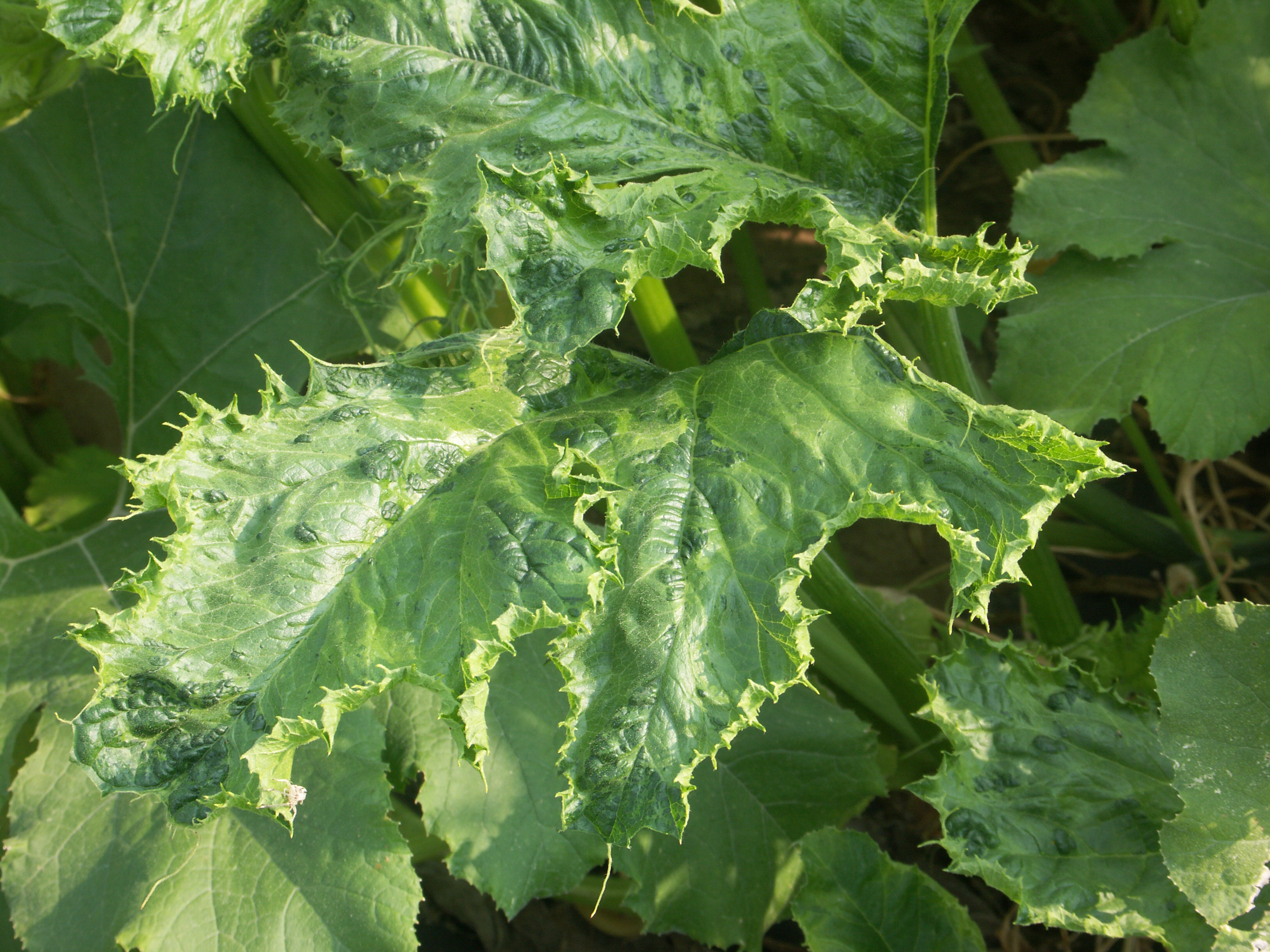 Figure 7. Pumpkin leaf with a poty virus.
 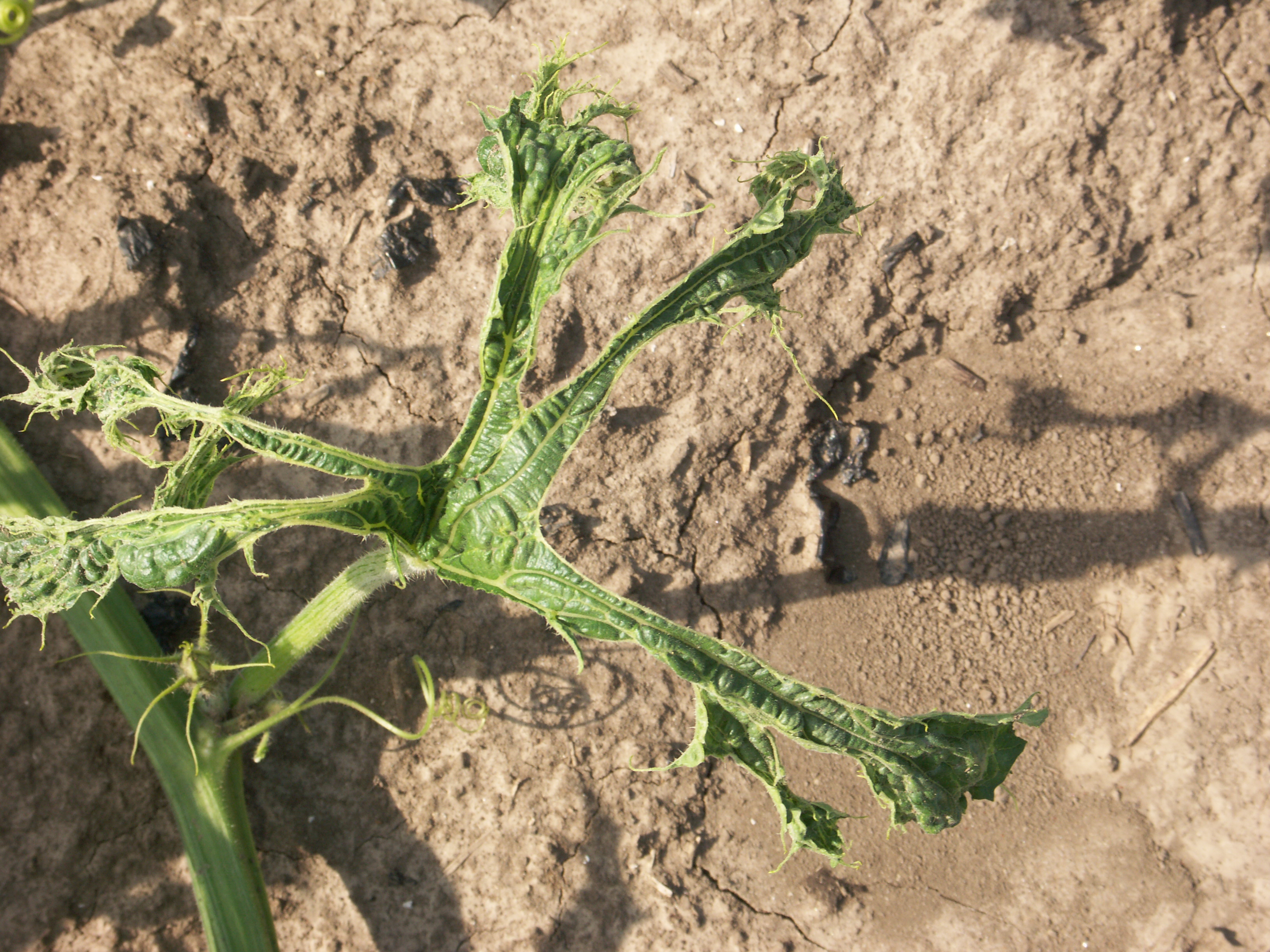 Figure 8. Pumpkin leaf with a poty virus.
 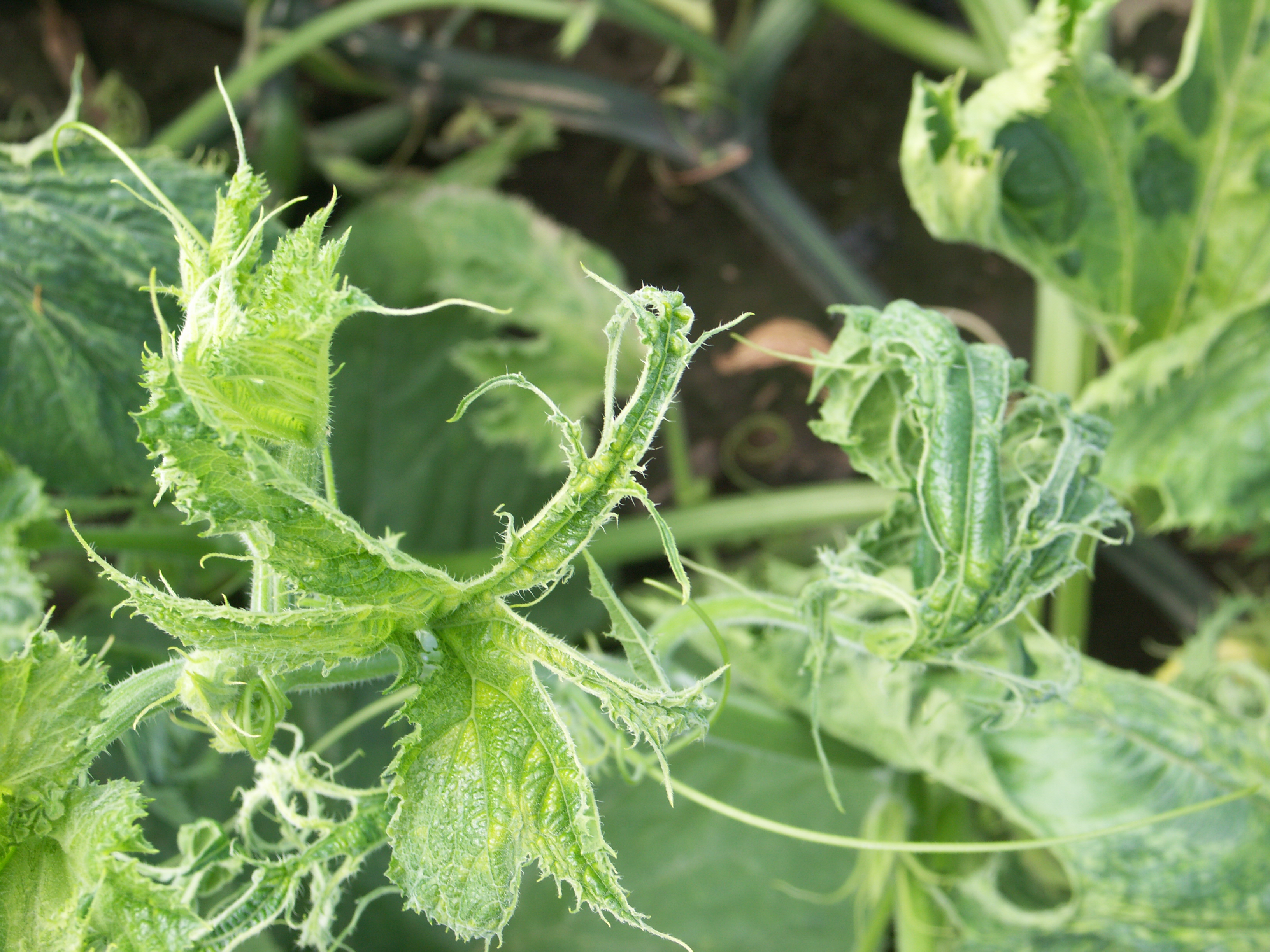 Figure 9. Pumpkin leaf with a poty virus.