

Annual Plant Art Competition
Every year the Department of Botany and Plant Pathology has a Plant Art Competition, highlighting all kinds of plant photos by our faculty, staff, post docs, graduate students, and undergraduate students. You can see this year's finalists below. The Community Favorite will be voted on this during Spring Fest, so stop by Lilly Hall on Saturday, April 15 to vote for your favorite!
2023 Spring Fest Community Favorite
"Emeralds and Sapphires" by Janna Beckerman.
Salvia and other members of the Lamiaceae (mint family) are favored by humans and hummingbirds.
Salvia and other members of the Lamiaceae (mint family) are favored by humans and hummingbirds.
Plant Art Finalists - 2023
 "Delicate Decay" by Daniel Raudabaugh.
Delicate fruit bodies of Coprinus lagopus. Saprophytic fungi are important for the carbon cycle allowing for the break-down of complex carbon sources and releasing locked up nutrients for the use of organisms including plants.
"Delicate Decay" by Daniel Raudabaugh.
Delicate fruit bodies of Coprinus lagopus. Saprophytic fungi are important for the carbon cycle allowing for the break-down of complex carbon sources and releasing locked up nutrients for the use of organisms including plants.  "When you see the Southern rust for the first time..." by Janna Beckerman.
Macro-photography. (1:1) image of rust infected oxalis, the alternate host of Southern rust.
"When you see the Southern rust for the first time..." by Janna Beckerman.
Macro-photography. (1:1) image of rust infected oxalis, the alternate host of Southern rust.  "A fern gametophyte (Green: nuclei; Magenta: cell walls)" by Yuan Geng.
This is 3D projection view of a living fern gametophyte. This image was created by using a confocal microscope.
"A fern gametophyte (Green: nuclei; Magenta: cell walls)" by Yuan Geng.
This is 3D projection view of a living fern gametophyte. This image was created by using a confocal microscope. 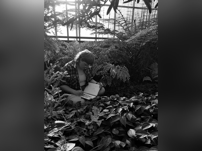 "Natural Habitat of a Botanist" by Julia Peterson.
This is a picture taken on a phone camera with a black and white filter. It is related in that this is one of Purdue's plant science students in the Lilly greenhouses that are to be demolished next winter.
"Natural Habitat of a Botanist" by Julia Peterson.
This is a picture taken on a phone camera with a black and white filter. It is related in that this is one of Purdue's plant science students in the Lilly greenhouses that are to be demolished next winter. 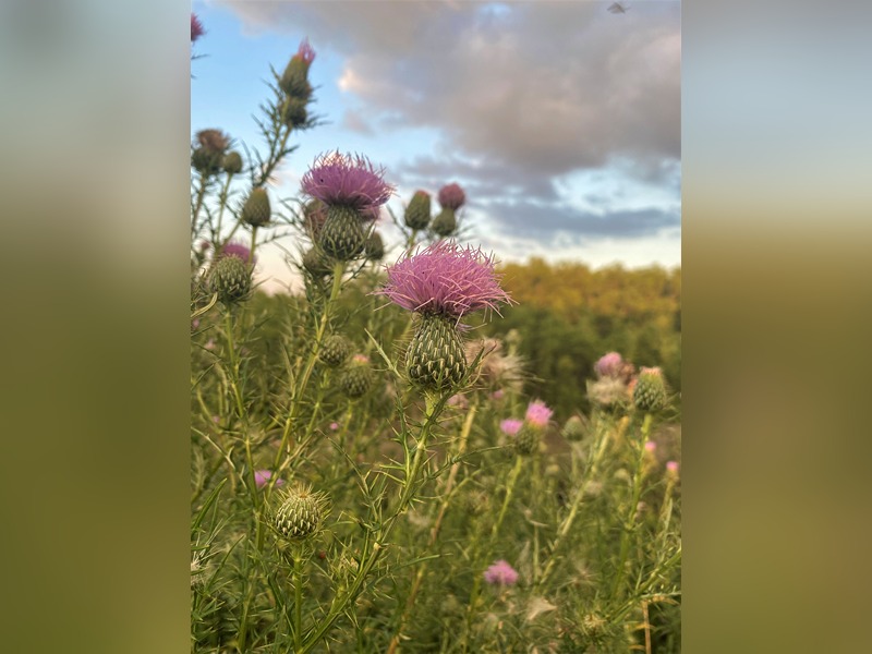 "Canada Thistle Flowering" by Marcelo Zimmer.
Canada thistle is a troublesome perennial weed that infests pastures, roadsides, and sometimes row crops.
Control requires the use of herbicides with systemic action such as synthetic auxins.
"Canada Thistle Flowering" by Marcelo Zimmer.
Canada thistle is a troublesome perennial weed that infests pastures, roadsides, and sometimes row crops.
Control requires the use of herbicides with systemic action such as synthetic auxins. 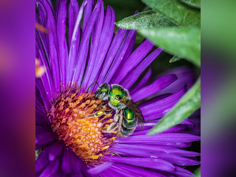 "Nature is metal" by Janna Beckerman.
Macrophotography (1:1)f16, 1/250. Asters are important fall pollen sources for migrating butterflies and hummingbirds, along with many bees. The halictid bees are responsible for over 80% of the pollination of flowers
"Nature is metal" by Janna Beckerman.
Macrophotography (1:1)f16, 1/250. Asters are important fall pollen sources for migrating butterflies and hummingbirds, along with many bees. The halictid bees are responsible for over 80% of the pollination of flowers 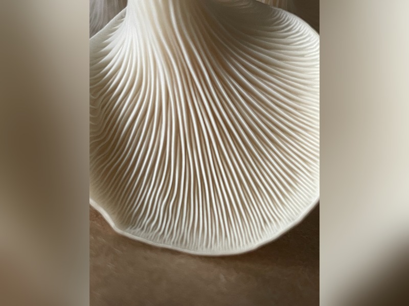 "Gills" by Morgan Murff.
Oyster mushroom gills from BTNY 207: The Microbial World
"Gills" by Morgan Murff.
Oyster mushroom gills from BTNY 207: The Microbial World 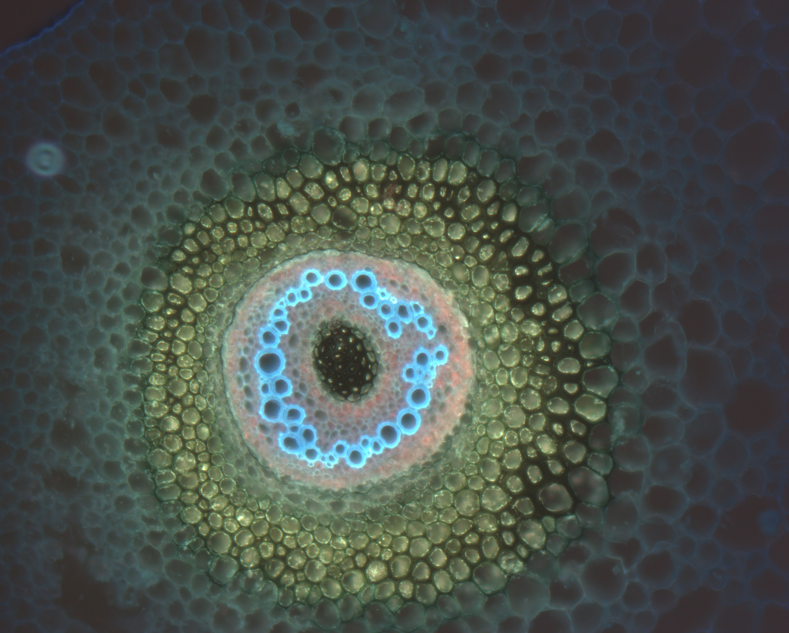 "Marsilea hirsuta L. - transversal section through the rhizome" by Yuliia Khoma
Marsilea hirsuta L., is a semi-aquatic fern. To obtain the material cross sections of the rhizome of M. hirsuta with a thickness of 20 μm were made on a microtome. Images of sections were taken at ×10 magnification using a fluorescence microscope (Zeiss Axio Scope).
"Marsilea hirsuta L. - transversal section through the rhizome" by Yuliia Khoma
Marsilea hirsuta L., is a semi-aquatic fern. To obtain the material cross sections of the rhizome of M. hirsuta with a thickness of 20 μm were made on a microtome. Images of sections were taken at ×10 magnification using a fluorescence microscope (Zeiss Axio Scope). 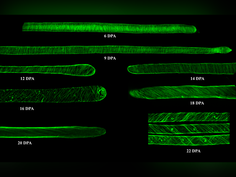 "The dynamic changes in wall microfibril orientation during cotton fiber development" by Heena Rani
How it was done?
Cotton bolls were dissected to isolate intact ovules, the fibers were detangled by treating the ovules with 1% (v/v) Nonidet-40 (80 °C, 20 min) followed by 0.025 M HCl (30 s). Ovules were then placed in 10 mM PBS (pH 7) to relax the fibers. Fibers were stained using calcofluor white stain for 10 min before imaging.
Material used:
Crop and Variety: Gossypium hirsutum (TM-1)
Stain: Calcofluor white stain (Fluorescent Brightener 28 (FB28))
Technique: Confocal laser scanning microscopy
Magnification: 63X
How it relates to the disciplines of plant biology:
Cotton fibers are single-celled trichomes, and their development can be divided into four different but partial overlapping stages: initiation, elongation, secondary cell wall biosynthesis, and maturation.
During the elongation phase (after 3 weeks of initiation), newly deposited cellulose microfibrils at the inner surface of the primary wall are oriented perpendicularly to the long axis of the cell. The patterns of cellulose microfibrils in the wall control the direction of cell expansion and the ultimate morphology of the fiber.
During secondary cell wall synthesis (begins at 15 to 22 DPA and continues for 30-40 days), a second, thick, multi-layered wall which is composed almost exclusively of cellulose microfibrils is laid between the cytoplasm and the primary cell wall. The quantity of cellulose deposited during secondary cell wall synthesis along with the degree of polymerization of cellulose molecules organization and orientation of crystalline microfibrils directly link to fiber quality.
"The dynamic changes in wall microfibril orientation during cotton fiber development" by Heena Rani
How it was done?
Cotton bolls were dissected to isolate intact ovules, the fibers were detangled by treating the ovules with 1% (v/v) Nonidet-40 (80 °C, 20 min) followed by 0.025 M HCl (30 s). Ovules were then placed in 10 mM PBS (pH 7) to relax the fibers. Fibers were stained using calcofluor white stain for 10 min before imaging.
Material used:
Crop and Variety: Gossypium hirsutum (TM-1)
Stain: Calcofluor white stain (Fluorescent Brightener 28 (FB28))
Technique: Confocal laser scanning microscopy
Magnification: 63X
How it relates to the disciplines of plant biology:
Cotton fibers are single-celled trichomes, and their development can be divided into four different but partial overlapping stages: initiation, elongation, secondary cell wall biosynthesis, and maturation.
During the elongation phase (after 3 weeks of initiation), newly deposited cellulose microfibrils at the inner surface of the primary wall are oriented perpendicularly to the long axis of the cell. The patterns of cellulose microfibrils in the wall control the direction of cell expansion and the ultimate morphology of the fiber.
During secondary cell wall synthesis (begins at 15 to 22 DPA and continues for 30-40 days), a second, thick, multi-layered wall which is composed almost exclusively of cellulose microfibrils is laid between the cytoplasm and the primary cell wall. The quantity of cellulose deposited during secondary cell wall synthesis along with the degree of polymerization of cellulose molecules organization and orientation of crystalline microfibrils directly link to fiber quality.